|
Ganglion Cyst
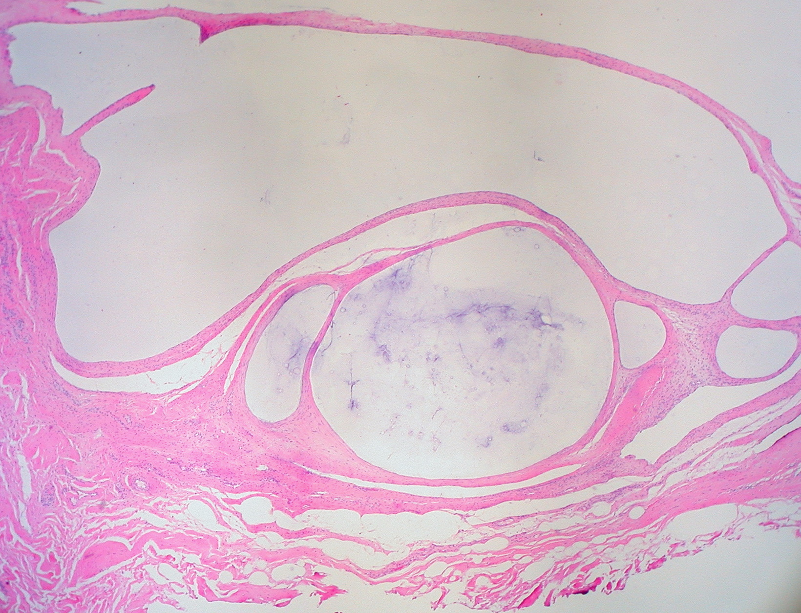
A ganglion cyst is a fluid-filled bump associated with a joint or tendon sheath. It most often occurs at the back of the wrist, followed by the front of the wrist. Onset is often over several months, typically with no further symptoms. Occasionally, pain or numbness may occur. Complications may include carpal tunnel syndrome. The cause is unknown. The underlying mechanism is believed to involve an outpouching of the synovial membrane. Risk factors include gymnastics activity. Diagnosis is typically based on examination with light shining through the lesion being supportive. Medical imaging may be done to rule out other potential causes. Treatment options include watchful waiting, splinting the affected joint, needle aspiration, or surgery. About half the time, they resolve on their own. About three per 10,000 people newly develop ganglion of the wrist or hand a year. They most commonly occur in young and middle-aged females. Presentation The average size of these cysts is 2.0 ... [...More Info...] [...Related Items...] OR: [Wikipedia] [Google] [Baidu] |
Ganglion
A ganglion is a group of neuron cell bodies in the peripheral nervous system. In the somatic nervous system this includes dorsal root ganglia and trigeminal ganglia among a few others. In the autonomic nervous system there are both sympathetic and parasympathetic ganglia which contain the cell bodies of postganglionic sympathetic and parasympathetic neurons respectively. A pseudoganglion looks like a ganglion, but only has nerve fibers and has no nerve cell bodies. Structure Ganglia are primarily made up of somata and dendritic structures which are bundled or connected. Ganglia often interconnect with other ganglia to form a complex system of ganglia known as a plexus. Ganglia provide relay points and intermediary connections between different neurological structures in the body, such as the peripheral and central nervous systems. Among vertebrates there are three major groups of ganglia: *Dorsal root ganglia (also known as the spinal ganglia) contain the cell bodies of se ... [...More Info...] [...Related Items...] OR: [Wikipedia] [Google] [Baidu] |
Watchful Waiting
Watchful waiting (also watch and wait or WAW) is an approach to a medical problem in which time is allowed to pass before medical intervention or therapy is used. During this time, repeated testing may be performed. Related terms include ''expectant management'', ''active surveillance'', and ''masterly inactivity''. The term ''masterly inactivity'' is also used in nonmedical contexts. A distinction can be drawn between ''watchful waiting'' and ''medical observation'', but some sources equate the terms. Usually, watchful waiting is an outpatient process and may have a duration of months or years. In contrast, medical observation is usually an inpatient process, often involving frequent or even continuous monitoring and may have a duration of hours or days. Medical uses Often watchful waiting is recommended in situations with a high likelihood of self-resolution if there is high uncertainty concerning the diagnosis, and the risks of intervention or therapy may outweigh the benefit ... [...More Info...] [...Related Items...] OR: [Wikipedia] [Google] [Baidu] |
Gastrocnemius
The gastrocnemius muscle (plural ''gastrocnemii'') is a superficial two-headed muscle that is in the back part of the lower leg of humans. It runs from its two heads just above the knee to the heel, a three joint muscle (knee, ankle and subtalar joints). The muscle is named via Latin, from Greek γαστήρ (''gaster'') 'belly' or 'stomach' and κνήμη (''knḗmē'') 'leg', meaning 'stomach of the leg' (referring to the bulging shape of the calf). Structure The gastrocnemius is located with the soleus in the posterior (back) compartment of the leg. The lateral head originates from the lateral condyle of the femur, while the medial head originates from the medial condyle of the femur. Its other end forms a common tendon with the soleus muscle; this tendon is known as the calcaneal tendon or Achilles tendon and inserts onto the posterior surface of the calcaneus, or heel bone. It is considered a superficial muscle as it is located directly under skin, and its shape may often b ... [...More Info...] [...Related Items...] OR: [Wikipedia] [Google] [Baidu] |
Cruciate Ligaments
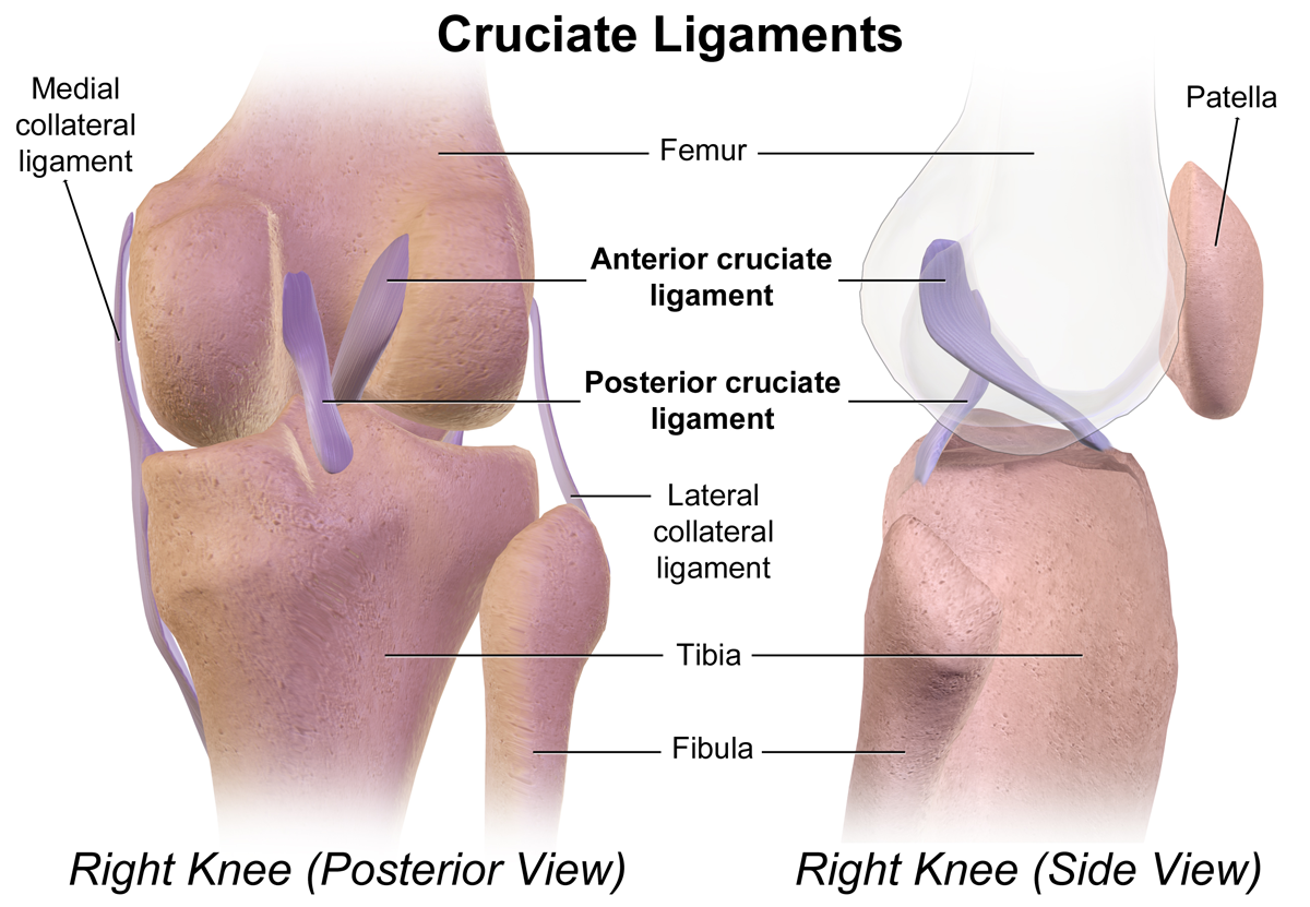
Cruciate ligaments (also cruciform ligaments) are pairs of ligaments arranged like a letter X. They occur in several joints of the body, such as the knee joint and the atlantoaxial joint, atlanto-axial joint. In a fashion similar to the cords in a toy Jacob's ladder (toy), Jacob's ladder, the crossed ligaments stabilize the joint while allowing a very large range of motion. Knee Structure Cruciate ligaments occur in the knee of humans and other bipedal animals and the corresponding Stifle joint, stifle of quadrupedal animals, and in the neck, fingers, and foot. * The cruciate ligaments of the knee are the anterior cruciate ligament (ACL) and the posterior cruciate ligament (PCL). These ligaments are two strong, rounded bands that extend from the head of the tibia to the intercondyloid notch of the femur. The ACL is lateral and the PCL is medial. They cross each other like the limbs of an X. They are named for their insertion into the tibia: the ACL attaches to the anterior ... [...More Info...] [...Related Items...] OR: [Wikipedia] [Google] [Baidu] |
Knee
In humans and other primates, the knee joins the thigh with the leg and consists of two joints: one between the femur and tibia (tibiofemoral joint), and one between the femur and patella (patellofemoral joint). It is the largest joint in the human body. The knee is a modified hinge joint, which permits flexion and extension as well as slight internal and external rotation. The knee is vulnerable to injury and to the development of osteoarthritis. It is often termed a ''compound joint'' having tibiofemoral and patellofemoral components. (The fibular collateral ligament is often considered with tibiofemoral components.) Structure The knee is a modified hinge joint, a type of synovial joint, which is composed of three functional compartments: the patellofemoral articulation, consisting of the patella, or "kneecap", and the patellar groove on the front of the femur through which it slides; and the medial and lateral tibiofemoral articulations linking the femur, or thigh bone ... [...More Info...] [...Related Items...] OR: [Wikipedia] [Google] [Baidu] |
Glasgow
Glasgow ( ; sco, Glesca or ; gd, Glaschu ) is the most populous city in Scotland and the fourth-most populous city in the United Kingdom, as well as being the 27th largest city by population in Europe. In 2020, it had an estimated population of 635,640. Straddling the border between historic Lanarkshire and Renfrewshire, the city now forms the Glasgow City Council area, one of the 32 council areas of Scotland, and is governed by Glasgow City Council. It is situated on the River Clyde in the country's West Central Lowlands. Glasgow has the largest economy in Scotland and the third-highest GDP per capita of any city in the UK. Glasgow's major cultural institutions – the Burrell Collection, Kelvingrove Art Gallery and Museum, the Royal Conservatoire of Scotland, the Royal Scottish National Orchestra, Scottish Ballet and Scottish Opera – enjoy international reputations. The city was the European Capital of Culture in 1990 and is notable for its architecture, cult ... [...More Info...] [...Related Items...] OR: [Wikipedia] [Google] [Baidu] |
Scapholunate Ligament
The scapholunate ligament is a ligament of the wrist. Rupture of the scapholunate ligament causes scapholunate instability, which, if untreated, will eventually cause a predictable pattern of wrist osteoarthritis called scapholunate advanced collapse (SLAC). Anatomy The scapholunate ligament is an intraarticular ligament binding the scaphoid and lunate bones of the wrist together. It is divided into three areas, dorsal, proximal and palmar, with the dorsal segment being the strongest part. It is the main stabilizer of the scaphoid. In contrast to the scapholunate ligament, the lunotriquetral ligament is more prominent on the palmar side. Instability Complete rupture of this ligament leads to wrist instability. The main type of such instability is dorsal intercalated segment instability (DISI) deformity, where the lunate angulates to the posterior side of the hand. A ''dynamic scapholunate instability'' is where the scapholunate ligament is completely ruptured, but secondary s ... [...More Info...] [...Related Items...] OR: [Wikipedia] [Google] [Baidu] |
Ankle
The ankle, or the talocrural region, or the jumping bone (informal) is the area where the foot and the leg meet. The ankle includes three joints: the ankle joint proper or talocrural joint, the subtalar joint, and the inferior tibiofibular joint. The movements produced at this joint are dorsiflexion and plantarflexion of the foot. In common usage, the term ankle refers exclusively to the ankle region. In medical terminology, "ankle" (without qualifiers) can refer broadly to the region or specifically to the talocrural joint. The main bones of the ankle region are the talus (in the foot), and the tibia and fibula (in the leg). The talocrural joint is a synovial hinge joint that connects the distal ends of the tibia and fibula in the lower limb with the proximal end of the talus. The articulation between the tibia and the talus bears more weight than that between the smaller fibula and the talus. Structure Region The ankle region is found at the junction of the leg and the f ... [...More Info...] [...Related Items...] OR: [Wikipedia] [Google] [Baidu] |
Foot
The foot ( : feet) is an anatomical structure found in many vertebrates. It is the terminal portion of a limb which bears weight and allows locomotion. In many animals with feet, the foot is a separate organ at the terminal part of the leg made up of one or more segments or bones, generally including claws or nails. Etymology The word "foot", in the sense of meaning the "terminal part of the leg of a vertebrate animal" comes from "Old English fot "foot," from Proto-Germanic *fot (source also of Old Frisian fot, Old Saxon fot, Old Norse fotr, Danish fod, Swedish fot, Dutch voet, Old High German fuoz, German Fuß, Gothic fotus "foot"), from PIE root *ped- "foot". The "plural form feet is an instance of i-mutation." Structure The human foot is a strong and complex mechanical structure containing 26 bones, 33 joints (20 of which are actively articulated), and more than a hundred muscles, tendons, and ligaments.Podiatry Channel, ''Anatomy of the foot and ankle'' The joints of the ... [...More Info...] [...Related Items...] OR: [Wikipedia] [Google] [Baidu] |
Appendicular Skeleton
The appendicular skeleton is the portion of the skeleton of vertebrates consisting of the bones that support the appendages. There are 126 bones. The appendicular skeleton includes the skeletal elements within the limbs, as well as supporting shoulder girdle and pelvic girdle. ''Encyclopædia Britannica''. Updated 24 August 2014. The word appendicular is the adjective of the noun ''appendage'', which itself means a part that is joined to something larger. The organization of the appendicular system Of the 206 bones in the human skeleton, the appendicular skeleton comprises 126. Functionally it is involved in locomotion (lower limbs) of the |
Extensor Carpi Radialis Brevis
In human anatomy, extensor carpi radialis brevis is a muscle in the forearm that acts to extend and abduct the wrist. It is shorter and thicker than its namesake extensor carpi radialis longus which can be found above the proximal end of the extensor carpi radialis brevis. Origin and insertion It arises from the lateral epicondyle of the humerus, by the common extensor tendon; from the radial collateral ligament of the elbow-joint; from a strong aponeurosis which covers its surface; and from the intermuscular septa between it and the adjacent muscles.''Gray's Anatomy'' 1918, see infobox The fibres end approximately at the middle of the forearm in the form of a flat tendon, which is closely connected with that of the extensor carpi radialis longus, and accompanies it to the wrist; it passes beneath the abductor pollicis longus and extensor pollicis brevis, beneath the extensor retinaculum, and inserts into the lateral dorsal surface of the base of the third metacarpal bone, with ... [...More Info...] [...Related Items...] OR: [Wikipedia] [Google] [Baidu] |
Finger
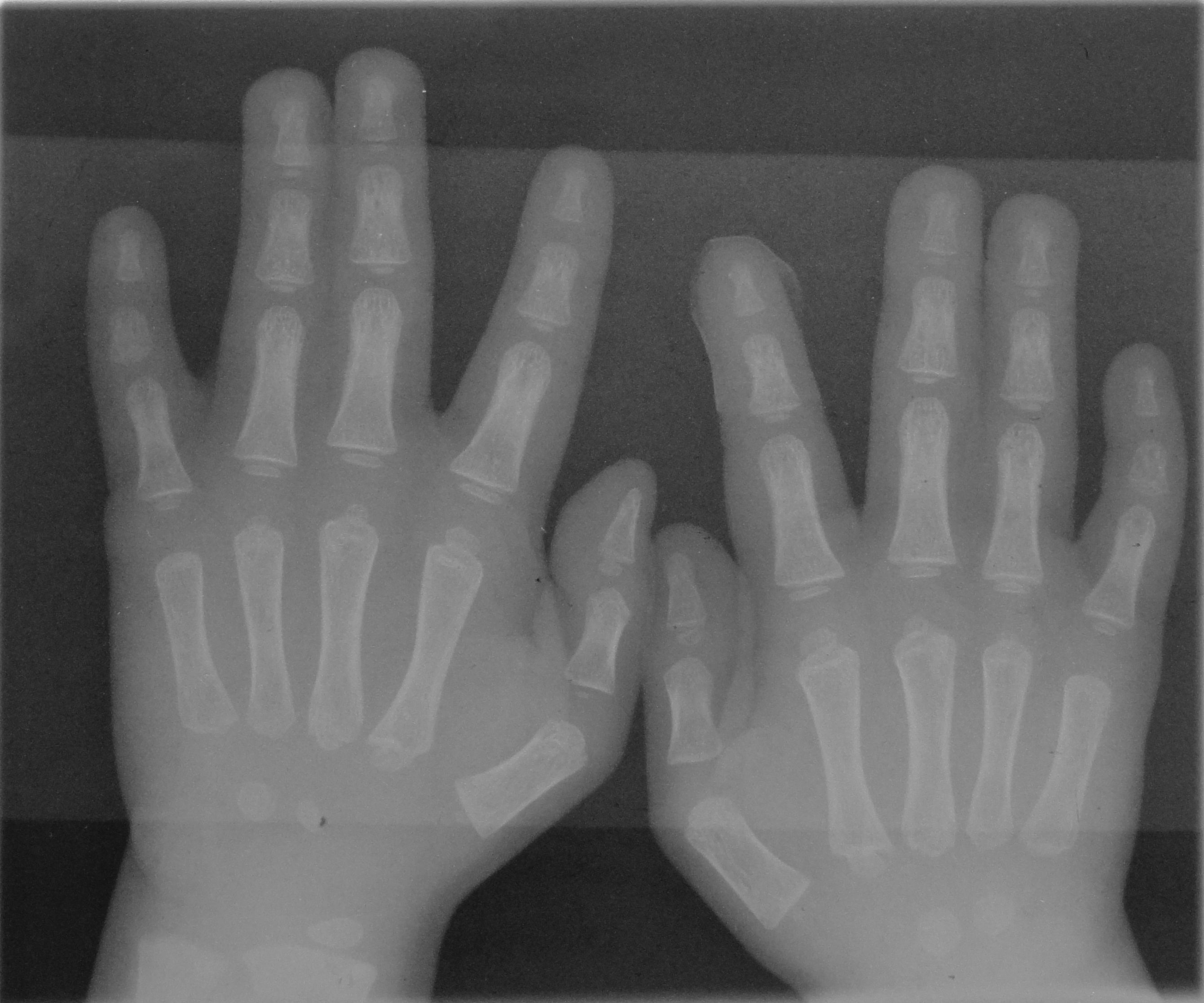
A finger is a limb of the body and a type of digit, an organ of manipulation and sensation found in the hands of most of the Tetrapods, so also with humans and other primates. Most land vertebrates have five fingers ( Pentadactyly). Chambers 1998 p. 603 Oxford Illustrated pp. 311, 380 Land vertebrate fingers The five-rayed anterior limbs of terrestrial vertebrates can be derived phylogenetically from the pectoral fins of fish. Within the taxa of the terrestrial vertebrates, the basic pentadactyl plan, and thus also the fingers and phalanges, undergo many variations. Morphologically the different fingers of terrestrial vertebrates are homolog. The wings of birds and those of bats are not homologous, they are analogue flight organs. However, the phalanges within them are homologous. Chimpanzees have lower limbs that are specialized for manipulation, and (arguably) have fingers on their lower limbs as well. In the case of Primates in general, the digits of the hand a ... [...More Info...] [...Related Items...] OR: [Wikipedia] [Google] [Baidu] |


.jpg)

