|
Codocyte
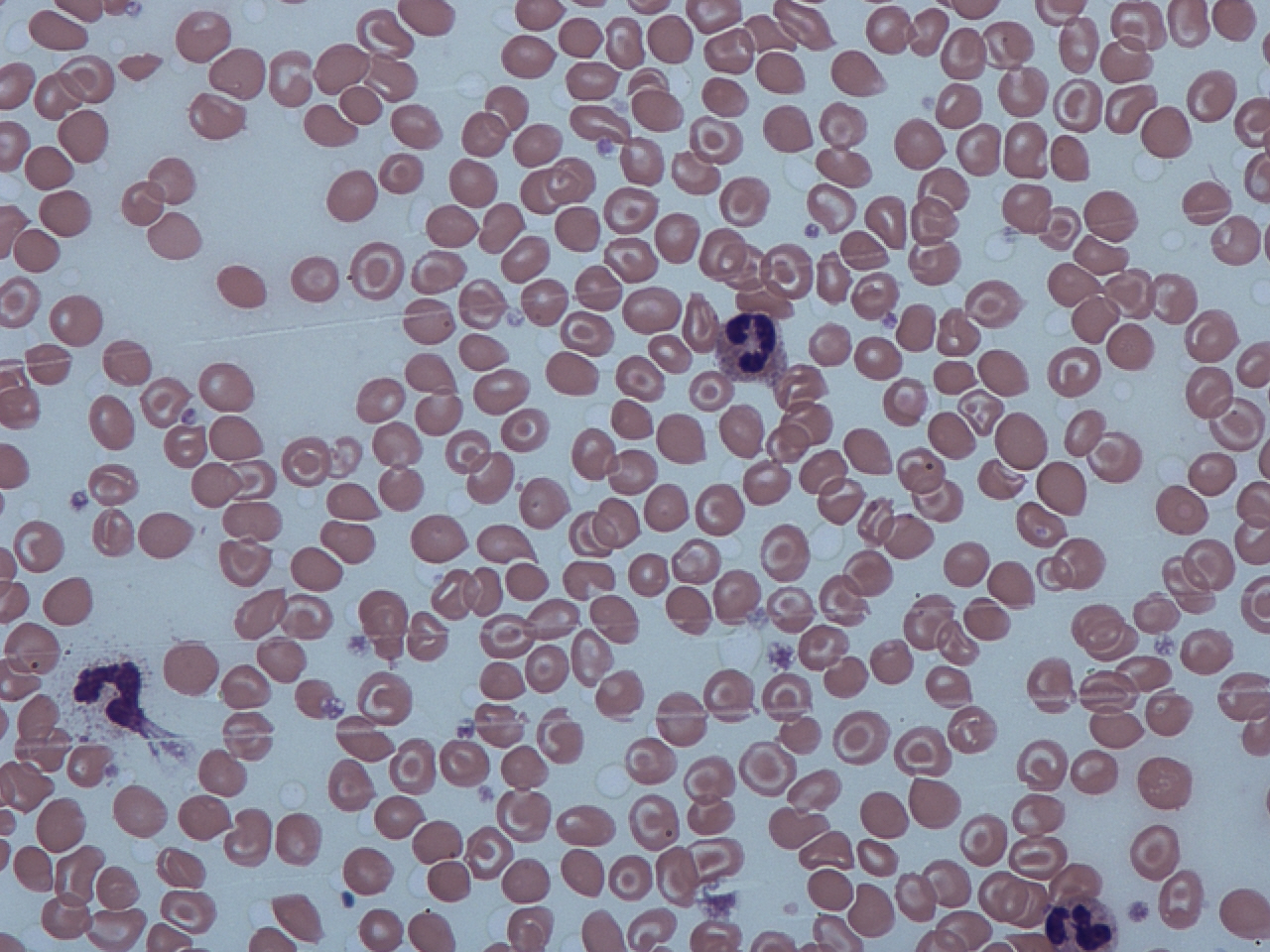
Codocytes, also known as target cells, are red blood cells that have the appearance of a shooting target with a bullseye. In optical microscopy these cells appear to have a dark center (a central, hemoglobinized area) surrounded by a white ring (an area of relative pallor), followed by dark outer (peripheral) second ring containing a band of hemoglobin. However, in electron microscopy they appear very thin and bell shaped (hence the name codo-: bell). Because of their thinness they are referred to as leptocytes. On routine smear morphology, some people like to make a distinction between leptocytes and codocytes- suggesting that in leptocytes the central spot is not completely detached from the peripheral ring, i.e. the pallor is in a C shape rather than a full ring. These cells are characterized by a disproportional increase in the ratio of surface membrane area to volume. This is also described as a "relative membrane excess." It is due to either increased red cell surface area ... [...More Info...] [...Related Items...] OR: [Wikipedia] [Google] [Baidu] |
Alpha-thalassemia
Alpha-thalassemia (α-thalassemia, α-thalassaemia) is a form of thalassemia involving the genes '' HBA1'' and '' HBA2''. Thalassemias are a group of inherited blood conditions which result in the impaired production of hemoglobin, the molecule that carries oxygen in the blood. Normal hemoglobin consists of two alpha chains and two beta chains; in alpha-thalassemia, there is a quantitative decrease in the amount of alpha chains, resulting in fewer normal hemoglobin molecules. Furthermore, alpha-thalassemia leads to the production of unstable beta globin molecules which cause increased red blood cell destruction. The degree of impairment is based on which clinical phenotype is present (how many genes are affected). Signs and symptoms The presentation of individuals with alpha-thalassemia consists of: Cause Alpha-thalassemias are most commonly inherited in a Mendelian recessive manner. They are also associated with deletions of chromosome 16p. Alpha thalassemia can also be a ... [...More Info...] [...Related Items...] OR: [Wikipedia] [Google] [Baidu] |
Beta-thalassemia
Beta thalassemias (β thalassemias) are a group of inherited blood disorders. They are forms of thalassemia caused by reduced or absent synthesis of the beta chains of hemoglobin that result in variable outcomes ranging from severe anemia to clinically asymptomatic individuals. Global annual incidence is estimated at one in 100,000. Beta thalassemias occur due to malfunctions in the hemoglobin subunit beta or HBB. The severity of the disease depends on the nature of the mutation. HBB blockage over time leads to decreased beta-chain synthesis. The body's inability to construct new beta-chains leads to the underproduction of HbA (adult hemoglobin). Reductions in HbA available overall to fill the red blood cells in turn leads to microcytic anemia. Microcytic anemia ultimately develops in respect to inadequate HBB protein for sufficient red blood cell functioning. Due to this factor, the patient may require blood transfusions to make up for the blockage in the beta-chains. Repeat ... [...More Info...] [...Related Items...] OR: [Wikipedia] [Google] [Baidu] |
Iron Deficiency Anemia
Iron-deficiency anemia is anemia caused by a lack of iron. Anemia is defined as a decrease in the number of red blood cells or the amount of hemoglobin in the blood. When onset is slow, symptoms are often vague such as feeling tired, weak, short of breath, or having decreased ability to exercise. Anemia that comes on quickly often has more severe symptoms, including confusion, feeling like one is going to pass out or increased thirst. Anemia is typically significant before a person becomes noticeably pale. Children with iron deficiency anemia may have problems with growth and development. There may be additional symptoms depending on the underlying cause. Iron-deficiency anemia is caused by blood loss, insufficient dietary intake, or poor absorption of iron from food. Sources of blood loss can include heavy periods, childbirth, uterine fibroids, stomach ulcers, colon cancer, and urinary tract bleeding. Poor absorption of iron from food may occur as a result of an inte ... [...More Info...] [...Related Items...] OR: [Wikipedia] [Google] [Baidu] |
Coeliac Disease
Coeliac disease (British English) or celiac disease (American English) is a long-term autoimmune disorder, primarily affecting the small intestine, where individuals develop intolerance to gluten, present in foods such as wheat, rye and barley. Classic symptoms include gastrointestinal problems such as chronic diarrhoea, abdominal distention, malabsorption, loss of appetite, and among children failure to grow normally. This often begins between six months and two years of age. Non-classic symptoms are more common, especially in people older than two years. There may be mild or absent gastrointestinal symptoms, a wide number of symptoms involving any part of the body, or no obvious symptoms. Coeliac disease was first described in childhood; however, it may develop at any age. It is associated with other autoimmune diseases, such as Type 1 diabetes mellitus and Hashimoto's thyroiditis, among others. Coeliac disease is caused by a reaction to gluten, a group of various prote ... [...More Info...] [...Related Items...] OR: [Wikipedia] [Google] [Baidu] |
Sickle Cell Anemia
Sickle cell disease (SCD) is a group of blood disorders typically inherited from a person's parents. The most common type is known as sickle cell anaemia. It results in an abnormality in the oxygen-carrying protein haemoglobin found in red blood cells. This leads to a rigid, sickle-like shape under certain circumstances. Problems in sickle cell disease typically begin around 5 to 6 months of age. A number of health problems may develop, such as attacks of pain (known as a sickle cell crisis), anemia, swelling in the hands and feet, bacterial infections and stroke. Long-term pain may develop as people get older. The average life expectancy in the developed world is 40 to 60 years. Sickle cell disease occurs when a person inherits two abnormal copies of the β-globin gene (''HBB'') that makes haemoglobin, one from each parent. This gene occurs in chromosome 11. Several subtypes exist, depending on the exact mutation in each haemoglobin gene. An attack can be set off by temp ... [...More Info...] [...Related Items...] OR: [Wikipedia] [Google] [Baidu] |
Autosplenectomy
An autosplenectomy (from'' 'auto-' ''self,'' '-splen-' ''spleen,'' ' -ectomy' ''removal) is a negative outcome of disease and occurs when a disease damages the spleen to such an extent that it becomes shrunken and non-functional. The spleen is an important immunological organ that acts as a filter for red blood cells, triggers phagocytosis of invaders, and mounts an immunological response when necessary. Lack of a spleen, called asplenia, can occur by autosplenectomy or the surgical counterpart, splenectomy. Asplenia can increase susceptibility to infection. Autosplenectomy can occur in cases of sickle-cell disease where the misshapen cells block blood flow to the spleen, causing scarring and eventual atrophy of the organ. Autosplenectomy is a rare condition that is linked to certain diseases but is not a common occurrence. It is also seen in systemic lupus erythematosus (SLE). Consequences Absence of effective splenic function or absence of the whole spleen (asplenia) is associate ... [...More Info...] [...Related Items...] OR: [Wikipedia] [Google] [Baidu] |
Macrophage
Macrophages (abbreviated as M φ, MΦ or MP) ( el, large eaters, from Greek ''μακρός'' (') = large, ''φαγεῖν'' (') = to eat) are a type of white blood cell of the immune system that engulfs and digests pathogens, such as cancer cells, microbes, cellular debris, and foreign substances, which do not have proteins that are specific to healthy body cells on their surface. The process is called phagocytosis, which acts to defend the host against infection and injury. These large phagocytes are found in essentially all tissues, where they patrol for potential pathogens by amoeboid movement. They take various forms (with various names) throughout the body (e.g., histiocytes, Kupffer cells, alveolar macrophages, microglia, and others), but all are part of the mononuclear phagocyte system. Besides phagocytosis, they play a critical role in nonspecific defense ( innate immunity) and also help initiate specific defense mechanisms (adaptive immunity) by recruiting othe ... [...More Info...] [...Related Items...] OR: [Wikipedia] [Google] [Baidu] |
Opsonin
Opsonins are extracellular proteins that, when bound to substances or cells, induce phagocytes to phagocytose the substances or cells with the opsonins bound. Thus, opsonins act as tags to label things in the body that should be phagocytosed (i.e. eaten) by phagocytes (cells that specialise in phagocytosis, i.e. cellular eating). Different types of things ("targets") can be tagged by opsonins for phagocytosis, including: pathogens (such as bacteria), cancer cells, aged cells, dead or dying cells (such as apoptotic cells), excess synapses, or protein aggregates (such as amyloid plaques). Opsonins help clear pathogens, as well as dead, dying and diseased cells. Opsonins were discovered and named "opsonins" in 1904 by Wright and Douglas, who found that incubating bacteria with blood plasma enabled phagocytes to phagocytose (and thereby destroy) the bacteria. They concluded that: “We have here conclusive proof that the blood fluids modify the bacteria in a manner which renders them ... [...More Info...] [...Related Items...] OR: [Wikipedia] [Google] [Baidu] |
Splenectomy
A splenectomy is the surgical procedure that partially or completely removes the spleen. The spleen is an important organ in regard to immunological function due to its ability to efficiently destroy encapsulated bacteria. Therefore, removal of the spleen runs the risk of overwhelming post-splenectomy infection, a medical emergency and rapidly fatal disease caused by the inability of the body's immune system to properly fight infection following splenectomy or asplenia. Common indications for splenectomy include trauma, tumors, splenomegaly or for hematological disease such as sickle cell anemia or thalassemia. Indications The spleen is an organ located in the abdomen next to the stomach. It is composed of red pulp which filters the blood, removing foreign material, damaged and worn out red blood cells. It also functions as a storage site for iron, red blood cells and platelets. The rest (~25%) of the spleen is known as the white pulp and functions like a large lymph no ... [...More Info...] [...Related Items...] OR: [Wikipedia] [Google] [Baidu] |
Target Cells And Spherocytes
Target may refer to: Physical items * Shooting target, used in marksmanship training and various shooting sports ** Bullseye (target), the goal one for which one aims in many of these sports ** Aiming point, in field artillery, fixed at a specific target * Color chart (or reference card), the reference target used in digital imaging for accurate color reproduction Places * Target, Allier, France * Target Lake, a lake in Minnesota Terms * Target market, marketing strategy ** Target audience, intended audience or readership of a publication, advertisement, or type of message * In mathematics, the target of a function is also called the codomain * Target (cricket), the total number of runs a team needs to win People * Target (rapper), stage name of Croatian hip-hop artist Nenad Šimun * DJ Target, stage name of English grime DJ Darren Joseph, member of Roll Deep * Gui-Jean-Baptiste Target (1733–1807), French lawyer Art and media * The Target, a comic book char ... [...More Info...] [...Related Items...] OR: [Wikipedia] [Google] [Baidu] |
Hemoglobin C
Hemoglobin C (abbreviated as HbC) is an abnormal hemoglobin in which glutamic acid residue at the 6th position of the β-globin chain is replaced with a lysine residue due to a point mutation in the '' HBB'' gene. People with one copy of the gene for hemoglobin C do not experience symptoms, but can pass the abnormal gene on to their children. Those with two copies of the gene are said to have hemoglobin C disease and can experience mild anemia. It is possible for a person to have both the gene for hemoglobin S (the form associated with sickle cell anemia) and the gene for hemoglobin C; this state is called hemoglobin SC disease, and is generally more severe than hemoglobin C disease, but milder than sickle cell anemia. HbC was discovered by Harvey Itano and James V. Neel in 1950 in two African-American families. It has since been established that it is most common among people in West Africa. It confers survival benefits as individuals with HbC are naturally resistant to malari ... [...More Info...] [...Related Items...] OR: [Wikipedia] [Google] [Baidu] |


