|
Collateral Circulation
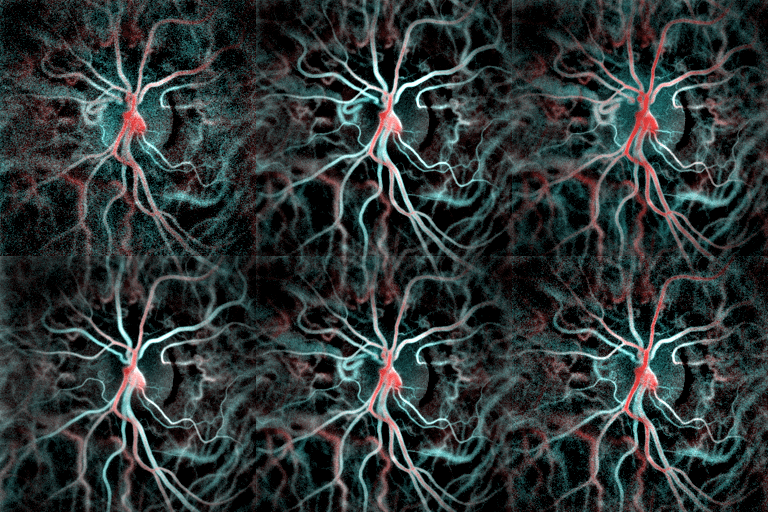
Collateral circulation is the alternate circulation around a blocked artery or vein via another path, such as nearby minor vessels. It may occur via preexisting vascular redundancy (analogous to engineered redundancy), as in the circle of Willis in the brain, or it may occur via new branches formed between adjacent blood vessels ( neovascularization), as in the eye after a retinal embolism or in the brain when moyamoya occurs. Its formation may be provoked by pathological conditions such as high vascular resistance or ischaemia. An example of the usefulness of collateral circulation is a systemic thromboembolism in cats. This is when a thrombotic embolus lodges above the external iliac artery (common iliac artery), blocking the external and internal iliac arteries and effectively shutting off all blood supply to the hind leg. Even though the main vessels to the leg are blocked, enough blood can get to the tissues in the leg via the collateral circulation to keep them aliv ... [...More Info...] [...Related Items...] OR: [Wikipedia] [Google] [Baidu] |
Circulatory System
The blood circulatory system is a system of organs that includes the heart, blood vessels, and blood which is circulated throughout the entire body of a human or other vertebrate. It includes the cardiovascular system, or vascular system, that consists of the heart and blood vessels (from Greek ''kardia'' meaning ''heart'', and from Latin ''vascula'' meaning ''vessels''). The circulatory system has two divisions, a systemic circulation or circuit, and a pulmonary circulation or circuit. Some sources use the terms ''cardiovascular system'' and ''vascular system'' interchangeably with the ''circulatory system''. The network of blood vessels are the great vessels of the heart including large elastic arteries, and large veins; other arteries, smaller arterioles, capillaries that join with venules (small veins), and other veins. The circulatory system is closed in vertebrates, which means that the blood never leaves the network of blood vessels. Some invertebrates such as ... [...More Info...] [...Related Items...] OR: [Wikipedia] [Google] [Baidu] |
Communicating Artery (other)
Communicating artery may refer to: *Anterior communicating artery (arteria communicans anterior) *Posterior communicating artery In human anatomy, the left and right posterior communicating arteries are arteries at the base of the brain that form part of the circle of Willis. Each posterior communicating artery connects the three cerebral arteries of the same side. Anterio ... (arteria communicans posterior) {{disambig ... [...More Info...] [...Related Items...] OR: [Wikipedia] [Google] [Baidu] |
Hemorrhoid
Hemorrhoids (or haemorrhoids), also known as piles, are vascular structures in the anal canal. In their normal state, they are cushions that help with stool control. They become a disease when swollen or inflamed; the unqualified term ''hemorrhoid'' is often used to refer to the disease. The signs and symptoms of hemorrhoids depend on the type present. Internal hemorrhoids often result in painless, bright red rectal bleeding when defecating. External hemorrhoids often result in pain and swelling in the area of the anus. If bleeding occurs, it is usually darker. Symptoms frequently get better after a few days. A skin tag may remain after the healing of an external hemorrhoid. While the exact cause of hemorrhoids remains unknown, a number of factors that increase pressure in the abdomen are believed to be involved. This may include constipation, diarrhea, and sitting on the toilet for long periods. Hemorrhoids are also more common during pregnancy. Diagnosis is made by ... [...More Info...] [...Related Items...] OR: [Wikipedia] [Google] [Baidu] |
Esophageal Varices
Esophageal varices are extremely dilated sub-mucosal veins in the lower third of the esophagus. They are most often a consequence of portal hypertension, commonly due to cirrhosis. People with esophageal varices have a strong tendency to develop severe bleeding which left untreated can be fatal. Esophageal varices are typically diagnosed through an esophagogastroduodenoscopy. Pathogenesis The upper two thirds of the esophagus are drained via the esophageal veins, which carry deoxygenated blood from the esophagus to the azygos vein, which in turn drains directly into the superior vena cava. These veins have no part in the development of esophageal varices. The lower one third of the esophagus is drained into the superficial veins lining the esophageal mucosa, which drain into the left gastric vein, which in turn drains directly into the portal vein. These superficial veins (normally only approximately 1 mm in diameter) become distended up to 1–2 cm in diameter ... [...More Info...] [...Related Items...] OR: [Wikipedia] [Google] [Baidu] |
Liver
The liver is a major organ only found in vertebrates which performs many essential biological functions such as detoxification of the organism, and the synthesis of proteins and biochemicals necessary for digestion and growth. In humans, it is located in the right upper quadrant of the abdomen, below the diaphragm. Its other roles in metabolism include the regulation of glycogen storage, decomposition of red blood cells, and the production of hormones. The liver is an accessory digestive organ that produces bile, an alkaline fluid containing cholesterol and bile acids, which helps the breakdown of fat. The gallbladder, a small pouch that sits just under the liver, stores bile produced by the liver which is later moved to the small intestine to complete digestion. The liver's highly specialized tissue, consisting mostly of hepatocytes, regulates a wide variety of high-volume biochemical reactions, including the synthesis and breakdown of small and complex molecu ... [...More Info...] [...Related Items...] OR: [Wikipedia] [Google] [Baidu] |
Hepatic Portal Vein
The portal vein or hepatic portal vein (HPV) is a blood vessel that carries blood from the gastrointestinal tract, gallbladder, pancreas and spleen to the liver. This blood contains nutrients and toxins extracted from digested contents. Approximately 75% of total liver blood flow is through the portal vein, with the remainder coming from the hepatic artery proper. The blood leaves the liver to the heart in the hepatic veins. The portal vein is not a true vein, because it conducts blood to capillary beds in the liver and not directly to the heart. It is a major component of the hepatic portal system, one of only two portal venous systems in the body – with the hypophyseal portal system being the other. The portal vein is usually formed by the confluence of the superior mesenteric, splenic veins, inferior mesenteric, left, right gastric veins and the pancreatic vein. Conditions involving the portal vein cause considerable illness and death. An important example of such ... [...More Info...] [...Related Items...] OR: [Wikipedia] [Google] [Baidu] |
Hepatic Cirrhosis
Cirrhosis, also known as liver cirrhosis or hepatic cirrhosis, and end-stage liver disease, is the impaired liver function caused by the formation of scar tissue known as fibrosis due to damage caused by liver disease. Damage causes tissue repair and subsequent formation of scar tissue, which over time can replace normal functioning tissue, leading to the impaired liver function of cirrhosis. The disease typically develops slowly over months or years. Early symptoms may include tiredness, weakness, loss of appetite, unexplained weight loss, nausea and vomiting, and discomfort in the right upper quadrant of the abdomen. As the disease worsens, symptoms may include itchiness, swelling in the lower legs, fluid build-up in the abdomen, jaundice, bruising easily, and the development of spider-like blood vessels in the skin. The fluid build-up in the abdomen may become spontaneously infected. More serious complications include hepatic encephalopathy, bleeding from dilated ... [...More Info...] [...Related Items...] OR: [Wikipedia] [Google] [Baidu] |
Choroid
The choroid, also known as the choroidea or choroid coat, is a part of the uvea, the vascular layer of the eye, and contains connective tissues, and lies between the retina and the sclera. The human choroid is thickest at the far extreme rear of the eye (at 0.2 mm), while in the outlying areas it narrows to 0.1 mm. The choroid provides oxygen and nourishment to the outer layers of the retina. Along with the ciliary body and iris, the choroid forms the uveal tract. The structure of the choroid is generally divided into four layers (classified in order of furthest away from the retina to closest): *Haller's layer - outermost layer of the choroid consisting of larger diameter blood vessels; * Sattler's layer - layer of medium diameter blood vessels; *Choriocapillaris - layer of capillaries; and * Bruch's membrane (synonyms: Lamina basalis, Complexus basalis, Lamina vitra) - innermost layer of the choroid. Blood supply There are two circulations of the eye: the ... [...More Info...] [...Related Items...] OR: [Wikipedia] [Google] [Baidu] |
Aqueous Humour
The aqueous humour is a transparent water-like fluid similar to plasma, but containing low protein concentrations. It is secreted from the ciliary body, a structure supporting the lens of the eyeball. It fills both the anterior and the posterior chambers of the eye, and is not to be confused with the vitreous humour, which is located in the space between the lens and the retina, also known as the posterior cavity or vitreous chamber. Blood cannot normally enter the eyeball. Structure Composition * Amino acids: transported by ciliary muscles *98% water * Electrolytes ( pH = 7.4 -one source gives 7.1) **Sodium = 142.09 **Potassium = 2.2 - 4.0 **Calcium = 1.8 **Magnesium = 1.1 **Chloride = 131.6 **HCO3- = 20.15 **Phosphate = 0.62 ** Osm = 304 *Ascorbic acid *Glutathione * Immunoglobulins Function *Maintains the intraocular pressure and inflates the globe of the eye. It is this hydrostatic pressure that keeps the eyeball in a roughly spherical shape and keeps the walls of the eye ... [...More Info...] [...Related Items...] OR: [Wikipedia] [Google] [Baidu] |
Glaucoma
Glaucoma is a group of eye diseases that result in damage to the optic nerve (or retina) and cause vision loss. The most common type is open-angle (wide angle, chronic simple) glaucoma, in which the drainage angle for fluid within the eye remains open, with less common types including closed-angle (narrow angle, acute congestive) glaucoma and normal-tension glaucoma. Open-angle glaucoma develops slowly over time and there is no pain. Peripheral vision may begin to decrease, followed by central vision, resulting in blindness if not treated. Closed-angle glaucoma can present gradually or suddenly. The sudden presentation may involve severe eye pain, blurred vision, mid-dilated pupil, redness of the eye, and nausea. Vision loss from glaucoma, once it has occurred, is permanent. Eyes affected by glaucoma are referred to as being glaucomatous. Risk factors for glaucoma include increasing age, high pressure in the eye, a family history of glaucoma, and use of steroid medicatio ... [...More Info...] [...Related Items...] OR: [Wikipedia] [Google] [Baidu] |
Central Retinal Vein Occlusion
Central retinal vein occlusion, also CRVO, is when the central retinal vein becomes occluded, usually through thrombosis. The central retinal vein is the venous equivalent of the central retinal artery and both may become occluded. Since the central retinal artery and vein are the sole source of blood supply and drainage for the retina, such occlusion can lead to severe damage to the retina and blindness, due to ischemia (restriction in blood supply) and edema (swelling). CRVO can cause ocular ischemic syndrome. Nonischemic CRVO is the milder form of the disease. It may progress to the more severe ischemic type. CRVO can also cause glaucoma. Diagnosis Despite the role of thrombosis in the development of CRVO, a systematic review found no increased prevalence of thrombophilia (an inherent propensity to thrombosis) in patients with retinal vascular occlusion. Treatment Treatment consists of Anti-VEGF drugs like Lucentis or intravitreal steroid implant (Ozurdex) and Pan-Retinal La ... [...More Info...] [...Related Items...] OR: [Wikipedia] [Google] [Baidu] |
Collateral Vein In Central Retinal Vein Occlusion
Collateral may refer to: Business and finance * Collateral (finance), a borrower's pledge of specific property to a lender, to secure repayment of a loan * Marketing collateral, in marketing and sales Arts, entertainment, and media * ''Collateral'' (album), an album by NERVO (2015) * ''Collateral'' (film), a thriller film starring Tom Cruise and Jamie Foxx (2004) * "Collateral" (''Justified''), an episode of the TV series ''Justified'' * ''Collateral'' (TV series), a four-part BBC television series (2018) Anatomy * Collateral ligament * a branch in an anatomical structure, e.g. the superior ulnar collateral artery or the prevertebral ganglia, also known as collateral ganglia * Collateral circulation, the alternate circulation around a blocked artery or vein via another path, such as nearby minor vessels See also * Collateral contract * Collateral damage * Collateral (kinship) * Collateral estoppel * Collateral management * Collateral source rule * Collateral succession * Co ... [...More Info...] [...Related Items...] OR: [Wikipedia] [Google] [Baidu] |


