Collateral circulation on:
[Wikipedia]
[Google]
[Amazon]
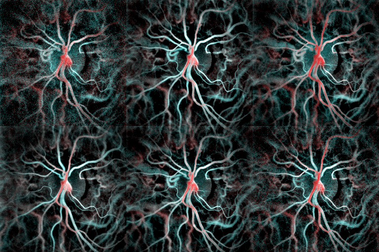
Collateral circulation is the alternate circulation around a blocked artery or vein via another path, such as nearby minor vessels.Dictionary.com collateral circulation
/ref> It may occur via preexisting vascular redundancy (analogous to engineered redundancy), as in the
 After
After
/ref> It may occur via preexisting vascular redundancy (analogous to engineered redundancy), as in the
circle of Willis
The circle of Willis (also called Willis' circle, loop of Willis, cerebral arterial circle, and Willis polygon) is a circulatory anastomosis that supplies blood to the brain and surrounding structures in reptiles, birds and mammals, including huma ...
in the brain, or it may occur via new branches formed between adjacent blood vessels (neovascularization
Neovascularization is the natural formation of new blood vessels ('' neo-'' + '' vascular'' + '' -ization''), usually in the form of functional microvascular networks, capable of perfusion by red blood cells, that form to serve as collateral circu ...
), as in the eye after a retina
The retina (from la, rete "net") is the innermost, light-sensitive layer of tissue of the eye of most vertebrates and some molluscs. The optics of the eye create a focused two-dimensional image of the visual world on the retina, which then ...
l embolism or in the brain when moyamoya
Moyamoya disease is a disease in which certain arteries in the brain are constricted. Blood flow is blocked by constriction and blood clots (thrombosis). A collateral circulation develops around the blocked vessels to compensate for the blockage, ...
occurs. Its formation may be provoked by pathological conditions such as high vascular resistance
Vascular resistance is the resistance that must be overcome to push blood through the circulatory system and create flow. The resistance offered by the systemic circulation is known as the systemic vascular resistance (SVR) or may sometimes be ca ...
or ischaemia
Ischemia American and British English spelling differences#ae and oe, or ischaemia is a restriction in blood supply to any tissue (biology), tissue, Skeletal muscle, muscle group, or Organ (biology), organ of the body, causing a shortage of oxyg ...
.
An example of the usefulness of collateral circulation is a systemic thromboembolism in cats. This is when a thrombotic embolus
An embolus (; plural emboli; from the Greek ἔμβολος "wedge", "plug") is an unattached mass that travels through the bloodstream and is capable of creating blockages. When an embolus occludes a blood vessel, it is called an embolism or emb ...
lodges above the external iliac artery
The external iliac arteries are two major arteries which bifurcate off the common iliac arteries anterior to the sacroiliac joint of the pelvis.
Structure
The external iliac artery arises from the bifurcation of the common iliac artery. Th ...
(common iliac artery), blocking the external and internal iliac arteries
The internal iliac artery (formerly known as the hypogastric artery) is the main artery of the pelvis.
Structure
The internal iliac artery supplies the walls and viscera of the pelvis, the buttock, the reproductive organs, and the medial compart ...
and effectively shutting off all blood supply to the hind leg. Even though the main vessels to the leg are blocked, enough blood can get to the tissues in the leg via the collateral circulation to keep them alive.
Brain
Blood flow to the brain in humans and some other animals is maintained via a network of collateral arteries thatanastomose
An anastomosis (, plural anastomoses) is a connection or opening between two things (especially cavities or passages) that are normally diverging or branching, such as between blood vessels, leaf veins, or streams. Such a connection may be normal ...
(join) in the circle of Willis
The circle of Willis (also called Willis' circle, loop of Willis, cerebral arterial circle, and Willis polygon) is a circulatory anastomosis that supplies blood to the brain and surrounding structures in reptiles, birds and mammals, including huma ...
, which lies at the base of the brain. In the circle of Willis so-called communicating arteries exist between the front (anterior) and back (posterior) parts of the circle of Willis, as well as between the left and right side of the circle of Willis.
Heart
Another example in humans and some other animals is after an acutemyocardial infarction
A myocardial infarction (MI), commonly known as a heart attack, occurs when blood flow decreases or stops to the coronary artery of the heart, causing damage to the heart muscle. The most common symptom is chest pain or discomfort which may ...
(heart attack). Collateral circulation in the heart tissue will sometimes bypass the blockage in the main artery and supply enough oxygenated blood to enable the cardiac tissue to survive and recover.
Eye
 After
After central retinal vein occlusion
Central retinal vein occlusion, also CRVO, is when the central retinal vein becomes occluded, usually through thrombosis. The central retinal vein is the venous equivalent of the central retinal artery and both may become occluded. Since the centra ...
, neovascularization
Neovascularization is the natural formation of new blood vessels ('' neo-'' + '' vascular'' + '' -ization''), usually in the form of functional microvascular networks, capable of perfusion by red blood cells, that form to serve as collateral circu ...
may restore some blood flow to the retina, but the new vessels' bulk also presents a risk of causing acute glaucoma
Glaucoma is a group of eye diseases that result in damage to the optic nerve (or retina) and cause vision loss. The most common type is open-angle (wide angle, chronic simple) glaucoma, in which the drainage angle for fluid within the eye rem ...
by blocking the drainage of aqueous humour
The aqueous humour is a transparent water-like fluid similar to plasma, but containing low protein concentrations. It is secreted from the ciliary body, a structure supporting the lens of the eyeball. It fills both the anterior and the posterio ...
. Collateral circulation is created (within months) around the blocked central vein via a generally winding path, usually from a branch vein to the choroid
The choroid, also known as the choroidea or choroid coat, is a part of the uvea, the vascular layer of the eye, and contains connective tissues, and lies between the retina and the sclera. The human choroid is thickest at the far extreme rear ...
.
Truncal venous system
Hepatic cirrhosis
Cirrhosis, also known as liver cirrhosis or hepatic cirrhosis, and end-stage liver disease, is the impaired liver function caused by the formation of scar tissue known as fibrosis due to damage caused by liver disease. Damage causes tissue repai ...
arising from congestion in the hepatic portal vein
The portal vein or hepatic portal vein (HPV) is a blood vessel that carries blood from the gastrointestinal tract, gallbladder, pancreas and spleen to the liver. This blood contains nutrients and toxins extracted from digested contents. Approx ...
may give rise to collateral circulation between branches of the portal and caval veins of the liver
The liver is a major Organ (anatomy), organ only found in vertebrates which performs many essential biological functions such as detoxification of the organism, and the Protein biosynthesis, synthesis of proteins and biochemicals necessary for ...
, or between the two caval veins. Consequences of newly established venous collaterals arising from portal hypertension include esophageal varices
Esophageal varices are extremely dilated sub-mucosal veins in the lower third of the esophagus. They are most often a consequence of portal hypertension, commonly due to cirrhosis. People with esophageal varices have a strong tendency to develop ...
and hemorrhoid
Hemorrhoids (or haemorrhoids), also known as piles, are vascular structures in the anal canal. In their normal state, they are cushions that help with stool control. They become a disease when swollen or inflamed; the unqualified term ''hemo ...
s (portocaval collateral circulation).
References
{{DEFAULTSORT:Collateral Circulation Angiology Fault tolerance