|
Chest Drainage Management
Chest drains are surgical drains placed within the pleural space to facilitate removal of unwanted substances (pneumothorax, air, hemothorax, blood, pleural effusion, fluid, etc.) in order to preserve respiratory system, respiratory functions and hemodynamic stability. Some chest drains may utilize a flutter valve to prevent retrograde flow, but those that do not have physical valves employ a trap (plumbing), water trap seal design, often aided by continuous suction from a suction (medicine), wall suction or a aspirator (medical device), portable vacuum pump. The active maintenance of an intrapleural negative pressure via chest drains builds the basis of chest drain management, as an intrapleural pressure lower than the surrounding atmosphere allows easier lung expansion and thus better pulmonary alveolus, alveolar ventilation (physiology), ventilation and gas exchange. History The so-called “central vacuum” was the first sub-atmospheric pressure device available. Sub-atmos ... [...More Info...] [...Related Items...] OR: [Wikipedia] [Google] [Baidu] |
Flutter Valve
A flutter valve (also known as the Heimlich valve after its inventor, Henry Heimlich) is a Check_valve, one-way valve used in respiratory medicine to prevent air from travelling back along a chest tube. One can also use a Chest_drainage_management, chest drainage management system, which typically enables vacuum to be applied along with quantifying the effluent. However, it is much larger with more tubing, which may encumber the patient. It is most commonly used to help remove air from a pneumothorax. The valve is usually designed as a rubber sleeve within a plastic case where the rubber sleeve is arranged so that when air passes through the valve one way the sleeve opens and lets the air through. However, when air is sucked back the other way, the sleeve closes off and no air is allowed backwards. This construction enables it to act as a one-way valve allowing air (or fluid) to flow only one way along the drainage tube. The end of the drainage tube is placed inside the patient ... [...More Info...] [...Related Items...] OR: [Wikipedia] [Google] [Baidu] |
Chest Drain
A chest tube (also chest drain, thoracic catheter, tube thoracostomy or intercostal drain) is a surgical drain that is inserted through the chest wall and into the pleural space or the mediastinum in order to remove clinically undesired substances such as air (pneumothorax), excess fluid (pleural effusion or hydrothorax), blood (hemothorax), chyle ( chylothorax) or pus (empyema) from the intrathoracic space. An intrapleural chest tube is also known as a Bülau drain or an intercostal catheter (ICC), and can either be a thin, flexible silicone tube (known as a "pigtail" drain), or a larger, semi-rigid, fenestrated plastic tube, which often involves a flutter valve or underwater seal. The concept of chest drainage was first advocated by Hippocrates when he described the treatment of empyema by means of incision, cautery and insertion of metal tubes. However, the technique was not widely used until the influenza epidemic of 1918 to evacuate post-pneumonic empyema, which was first d ... [...More Info...] [...Related Items...] OR: [Wikipedia] [Google] [Baidu] |
Ventilation (physiology)
Breathing (or ventilation) is the process of moving air into and from the lungs to facilitate gas exchange with the internal environment, mostly to flush out carbon dioxide and bring in oxygen. All aerobic creatures need oxygen for cellular respiration, which extracts energy from the reaction of oxygen with molecules derived from food and produces carbon dioxide as a waste product. Breathing, or "external respiration", brings air into the lungs where gas exchange takes place in the alveoli through diffusion. The body's circulatory system transports these gases to and from the cells, where "cellular respiration" takes place. The breathing of all vertebrates with lungs consists of repetitive cycles of inhalation and exhalation through a highly branched system of tubes or airways which lead from the nose to the alveoli. The number of respiratory cycles per minute is the breathing or respiratory rate, and is one of the four primary vital signs of life. Under normal conditions th ... [...More Info...] [...Related Items...] OR: [Wikipedia] [Google] [Baidu] |
Sternum
The sternum or breastbone is a long flat bone located in the central part of the chest. It connects to the ribs via cartilage and forms the front of the rib cage, thus helping to protect the heart, lungs, and major blood vessels from injury. Shaped roughly like a necktie, it is one of the largest and longest flat bones of the body. Its three regions are the manubrium, the body, and the xiphoid process. The word "sternum" originates from the Ancient Greek στέρνον (stérnon), meaning "chest". Structure The sternum is a narrow, flat bone, forming the middle portion of the front of the chest. The top of the sternum supports the clavicles (collarbones) and its edges join with the costal cartilages of the first two pairs of ribs. The inner surface of the sternum is also the attachment of the sternopericardial ligaments. Its top is also connected to the sternocleidomastoid muscle. The sternum consists of three main parts, listed from the top: * Manubrium * Body (gladiolus) * ... [...More Info...] [...Related Items...] OR: [Wikipedia] [Google] [Baidu] |
Mediastinum
The mediastinum (from ) is the central compartment of the thoracic cavity. Surrounded by loose connective tissue, it is an undelineated region that contains a group of structures within the thorax, namely the heart and its vessels, the esophagus, the trachea, the phrenic nerve, phrenic and cardiac nerves, the thoracic duct, the thymus and the lymph nodes of the central chest. Anatomy The mediastinum lies within the thorax and is enclosed on the right and left by pulmonary pleurae, pleurae. It is surrounded by the chest wall in front, the lungs to the sides and the Spine (anatomy), spine at the back. It extends from the sternum in front to the vertebral column behind. It contains all the organs of the thorax except the lungs. It is continuous with the loose connective tissue of the neck. The mediastinum can be divided into an upper (or superior) and lower (or inferior) part: * The superior mediastinum starts at the superior thoracic aperture and ends at the #Thoracic plane, t ... [...More Info...] [...Related Items...] OR: [Wikipedia] [Google] [Baidu] |
Cardiac Surgery
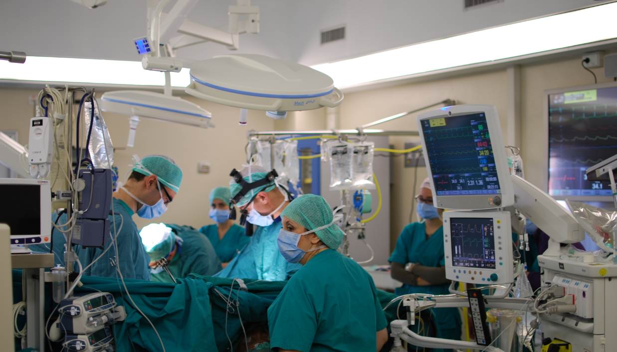
Cardiac surgery, or cardiovascular surgery, is surgery on the heart or great vessels performed by cardiac surgeons. It is often used to treat complications of ischemic heart disease (for example, with coronary artery bypass grafting); to correct congenital heart disease; or to treat valvular heart disease from various causes, including endocarditis, Rheumatic fever, rheumatic heart disease, and atherosclerosis. It also includes heart transplantation. History 19th century The earliest operations on the pericardium (the sac that surrounds the heart) took place in the 19th century and were performed by Francisco Romero (surgeon), Francisco Romero (1801) in the city of Almería (Spain), Dominique Jean Larrey (1810), Henry Dalton (1891), and Daniel Hale Williams (1893). The first surgery on the heart itself was performed by Axel Cappelen on 4 September 1895 at Rikshospitalet in Kristiania, now Oslo. Cappelen ligature (medicine), ligated a bleeding coronary circulation, coronary ... [...More Info...] [...Related Items...] OR: [Wikipedia] [Google] [Baidu] |
Pleural Empyema
Pleural empyema is a collection of pus in the pleural cavity caused by microorganisms, usually bacteria. Often it happens in the context of a pneumonia, injury, or chest surgery. It is one of the various kinds of pleural effusion. There are three stages: exudative, when there is an increase in pleural fluid with or without the presence of pus; fibrinopurulent, when fibrous septa form localized pus pockets; and the final organizing stage, when there is scarring of the pleura membranes with possible inability of the lung to expand. Simple pleural effusions occur in up to 40% of bacterial pneumonias. They are usually small and resolve with appropriate antibiotic therapy. If however an empyema develops additional intervention is required. Signs and symptoms The clinical presentation of both the adult and pediatric patient with pleural empyema depends upon several factors, including the causative micro-organism. Most cases present themselves in the setting of a pneumonia, although up ... [...More Info...] [...Related Items...] OR: [Wikipedia] [Google] [Baidu] |
Pulmonology
Pulmonology (, , from Latin ''pulmō, -ōnis'' "lung" and the Greek suffix "study of"), pneumology (, built on Greek πνεύμων "lung") or pneumonology () is a medical specialty that deals with diseases involving the respiratory tract.ACP: Pulmonology: Internal Medicine Subspecialty . Acponline.org. Retrieved on 2011-09-30. It is also known as respirology, respiratory medicine, or chest medicine in some countries and areas. Pulmonology is considered a branch of internal medicine, and is related to intensive care medicine ...
[...More Info...] [...Related Items...] OR: [Wikipedia] [Google] [Baidu] |
Gravity
In physics, gravity () is a fundamental interaction which causes mutual attraction between all things with mass or energy. Gravity is, by far, the weakest of the four fundamental interactions, approximately 1038 times weaker than the strong interaction, 1036 times weaker than the electromagnetic force and 1029 times weaker than the weak interaction. As a result, it has no significant influence at the level of subatomic particles. However, gravity is the most significant interaction between objects at the macroscopic scale, and it determines the motion of planets, stars, galaxies, and even light. On Earth, gravity gives weight to physical objects, and the Moon's gravity is responsible for sublunar tides in the oceans (the corresponding antipodal tide is caused by the inertia of the Earth and Moon orbiting one another). Gravity also has many important biological functions, helping to guide the growth of plants through the process of gravitropism and influencing the circ ... [...More Info...] [...Related Items...] OR: [Wikipedia] [Google] [Baidu] |
Bülau
A chest tube (also chest drain, thoracic catheter, tube thoracostomy or intercostal drain) is a surgical drain that is inserted through the chest wall and into the pleural space or the mediastinum in order to remove clinically undesired substances such as air (pneumothorax), excess fluid (pleural effusion or hydrothorax), blood (hemothorax), chyle (chylothorax) or pus (empyema) from the intrathoracic space. An intrapleural chest tube is also known as a Bülau drain or an intercostal catheter (ICC), and can either be a thin, flexible silicone tube (known as a "pigtail" drain), or a larger, semi-rigid, fenestrated plastic tube, which often involves a flutter valve or underwater seal. The concept of chest drainage was first advocated by Hippocrates when he described the treatment of empyema by means of incision, cautery and insertion of metal tubes. However, the technique was not widely used until the influenza epidemic of 1918 to evacuate post-pneumonic empyema, which was first do ... [...More Info...] [...Related Items...] OR: [Wikipedia] [Google] [Baidu] |
Pleural Cavity
The pleural cavity, pleural space, or interpleural space is the potential space between the pleurae of the pleural sac that surrounds each lung. A small amount of serous pleural fluid is maintained in the pleural cavity to enable lubrication between the membranes, and also to create a pressure gradient. The serous membrane that covers the surface of the lung is the visceral pleura and is separated from the outer membrane the parietal pleura by just the film of pleural fluid in the pleural cavity. The visceral pleura follows the fissures of the lung and the root of the lung structures. The parietal pleura is attached to the mediastinum, the upper surface of the diaphragm, and to the inside of the ribcage. Structure In humans, the left and right lungs are completely separated by the mediastinum, and there is no communication between their pleural cavities. Therefore, in cases of a unilateral pneumothorax, the contralateral lung will remain functioning normally unless there is ... [...More Info...] [...Related Items...] OR: [Wikipedia] [Google] [Baidu] |
Catheter
In medicine, a catheter (/ˈkæθətər/) is a thin tube made from medical grade materials serving a broad range of functions. Catheters are medical devices that can be inserted in the body to treat diseases or perform a surgical procedure. Catheters are manufactured for specific applications, such as cardiovascular, urological, gastrointestinal, neurovascular and ophthalmic procedures. The process of inserting a catheter is ''catheterization''. In most uses, a catheter is a thin, flexible tube (''soft'' catheter) though catheters are available in varying levels of stiffness depending on the application. A catheter left inside the body, either temporarily or permanently, may be referred to as an "indwelling catheter" (for example, a peripherally inserted central catheter). A permanently inserted catheter may be referred to as a "permcath" (originally a trademark). Catheters can be inserted into a body cavity, duct, or vessel, brain, skin or adipose tissue. Functionally, they all ... [...More Info...] [...Related Items...] OR: [Wikipedia] [Google] [Baidu] |


