Mediastinum on:
[Wikipedia]
[Google]
[Amazon]
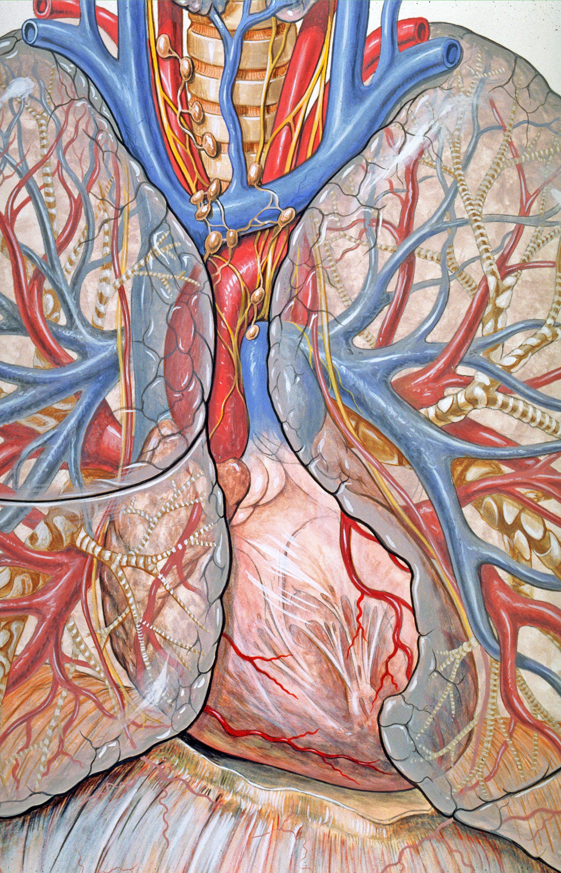
The mediastinum (from ) is the central compartment of the thoracic cavity. Surrounded by loose connective tissue, it is an undelineated region that contains a group of structures within the
 The mediastinum lies within the
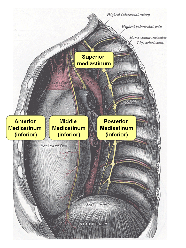
  The mediastinum can be divided into an upper (or superior) and lower (or inferior) part:
* The superior mediastinum starts at the superior thoracic aperture and ends at the thoracic plane.
* The inferior mediastinum from this level to the
A number of important anatomical structures and transitions occur at the level of the thoracic plane, including: * The carinal bifurcation of the

 ;Contents
* muscles
** origins of the Sternohyoidei and Sternothyreoidei
** lower ends of the Longi colli
* arteries
**
thorax
The thorax or chest is a part of the anatomy of humans, mammals, and other tetrapod animals located between the neck and the abdomen. In insects, crustaceans, and the extinct trilobites, the thorax is one of the three main divisions of the c ...
, namely the heart
The heart is a muscular Organ (biology), organ in most animals. This organ pumps blood through the blood vessels of the circulatory system. The pumped blood carries oxygen and nutrients to the body, while carrying metabolic waste such as ca ...
and its vessels, the esophagus
The esophagus (American English) or oesophagus (British English; both ), non-technically known also as the food pipe or gullet, is an organ in vertebrates through which food passes, aided by peristaltic contractions, from the pharynx to t ...
, the trachea
The trachea, also known as the windpipe, is a cartilaginous tube that connects the larynx to the bronchi of the lungs, allowing the passage of air, and so is present in almost all air- breathing animals with lungs. The trachea extends from t ...
, the phrenic and cardiac nerves, the thoracic duct
In human anatomy, the thoracic duct is the larger of the two lymph ducts of the lymphatic system. It is also known as the ''left lymphatic duct'', ''alimentary duct'', ''chyliferous duct'', and ''Van Hoorne's canal''. The other duct is the rig ...
, the thymus
The thymus is a specialized primary lymphoid organ of the immune system. Within the thymus, thymus cell lymphocytes or '' T cells'' mature. T cells are critical to the adaptive immune system, where the body adapts to specific foreign invaders ...
and the lymph nodes
A lymph node, or lymph gland, is a kidney-shaped organ of the lymphatic system and the adaptive immune system. A large number of lymph nodes are linked throughout the body by the lymphatic vessels. They are major sites of lymphocytes that includ ...
of the central chest.
Anatomy
thorax
The thorax or chest is a part of the anatomy of humans, mammals, and other tetrapod animals located between the neck and the abdomen. In insects, crustaceans, and the extinct trilobites, the thorax is one of the three main divisions of the c ...
and is enclosed on the right and left by pleurae. It is surrounded by the chest wall in front, the lungs to the sides and the spine
Spine or spinal may refer to:
Science Biology
* Vertebral column, also known as the backbone
* Dendritic spine, a small membranous protrusion from a neuron's dendrite
* Thorns, spines, and prickles, needle-like structures in plants
* Spine (zoolo ...
at the back. It extends from the sternum in front to the vertebral column
The vertebral column, also known as the backbone or spine, is part of the axial skeleton. The vertebral column is the defining characteristic of a vertebrate in which the notochord (a flexible rod of uniform composition) found in all chordate ...
behind. It contains all the organs of the thorax except the lungs. It is continuous with the loose connective tissue of the neck.
 The mediastinum can be divided into an upper (or superior) and lower (or inferior) part:
* The superior mediastinum starts at the superior thoracic aperture and ends at the thoracic plane.
* The inferior mediastinum from this level to the
 diaphragm
Diaphragm may refer to:
Anatomy
* Thoracic diaphragm, a thin sheet of muscle between the thorax and the abdomen
* Pelvic diaphragm or pelvic floor, a pelvic structure
* Urogenital diaphragm or triangular ligament, a pelvic structure
Other
* Diap ...
. This lower part is subdivided into three regions, all relative to the pericardium
The pericardium, also called pericardial sac, is a double-walled sac containing the heart and the roots of the great vessels. It has two layers, an outer layer made of strong connective tissue (fibrous pericardium), and an inner layer made ...
– the anterior mediastinum being in front of the pericardium, the ''middle mediastinum'' contains the pericardium and its contents, and the ''posterior mediastinum'' being behind the pericardium.
Anatomists, surgeon
In modern medicine, a surgeon is a medical professional who performs surgery. Although there are different traditions in different times and places, a modern surgeon usually is also a licensed physician or received the same medical training as ...
s, and clinical radiologists
Radiology ( ) is the medical discipline that uses medical imaging to diagnose diseases and guide their treatment, within the bodies of humans and other animals. It began with radiography (which is why its name has a root referring to radiati ...
compartmentalize the mediastinum differently. For instance, in the radiological scheme of Felson, there are only three compartments (anterior, middle, and posterior), and the heart is part of the middle (inferior) mediastinum.
Thoracic plane
The transverse thoracic plane, thoracic plane, plane of Louis or plane of Ludwig is an importantanatomical plane
An anatomical plane is a hypothetical plane used to transect the body, in order to describe the location of structures or the direction of movements. In human and animal anatomy, three principal planes are used:
* The sagittal plane or latera ...
at the level of the sternal angle and the T4/T5 intervertebral disc
An intervertebral disc (or intervertebral fibrocartilage) lies between adjacent vertebrae in the vertebral column. Each disc forms a fibrocartilaginous joint (a symphysis), to allow slight movement of the vertebrae, to act as a ligament to h ...
. It serves as an imaginary boundary that separates the superior and inferior mediastinum.Thoracic Wall, Pleura, and Pericardium – Dissector AnswersA number of important anatomical structures and transitions occur at the level of the thoracic plane, including: * The carinal bifurcation of the
trachea
The trachea, also known as the windpipe, is a cartilaginous tube that connects the larynx to the bronchi of the lungs, allowing the passage of air, and so is present in almost all air- breathing animals with lungs. The trachea extends from t ...
into the left and right main bronchi.
* The left recurrent laryngeal nerve branching off the left vagus nerve
The vagus nerve, also known as the tenth cranial nerve, cranial nerve X, or simply CN X, is a cranial nerve that interfaces with the parasympathetic control of the heart, lungs, and digestive tract. It comprises two nerves—the left and righ ...
and hooking under the ligamentum arteriosum
The ligamentum arteriosum (arterial ligament), also known as the Ligament of Botallo or Harvey's ligament, is a small ligament attaching the aorta to the pulmonary artery. It serves no function in adults but is the remnant of the ductus arteriosus ...
between the aortic arch
The aortic arch, arch of the aorta, or transverse aortic arch () is the part of the aorta between the ascending and descending aorta. The arch travels backward, so that it ultimately runs to the left of the trachea.
Structure
The aorta begins ...
above and the pulmonary trunk
A pulmonary artery is an artery in the pulmonary circulation that carries deoxygenated blood from the right side of the heart to the lungs. The largest pulmonary artery is the ''main pulmonary artery'' or ''pulmonary trunk'' from the heart, and ...
below.
* The starting of the cardiac plexus.
* The azygos vein arching over the right main bronchus and joining into the superior vena cava.
* The thoracic duct
In human anatomy, the thoracic duct is the larger of the two lymph ducts of the lymphatic system. It is also known as the ''left lymphatic duct'', ''alimentary duct'', ''chyliferous duct'', and ''Van Hoorne's canal''. The other duct is the rig ...
crossing the midline from right to left behind the esophagus
The esophagus (American English) or oesophagus (British English; both ), non-technically known also as the food pipe or gullet, is an organ in vertebrates through which food passes, aided by peristaltic contractions, from the pharynx to t ...
* The end of the pretracheal and prevertebral fasciae.
Superior
The superior mediastinum is bounded: * ''superiorly'' by the thoracic inlet, the upper opening of thethorax
The thorax or chest is a part of the anatomy of humans, mammals, and other tetrapod animals located between the neck and the abdomen. In insects, crustaceans, and the extinct trilobites, the thorax is one of the three main divisions of the c ...
;
* ''inferiorly'' by the transverse thoracic plane. which is an imaginary plane passing from the sternal angle anteriorly to the lower border of the body of the 4th thoracic vertebra posteriorly;
* ''laterally'' by the pleurae;
* ''anteriorly'' by the manubrium of the sternum;
* ''posteriorly'' by the first four thoracic vertebral bodies
The spinal column, a defining synapomorphy shared by nearly all vertebrates,Hagfish are believed to have secondarily lost their spinal column is a moderately flexible series of vertebrae (singular vertebra), each constituting a characteristic i ...
.

 ;Contents
* muscles
** origins of the Sternohyoidei and Sternothyreoidei
** lower ends of the Longi colli
* arteries
**
 aortic arch
The aortic arch, arch of the aorta, or transverse aortic arch () is the part of the aorta between the ascending and descending aorta. The arch travels backward, so that it ultimately runs to the left of the trachea.
Structure
The aorta begins ...
** brachiocephalic artery
** thoracic portions of the left common carotid
In anatomy, the left and right common carotid arteries (carotids) (Entry "carotid"
in
left subclavian * veins **
File:Gray968.png , A transverse section of the
 The mediastinum is frequently the site of involvement of various tumors:
* ''Anterior mediastinum'': substernal
in
left subclavian * veins **
brachiocephalic veins
The left and right brachiocephalic veins (previously called innominate veins) are major veins in the upper chest, formed by the union of each corresponding internal jugular vein and subclavian vein. This is at the level of the sternoclavicular ...
and
** upper half of the superior vena cava
** left highest intercostal vein
* nerves
** vagus nerve
The vagus nerve, also known as the tenth cranial nerve, cranial nerve X, or simply CN X, is a cranial nerve that interfaces with the parasympathetic control of the heart, lungs, and digestive tract. It comprises two nerves—the left and righ ...
** cardiac nerve
** superficial and deep cardiac plexuses
** phrenic nerve
The phrenic nerve is a mixed motor/sensory nerve which originates from the C3-C5 spinal nerves in the neck. The nerve is important for breathing because it provides exclusive motor control of the diaphragm, the primary muscle of respiration. ...
** left recurrent laryngeal nerve
The recurrent laryngeal nerve (RLN) is a branch of the vagus nerve ( cranial nerve X) that supplies all the intrinsic muscles of the larynx, with the exception of the cricothyroid muscles. There are two recurrent laryngeal nerves, right and ...
* trachea
The trachea, also known as the windpipe, is a cartilaginous tube that connects the larynx to the bronchi of the lungs, allowing the passage of air, and so is present in almost all air- breathing animals with lungs. The trachea extends from t ...
with paratracheal and tracheobronchial lymph nodes
* esophagus
The esophagus (American English) or oesophagus (British English; both ), non-technically known also as the food pipe or gullet, is an organ in vertebrates through which food passes, aided by peristaltic contractions, from the pharynx to t ...
* thoracic duct
In human anatomy, the thoracic duct is the larger of the two lymph ducts of the lymphatic system. It is also known as the ''left lymphatic duct'', ''alimentary duct'', ''chyliferous duct'', and ''Van Hoorne's canal''. The other duct is the rig ...
* remains of the thymus
The thymus is a specialized primary lymphoid organ of the immune system. Within the thymus, thymus cell lymphocytes or '' T cells'' mature. T cells are critical to the adaptive immune system, where the body adapts to specific foreign invaders ...
* some lymph glands
* anterior longitudinal ligament
Inferior
;Anterior mediastinum Is bounded: * ''laterally'' by the pleurae; * ''posteriorly'' by thepericardium
The pericardium, also called pericardial sac, is a double-walled sac containing the heart and the roots of the great vessels. It has two layers, an outer layer made of strong connective tissue (fibrous pericardium), and an inner layer made ...
;
* ''anteriorly'' by the sternum , the left transversus thoracis and the fifth, sixth, and seventh left costal cartilages.
;Contents
* A quantity of loose areolar tissue
* Some lymphatic vessels which ascend from the convex surface of the liver
The liver is a major organ only found in vertebrates which performs many essential biological functions such as detoxification of the organism, and the synthesis of proteins and biochemicals necessary for digestion and growth. In humans, it i ...
* Two or three anterior mediastinal lymph nodes
A lymph node, or lymph gland, is a kidney-shaped organ of the lymphatic system and the adaptive immune system. A large number of lymph nodes are linked throughout the body by the lymphatic vessels. They are major sites of lymphocytes that includ ...
* The small mediastinal branches of the internal thoracic artery
* Thymus
The thymus is a specialized primary lymphoid organ of the immune system. Within the thymus, thymus cell lymphocytes or '' T cells'' mature. T cells are critical to the adaptive immune system, where the body adapts to specific foreign invaders ...
(involuted in adults)
*superior and inferior sternopericardial ligaments
;Middle mediastinum
Bounded: pericardial sac – It contains the vital organs and is classified into the serous and fibrous pericardium.
;Contents
* the heart
The heart is a muscular Organ (biology), organ in most animals. This organ pumps blood through the blood vessels of the circulatory system. The pumped blood carries oxygen and nutrients to the body, while carrying metabolic waste such as ca ...
enclosed in the pericardium
The pericardium, also called pericardial sac, is a double-walled sac containing the heart and the roots of the great vessels. It has two layers, an outer layer made of strong connective tissue (fibrous pericardium), and an inner layer made ...
* the ascending aorta
The ascending aorta (AAo) is a portion of the aorta commencing at the upper part of the base of the left ventricle, on a level with the lower border of the third costal cartilage behind the left half of the sternum.
Structure
It passes obliqu ...
* the lower half of the superior vena cava with the azygos vein opening into it
* the bifurcation of the trachea and the two bronchi
* the pulmonary trunk
A pulmonary artery is an artery in the pulmonary circulation that carries deoxygenated blood from the right side of the heart to the lungs. The largest pulmonary artery is the ''main pulmonary artery'' or ''pulmonary trunk'' from the heart, and ...
dividing into its two branches
* the right and left pulmonary veins
* the phrenic nerves
* some bronchial lymphatic glands
A lymph node, or lymph gland, is a kidney-shaped organ of the lymphatic system and the adaptive immune system. A large number of lymph nodes are linked throughout the body by the lymphatic vessels. They are major sites of lymphocytes that includ ...
* pericardiocophrenic vessels
;Posterior mediastinum
Is bounded:
* Anteriorly by (from above downwards): bifurcation of trachea
The trachea, also known as the windpipe, is a cartilaginous tube that connects the larynx to the bronchi of the lungs, allowing the passage of air, and so is present in almost all air- breathing animals with lungs. The trachea extends from t ...
; pulmonary vessels; fibrous pericardium and posterior sloping surface of diaphragm
Diaphragm may refer to:
Anatomy
* Thoracic diaphragm, a thin sheet of muscle between the thorax and the abdomen
* Pelvic diaphragm or pelvic floor, a pelvic structure
* Urogenital diaphragm or triangular ligament, a pelvic structure
Other
* Diap ...
* Inferiorly by the thoracic surface of the diaphragm
Diaphragm may refer to:
Anatomy
* Thoracic diaphragm, a thin sheet of muscle between the thorax and the abdomen
* Pelvic diaphragm or pelvic floor, a pelvic structure
* Urogenital diaphragm or triangular ligament, a pelvic structure
Other
* Diap ...
(below);
* Superiorly by the transverse thoracic plane;
* Posteriorly by the bodies of the vertebral column
The vertebral column, also known as the backbone or spine, is part of the axial skeleton. The vertebral column is the defining characteristic of a vertebrate in which the notochord (a flexible rod of uniform composition) found in all chordate ...
from the lower border of the fifth to the twelfth thoracic vertebra (behind);
* Laterally by the mediastinal pleura (on either side).
* artery
** thoracic part of the descending aorta
* veins
** azygos vein
** the hemiazygos vein and the accessory hemiazygos vein
* nerves
** vagus nerve
The vagus nerve, also known as the tenth cranial nerve, cranial nerve X, or simply CN X, is a cranial nerve that interfaces with the parasympathetic control of the heart, lungs, and digestive tract. It comprises two nerves—the left and righ ...
** splanchnic nerves
The splanchnic nerves are paired visceral nerves (nerves that contribute to the innervation of the internal organs), carrying fibers of the autonomic nervous system ( visceral efferent fibers) as well as sensory fibers from the organs ( visceral a ...
** sympathetic chain
* esophagus
The esophagus (American English) or oesophagus (British English; both ), non-technically known also as the food pipe or gullet, is an organ in vertebrates through which food passes, aided by peristaltic contractions, from the pharynx to t ...
* thoracic duct
In human anatomy, the thoracic duct is the larger of the two lymph ducts of the lymphatic system. It is also known as the ''left lymphatic duct'', ''alimentary duct'', ''chyliferous duct'', and ''Van Hoorne's canal''. The other duct is the rig ...
* some lymph glands
thorax
The thorax or chest is a part of the anatomy of humans, mammals, and other tetrapod animals located between the neck and the abdomen. In insects, crustaceans, and the extinct trilobites, the thorax is one of the three main divisions of the c ...
, showing the contents of the middle and the posterior mediastinum.
Clinical significance
 The mediastinum is frequently the site of involvement of various tumors:
* ''Anterior mediastinum'': substernal
 thyroid
The thyroid, or thyroid gland, is an endocrine gland in vertebrates. In humans it is in the neck and consists of two connected lobes. The lower two thirds of the lobes are connected by a thin band of tissue called the thyroid isthmus. The ...
goiters, lymphoma
Lymphoma is a group of blood and lymph tumors that develop from lymphocytes (a type of white blood cell). In current usage the name usually refers to just the cancerous versions rather than all such tumours. Signs and symptoms may include en ...
, thymoma
A thymoma is a tumor originating from the epithelial cells of the thymus that is considered a rare malignancy. Thymomas are frequently associated with neuromuscular disorders such as myasthenia gravis; thymoma is found in 20% of patients with ...
, and teratoma
A teratoma is a tumor made up of several different types of tissue, such as hair, muscle, teeth, or bone. Teratomata typically form in the ovary, testicle, or coccyx.
Symptoms
Symptoms may be minimal if the tumor is small. A testicular ter ...
.
* ''Middle mediastinum'': lymphadenopathy, metastatic disease such as from small cell carcinoma from the lung.
* ''Posterior mediastinum'': Neurogenic tumors, either from the nerve sheath (mostly benign) or elsewhere (mostly malignant).
Mediastinitis is inflammation
Inflammation (from la, inflammatio) is part of the complex biological response of body tissues to harmful stimuli, such as pathogens, damaged cells, or irritants, and is a protective response involving immune cells, blood vessels, and molec ...
of the tissues in the mediastinum, usually bacteria
Bacteria (; singular: bacterium) are ubiquitous, mostly free-living organisms often consisting of one biological cell. They constitute a large domain of prokaryotic microorganisms. Typically a few micrometres in length, bacteria were am ...
l and due to rupture of organs in the mediastinum. As the infection can progress very quickly, this is a serious condition.
Pneumomediastinum is the presence of air in the mediastinum, which in some cases can lead to pneumothorax, pneumoperitoneum, and pneumopericardium if left untreated. However, that does not always occur and sometimes those conditions are actually the cause, not the result, of pneumomediastinum. These conditions frequently accompany Boerhaave syndrome, or spontaneous esophageal rupture.
Widening
''Widened mediastinum/mediastinal widening'' is where the mediastinum has a width greater than 6 cm on an upright PA chest X-ray or 8 cm on supine AP chest film. A widened mediastinum can be indicative of several pathologies: *aortic aneurysm
An aortic aneurysm is an enlargement (dilatation) of the aorta to greater than 1.5 times normal size. They usually cause no symptoms except when ruptured. Occasionally, there may be abdominal, back, or leg pain. The prevalence of abdominal aorti ...
* aortic dissection
* aortic unfolding Aortic unfolding is an abnormality visible on a chest X-ray, that shows widening of the mediastinum which may mimic the appearance of a thoracic aortic aneurysm.
With aging, the ascending portion of the thoracic aorta increases in length by approx ...
* aortic rupture
Aortic rupture is the rupture or breakage of the aorta, the largest artery in the body. Aortic rupture is a rare, extremely dangerous condition. The most common cause is an abdominal aortic aneurysm that has ruptured spontaneously. Aortic rupture ...
* hilar lymphadenopathy
* anthrax inhalation - a widened mediastinum was found in 7 of the first 10 victims infected by anthrax (''Bacillus anthracis'') in 2001.
* esophageal rupture - presents usually with pneumomediastinum and pleural effusion. It is diagnosed with water-soluble swallowed contrast.
* mediastinal mass
* mediastinitis
* cardiac tamponade
*pericardial effusion
A pericardial effusion is an abnormal accumulation of fluid in the pericardial cavity. The pericardium is a two-part membrane surrounding the heart: the outer fibrous connective membrane and an inner two-layered serous membrane. The two layers of t ...
* thoracic vertebrae fractures in trauma patients.
See also
*Mediastinum testis
The mediastinum testis is a network of fibrous connective tissue that extends from the top to near the bottom of each testis. It is wider above than below.
Numerous imperfect septa are given off from its front and sides, which radiate toward the ...
(unrelated structure in the scrotum
The scrotum or scrotal sac is an anatomical male reproductive structure located at the base of the penis that consists of a suspended dual-chambered sac of skin and smooth muscle. It is present in most terrestrial male mammals. The scrotum co ...
)
* Mediastinal germ cell tumor
* Mediastinitis
* Mediastinal tumor
References
External links
* – "Divisions of the mediastinum." * – "The anatomical divisions of the inferior mediastinum." * – "Subdivisions of the Thoracic Cavity" {{Authority control Thoracic cavity