|
CTGF
CTGF, also known as CCN2 or connective tissue growth factor, is a matricellular protein of the CCN family of extracellular matrix-associated heparin-binding proteins (see also CCN intercellular signaling protein). CTGF has important roles in many biological processes, including cell adhesion, migration, proliferation, angiogenesis, skeletal development, and tissue wound repair, and is critically involved in fibrotic disease and several forms of cancers. Structure and binding partners Members of the CCN protein family, including CTGF, are structurally characterized by having four conserved, cysteine-rich domains. These domains are, from N- to C-termini, the insulin-like growth factor binding protein ( IGFBP) domain, the von Willebrand type C repeats ( vWC) domain, the thrombospondin type 1 repeat (TSR) domain, and a C-terminal domain (CT) with a cysteine knot motif. CTGF exerts its functions by binding to various cell surface receptors in a context-dependent manner, including i ... [...More Info...] [...Related Items...] OR: [Wikipedia] [Google] [Baidu] |
CCN Intercellular Signaling Protein
CCN proteins are a family of extracellular matrix (ECM)-associated proteins involved in intercellular signaling. Due to their dynamic role within the ECM they are considered matricellular proteins. Background The acronym CCN is derived from the first three members of the family discovered, namely CYR61 (cysteine-rich angiogenic protein 61 or CCN1), CTGF (connective tissue growth factor or CCN2), and NOV (nephroblastoma overexpressed or CCN3). Together with three Wnt-induced secreted proteins, they comprise the CCN family of matricellular proteins. These proteins have now been renamed CCN1-6 by international consensus. Members of the CCN protein family are characterized by having four conserved cysteine-rich domains, which include the insulin-like growth factor-binding domain (IGFBP), the Von Willebrand factor type C domain (VWC), the thrombospondin type 1 repeat (TSR), and a C-terminal domain (CT) with a cysteine knot motif. CCN proteins have been shown to play important roles in m ... [...More Info...] [...Related Items...] OR: [Wikipedia] [Google] [Baidu] |
Von Willebrand Factor Type C Domain
Von Willebrand factor, type C (VWFC or VWC)is a protein domain is found in various blood plasma proteins: complement factors B, C2, CR3 and CR4; the integrins (I-domains); collagen types VI, VII, XII and XIV; and other extracellular proteins. Function Although the majority of VWA-containing proteins are extracellular, the most ancient ones present in all eukaryotes are all intracellular proteins involved in functions such as transcription, DNA repair, ribosomal and membrane transport and the proteasome. A common feature appears to be involvement in multiprotein complexes. Proteins that incorporate vWF domains participate in numerous biological events (e.g. cell adhesion, migration, homing, pattern formation, and signal transduction), involving interaction with a large array of ligands. Mutation effects A number of human diseases arise from mutations in VWA domains. The domain is named after the von Willebrand factor (VWF) type C repeat which is found in multidomain protein/mul ... [...More Info...] [...Related Items...] OR: [Wikipedia] [Google] [Baidu] |
Fibrosis
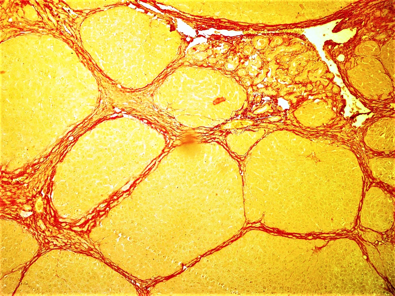
Fibrosis, also known as fibrotic scarring, is a pathological wound healing in which connective tissue replaces normal parenchymal tissue to the extent that it goes unchecked, leading to considerable tissue remodelling and the formation of permanent scar tissue. Repeated injuries, chronic inflammation and repair are susceptible to fibrosis where an accidental excessive accumulation of extracellular matrix components, such as the collagen is produced by fibroblasts, leading to the formation of a permanent fibrotic scar. In response to injury, this is called scarring, and if fibrosis arises from a single cell line, this is called a fibroma. Physiologically, fibrosis acts to deposit connective tissue, which can interfere with or totally inhibit the normal architecture and function of the underlying organ or tissue. Fibrosis can be used to describe the pathological state of excess deposition of fibrous tissue, as well as the process of connective tissue deposition in healing. Define ... [...More Info...] [...Related Items...] OR: [Wikipedia] [Google] [Baidu] |
Matricellular Protein
A matricellular protein is a dynamically expressed non-structural protein that is present in the extracellular matrix (ECM). Rather than serving as stable structural elements in the ECM, these proteins are rapidly turned over and have regulatory roles. They characteristically contain binding sites for ECM structural proteins and cell surface receptors, and may sequester and modulate activities of specific growth factors. Examples of matricellular proteins include the CCN family of proteins (also known as CCN intercellular signaling protein), fibulins, osteopontin, periostin, SPARC family members, tenascin(s), and thrombospondins. Many of these proteins have important functions in wound healing and tissue repair. See also * CCN protein CCN proteins are a family of extracellular matrix (ECM)-associated proteins involved in intercellular signaling. Due to their dynamic role within the ECM they are considered matricellular proteins. Background The acronym CCN is derived from the fi ... [...More Info...] [...Related Items...] OR: [Wikipedia] [Google] [Baidu] |
Fibronectin
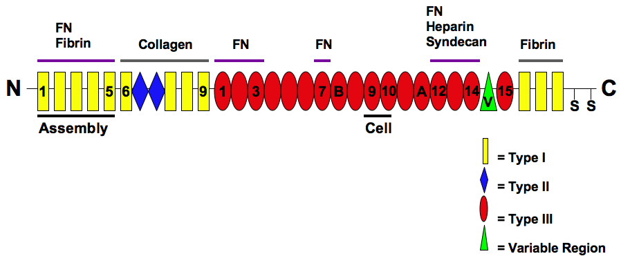
Fibronectin is a high-molecular weight (~500-~600 kDa) glycoprotein of the extracellular matrix that binds to membrane-spanning receptor proteins called integrins. Fibronectin also binds to other extracellular matrix proteins such as collagen, fibrin, and heparan sulfate proteoglycans (e.g. syndecans). Fibronectin exists as a protein dimer, consisting of two nearly identical monomers linked by a pair of disulfide bonds. The fibronectin protein is produced from a single gene, but alternative splicing of its pre-mRNA leads to the creation of several isoforms. Two types of fibronectin are present in vertebrates: * soluble plasma fibronectin (formerly called "cold-insoluble globulin", or CIg) is a major protein component of blood plasma (300 μg/ml) and is produced in the liver by hepatocytes. * insoluble cellular fibronectin is a major component of the extracellular matrix. It is secreted by various cells, primarily fibroblasts, as a soluble protein dimer and is the ... [...More Info...] [...Related Items...] OR: [Wikipedia] [Google] [Baidu] |
TGF-β
Transforming growth factor beta (TGF-β) is a multifunctional cytokine belonging to the transforming growth factor superfamily that includes three different mammalian isoforms (TGF-β 1 to 3, HGNC symbols TGFB1, TGFB2, TGFB3) and many other signaling proteins. TGFB proteins are produced by all white blood cell lineages. Activated TGF-β complexes with other factors to form a serine/threonine kinase complex that binds to TGF-β receptors. TGF-β receptors are composed of both type 1 and type 2 receptor subunits. After the binding of TGF-β, the type 2 receptor kinase phosphorylates and activates the type 1 receptor kinase that activates a signaling cascade. This leads to the activation of different downstream substrates and regulatory proteins, inducing transcription of different target genes that function in differentiation, chemotaxis, proliferation, and activation of many immune cells. TGF-β is secreted by many cell types, including macrophages, in a latent form in whi ... [...More Info...] [...Related Items...] OR: [Wikipedia] [Google] [Baidu] |
Wound Healing
Wound healing refers to a living organism's replacement of destroyed or damaged tissue by newly produced tissue. In undamaged skin, the epidermis (surface, epithelial layer) and dermis (deeper, connective layer) form a protective barrier against the external environment. When the barrier is broken, a regulated sequence of biochemical events is set into motion to repair the damage. This process is divided into predictable phases: blood clotting ( hemostasis), inflammation Inflammation (from la, wikt:en:inflammatio#Latin, inflammatio) is part of the complex biological response of body tissues to harmful stimuli, such as pathogens, damaged cells, or Irritation, irritants, and is a protective response involving im ..., tissue growth ( cell proliferation), and tissue remodeling (maturation and cell differentiation). Blood clotting may be considered to be part of the inflammation stage instead of a separate stage. The wound healing process is not only complex but fragile, a ... [...More Info...] [...Related Items...] OR: [Wikipedia] [Google] [Baidu] |
Ovulation
Ovulation is the release of eggs from the ovaries. In women, this event occurs when the ovarian follicles rupture and release the secondary oocyte ovarian cells. After ovulation, during the luteal phase, the egg will be available to be fertilized by sperm. In addition, the uterine lining ( endometrium) is thickened to be able to receive a fertilized egg. If no conception occurs, the uterine lining as well as the egg will be shed during menstruation. Process Ovulation occurs about midway through the menstrual cycle, after the follicular phase. The days in which a person is most fertile can be calculated based on the date of the last menstrual period and the length of a typical menstrual cycle. The few days surrounding ovulation (from approximately days 10 to 18 of a 28-day cycle), constitute the most fertile phase. The time from the beginning of the last menstrual period (LMP) until ovulation is, on average, 14.6 days, but with substantial variation among females and be ... [...More Info...] [...Related Items...] OR: [Wikipedia] [Google] [Baidu] |
Ovarian Follicle
An ovarian follicle is a roughly spheroid cellular aggregation set found in the ovaries. It secretes hormones that influence stages of the menstrual cycle. At the time of puberty, women have approximately 200,000 to 300,000 follicles, each with the potential to release an egg cell (ovum) at ovulation for fertilization. These eggs are developed once every menstrual cycle with around 450–500 being ovulated during a woman's reproductive lifetime. Structure Ovarian follicles are the basic units of female reproductive biology. Each of them contains a single oocyte (immature ovum or egg cell). These structures are periodically initiated to grow and develop, culminating in ovulation of usually a single competent oocyte in humans. They also consist of granulosa cells and theca of follicle. Oocyte Once a month, one of the ovaries releases a mature egg (ovum), known as an oocyte. The nucleus of such an oocyte is called a ''germinal vesicle (see picture).'' Cumulus oophorus Cu ... [...More Info...] [...Related Items...] OR: [Wikipedia] [Google] [Baidu] |
Pancreatic Beta Cell
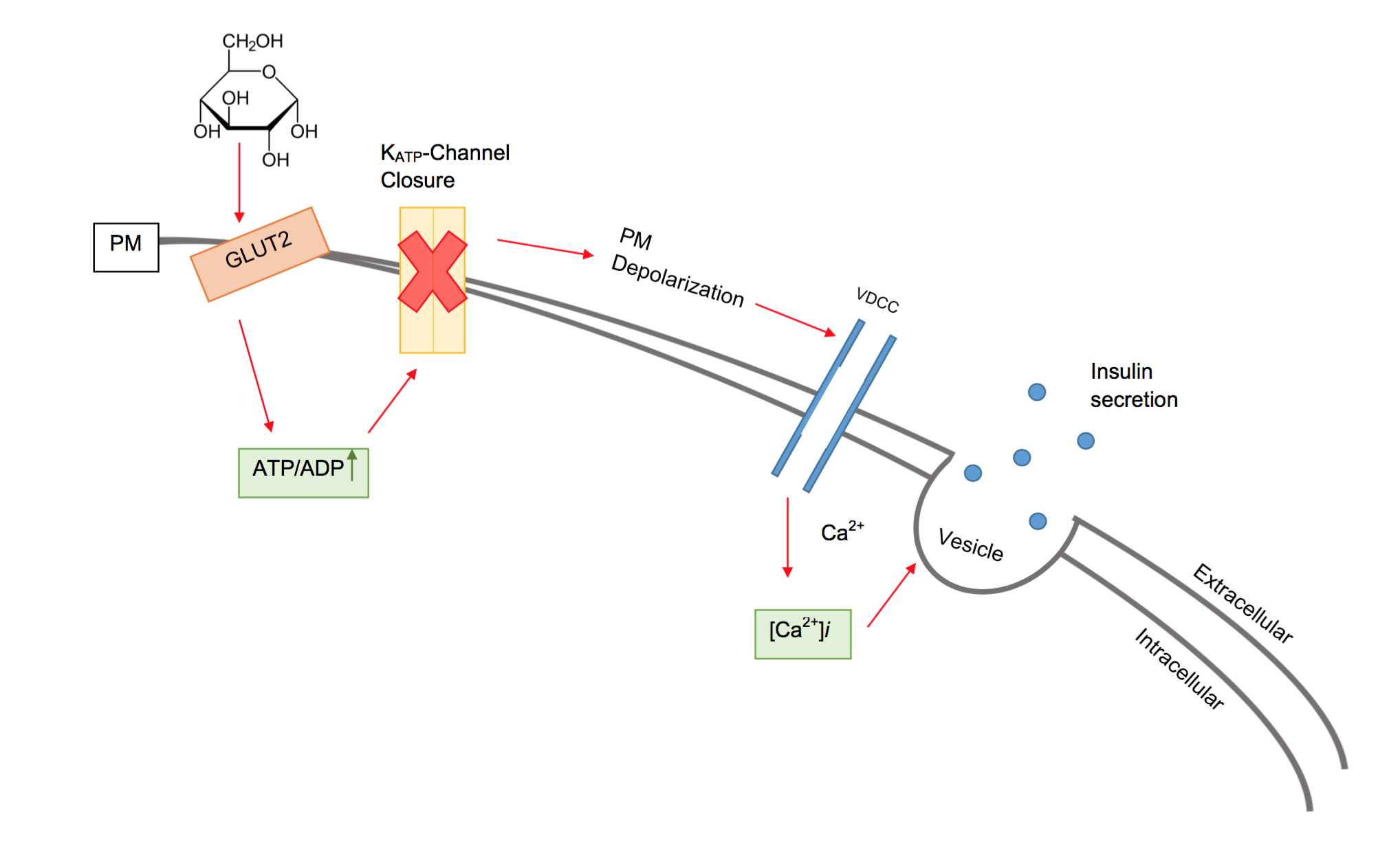
Beta cells (β-cells) are a type of cell found in pancreatic islets that synthesize and secrete insulin and amylin. Beta cells make up 50–70% of the cells in human islets. In patients with Type 1 diabetes, beta-cell mass and function are diminished, leading to insufficient insulin secretion and hyperglycemia. Function The primary function of a beta cell is to produce and release insulin and amylin. Both are hormones which reduce blood glucose levels by different mechanisms. Beta cells can respond quickly to spikes in blood glucose concentrations by secreting some of their stored insulin and amylin while simultaneously producing more. Primary cilia on beta cells regulate their function and energy metabolism. Cilia deletion can lead to islet dysfunction and type 2 diabetes. Insulin synthesis Beta cells are the only site of insulin synthesis in mammals. As glucose stimulates insulin secretion, it simultaneously increases proinsulin biosynthesis, mainly through translational c ... [...More Info...] [...Related Items...] OR: [Wikipedia] [Google] [Baidu] |
Chondrodysplasia
Osteochondrodysplasia is a general term for a disorder of the development (dysplasia) of bone ("osteo") and cartilage ("chondro"). Osteochondrodysplasias are rare diseases. About 1 in 5,000 babies are born with some type of skeletal dysplasia. Nonetheless, if taken collectively, genetic skeletal dysplasias or osteochondrodysplasias comprise a recognizable group of genetically determined disorders with generalized skeletal affection. Osteochondrodysplasias can result in marked functional limitation and even mortality. Osteochondrodysplasias subtypes can overlap in clinical aspects, therefore plain radiography is absolutely necessary to establish an accurate diagnosis. Magnetic resonance imaging can provide further diagnostic insights and guide treatment strategies especially in cases of spinal involvement. Early diagnosis, and timely management of skeletal dysplasia are important to combat functional deterioration. Types Achondroplasia ''Achondroplasia'' is a type of autosomal ... [...More Info...] [...Related Items...] OR: [Wikipedia] [Google] [Baidu] |


