|
CCN Intercellular Signaling Protein
CCN proteins are a family of extracellular matrix (ECM)-associated proteins involved in intercellular signaling. Due to their dynamic role within the ECM they are considered matricellular proteins. Background The acronym CCN is derived from the first three members of the family discovered, namely CYR61 (cysteine-rich angiogenic protein 61 or CCN1), CTGF (connective tissue growth factor or CCN2), and NOV (nephroblastoma overexpressed or CCN3). Together with three Wnt-induced secreted proteins, they comprise the CCN family of matricellular proteins. These proteins have now been renamed CCN1-6 by international consensus. Members of the CCN protein family are characterized by having four conserved cysteine-rich domains, which include the insulin-like growth factor-binding domain (IGFBP), the Von Willebrand factor type C domain (VWC), the thrombospondin type 1 repeat (TSR), and a C-terminal domain (CT) with a cysteine knot motif. CCN proteins have been shown to play important roles in ... [...More Info...] [...Related Items...] OR: [Wikipedia] [Google] [Baidu] |
Extracellular Matrix
In biology, the extracellular matrix (ECM), also called intercellular matrix, is a three-dimensional network consisting of extracellular macromolecules and minerals, such as collagen, enzymes, glycoproteins and hydroxyapatite that provide structural and biochemical support to surrounding cells. Because multicellularity evolved independently in different multicellular lineages, the composition of ECM varies between multicellular structures; however, cell adhesion, cell-to-cell communication and differentiation are common functions of the ECM. The animal extracellular matrix includes the interstitial matrix and the basement membrane. Interstitial matrix is present between various animal cells (i.e., in the intercellular spaces). Gels of polysaccharides and fibrous proteins fill the Interstitial fluid, interstitial space and act as a compression buffer against the stress placed on the ECM. Basement membranes are sheet-like depositions of ECM on which various epithelial cells rest ... [...More Info...] [...Related Items...] OR: [Wikipedia] [Google] [Baidu] |
Cell Adhesion
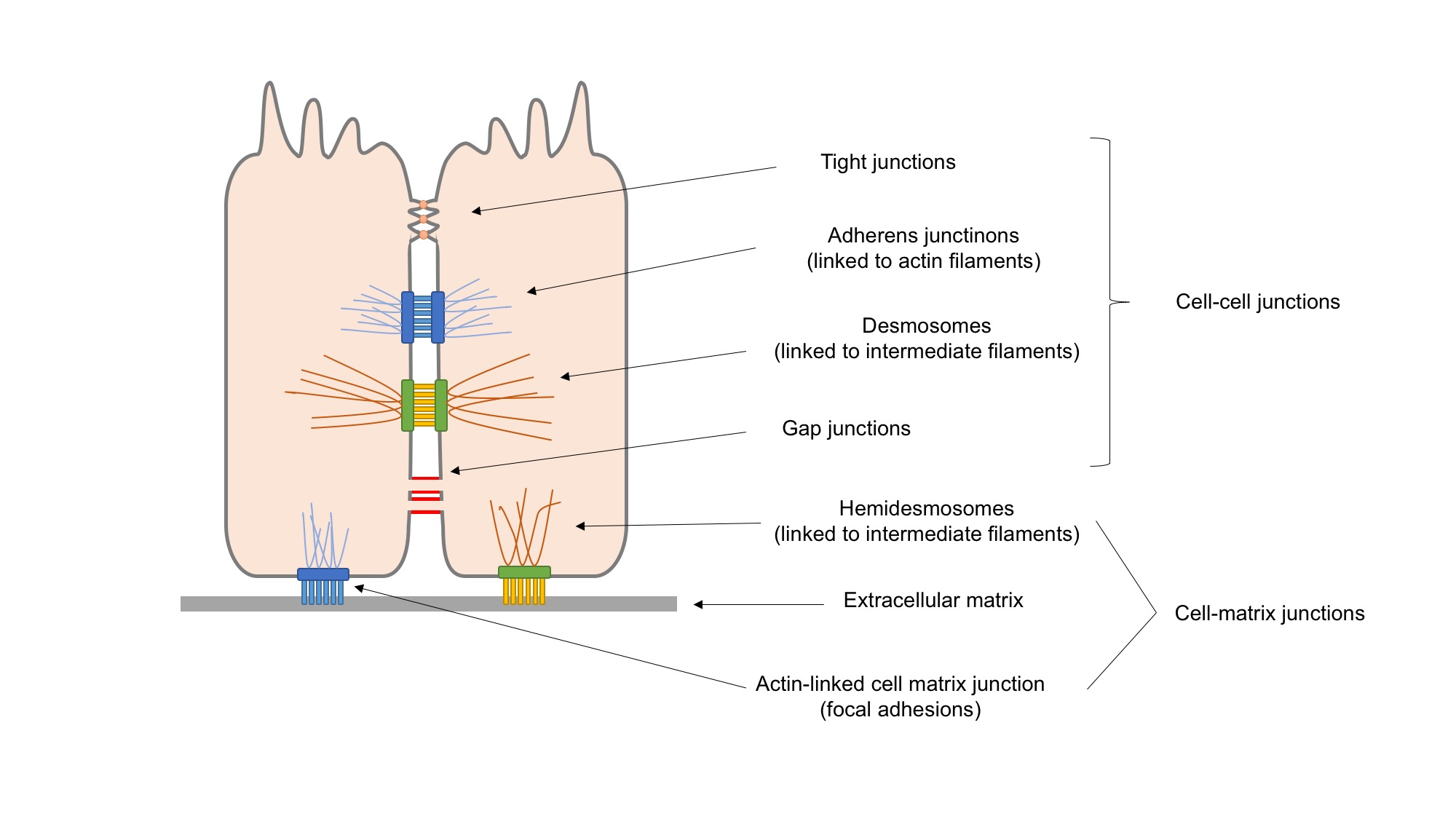
Cell adhesion is the process by which cells interact and attach to neighbouring cells through specialised molecules of the cell surface. This process can occur either through direct contact between cell surfaces such as cell junctions or indirect interaction, where cells attach to surrounding extracellular matrix, a gel-like structure containing molecules released by cells into spaces between them. Cells adhesion occurs from the interactions between cell-adhesion molecules (CAMs), transmembrane proteins located on the cell surface. Cell adhesion links cells in different ways and can be involved in signal transduction for cells to detect and respond to changes in the surroundings. Other cellular processes regulated by cell adhesion include cell migration and tissue development in multicellular organisms. Alterations in cell adhesion can disrupt important cellular processes and lead to a variety of diseases, including cancer and arthritis. Cell adhesion is also essential for in ... [...More Info...] [...Related Items...] OR: [Wikipedia] [Google] [Baidu] |
WISP2
WNT1-inducible-signaling pathway protein 2, or WISP-2 (also named CCN5) is a matricellular protein that in humans is encoded by the ''WISP2'' gene. Function The CCN family of proteins regulates diverse cellular functions, including cell adhesion, migration, proliferation, differentiation. Structure WISP-2 is a member of the CCN family (CCN intercellular signaling protein) of secreted, extracellular matrix (ECM)-associated signaling matricellular proteins. The CCN acronym is derived from the first three members of the family identified, namely CYR61 (CCN1), CTGF (connective tissue growth factor, or CCN2), and NOV. These proteins, together with WISP1/CCN4, WISP2 (CCN5, this gene), and WISP3 (CCN6) comprise the six-member CCN family in vertebrates. CCN proteins characteristically contain an N-terminal secretory signal peptide followed by four structurally distinct domains with homologies to insulin-like growth factor binding protein (IGFBP), von Willebrand type C repeats ( v ... [...More Info...] [...Related Items...] OR: [Wikipedia] [Google] [Baidu] |
WISP1
WNT1-inducible-signaling pathway protein 1 (WISP-1), also known as CCN4, is a matricellular protein that in humans is encoded by the ''WISP1'' gene. Structure WISP-1 is highly homologous to CYR61 (CCN1) and CTGF (CCN2), and is a member of the CCN family of secreted, extracellular matrix (ECM)-associated signaling proteins (CCN intercellular signaling protein). The CCN family of proteins shares a common molecular protein structure, characterized by an N-terminal secretory signal peptide followed by four distinct domains with homologies to insulin-like growth factor binding protein (IGFBP), von Willebrand type C repeats ( vWC), thrombospondin type 1 repeat (TSR), and a cysteine knot motif within the C-terminal (CT) domain. This family of proteins regulates diverse cellular functions, including cell adhesion, migration, proliferation, differentiation, and survival. Role in bone development WISP-1 promotes mesenchymal cell proliferation and osteoblastic differentiation, and re ... [...More Info...] [...Related Items...] OR: [Wikipedia] [Google] [Baidu] |
Tumorigenesis
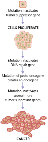
Carcinogenesis, also called oncogenesis or tumorigenesis, is the formation of a cancer, whereby normal cells are transformed into cancer cells. The process is characterized by changes at the cellular, genetic, and epigenetic levels and abnormal cell division. Cell division is a physiological process that occurs in almost all tissues and under a variety of circumstances. Normally, the balance between proliferation and programmed cell death, in the form of apoptosis, is maintained to ensure the integrity of tissues and organs. According to the prevailing accepted theory of carcinogenesis, the somatic mutation theory, mutations in DNA and epimutations that lead to cancer disrupt these orderly processes by interfering with the programming regulating the processes, upsetting the normal balance between proliferation and cell death. This results in uncontrolled cell division and the evolution of those cells by natural selection in the body. Only certain mutations lead to cancer ... [...More Info...] [...Related Items...] OR: [Wikipedia] [Google] [Baidu] |
Fibrosis
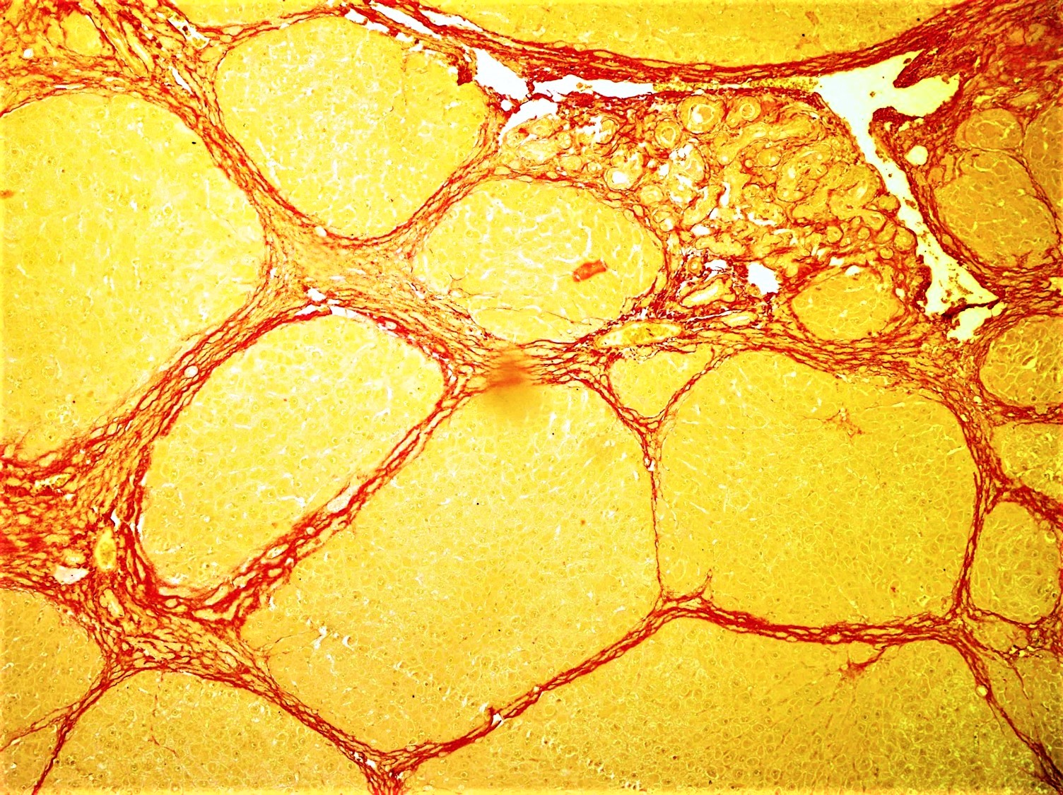
Fibrosis, also known as fibrotic scarring, is a pathological wound healing in which connective tissue replaces normal parenchymal tissue to the extent that it goes unchecked, leading to considerable tissue remodelling and the formation of permanent scar tissue. Repeated injuries, chronic inflammation and repair are susceptible to fibrosis where an accidental excessive accumulation of extracellular matrix components, such as the collagen is produced by fibroblasts, leading to the formation of a permanent fibrotic scar. In response to injury, this is called scarring, and if fibrosis arises from a single cell line, this is called a fibroma. Physiologically, fibrosis acts to deposit connective tissue, which can interfere with or totally inhibit the normal architecture and function of the underlying organ or tissue. Fibrosis can be used to describe the pathological state of excess deposition of fibrous tissue, as well as the process of connective tissue deposition in healing. Define ... [...More Info...] [...Related Items...] OR: [Wikipedia] [Google] [Baidu] |
Angiogenesis
Angiogenesis is the physiological process through which new blood vessels form from pre-existing vessels, formed in the earlier stage of vasculogenesis. Angiogenesis continues the growth of the vasculature by processes of sprouting and splitting. Vasculogenesis is the embryonic formation of endothelial cells from mesoderm cell precursors, and from neovascularization, although discussions are not always precise (especially in older texts). The first vessels in the developing embryo form through vasculogenesis, after which angiogenesis is responsible for most, if not all, blood vessel growth during development and in disease. Angiogenesis is a normal and vital process in growth and development, as well as in wound healing and in the formation of granulation tissue. However, it is also a fundamental step in the transition of tumors from a benign state to a malignant one, leading to the use of angiogenesis inhibitors in the treatment of cancer. The essential role of angiogenesis in ... [...More Info...] [...Related Items...] OR: [Wikipedia] [Google] [Baidu] |
Senescence
Senescence () or biological aging is the gradual deterioration of functional characteristics in living organisms. The word ''senescence'' can refer to either cellular senescence or to senescence of the whole organism. Organismal senescence involves an increase in death rates and/or a decrease in fecundity with increasing age, at least in the latter part of an organism's life cycle. Senescence is the inevitable fate of almost all multicellular organisms with germ-soma separation, but it can be delayed. The discovery, in 1934, that calorie restriction can extend lifespan by 50% in rats, and the existence of species having negligible senescence and potentially immortal organisms such as '' Hydra'', have motivated research into delaying senescence and thus age-related diseases. Rare human mutations can cause accelerated aging diseases. Environmental factors may affect aging – for example, overexposure to ultraviolet radiation accelerates skin aging. Different parts of the body ... [...More Info...] [...Related Items...] OR: [Wikipedia] [Google] [Baidu] |
Apoptosis
Apoptosis (from grc, ἀπόπτωσις, apóptōsis, 'falling off') is a form of programmed cell death that occurs in multicellular organisms. Biochemical events lead to characteristic cell changes (morphology) and death. These changes include blebbing, cell shrinkage, nuclear fragmentation, chromatin condensation, DNA fragmentation, and mRNA decay. The average adult human loses between 50 and 70 billion cells each day due to apoptosis. For an average human child between eight and fourteen years old, approximately twenty to thirty billion cells die per day. In contrast to necrosis, which is a form of traumatic cell death that results from acute cellular injury, apoptosis is a highly regulated and controlled process that confers advantages during an organism's life cycle. For example, the separation of fingers and toes in a developing human embryo occurs because cells between the digits undergo apoptosis. Unlike necrosis, apoptosis produces cell fragments called apoptotic ... [...More Info...] [...Related Items...] OR: [Wikipedia] [Google] [Baidu] |
Cell Proliferation
Cell proliferation is the process by which ''a cell grows and divides to produce two daughter cells''. Cell proliferation leads to an exponential increase in cell number and is therefore a rapid mechanism of tissue growth. Cell proliferation requires both cell growth and cell division to occur at the same time, such that the average size of cells remains constant in the population. Cell division can occur without cell growth, producing many progressively smaller cells (as in cleavage of the zygote), while cell growth can occur without cell division to produce a single larger cell (as in growth of neurons). Thus, cell proliferation is not synonymous with either cell growth or cell division, despite the fact that these terms are sometimes used interchangeably. Stem cells undergo cell proliferation to produce proliferating "transit amplifying" daughter cells that later differentiate to construct tissues during normal development and tissue growth, during tissue regeneration aft ... [...More Info...] [...Related Items...] OR: [Wikipedia] [Google] [Baidu] |
Cysteine Knot
A cystine knot is a protein structural motif containing three disulfide bridges (formed from pairs of cysteine residues). The sections of polypeptide that occur between two of them form a loop through which a third disulfide bond passes, forming a rotaxane substructure. The cystine knot motif stabilizes protein structure and is conserved in proteins across various species. There are three types of cystine knot, which differ in the topology of the disulfide bonds: * The growth factor cystine knot (GFCK) * inhibitor cystine knot (ICK) common in spider and snail toxins * Cyclic Cystine Knot, or cyclotide The growth factor cystine knot was first observed in the structure of nerve growth factor (NGF), solved by X-ray crystallography and published in 1991 by Tom Blundell in Nature (journal), Nature.; The GFCK is present in four superfamilies. These include nerve growth factor, transforming growth factor beta (TGF-β), platelet-derived growth factor, and glycoprotein hormones including ... [...More Info...] [...Related Items...] OR: [Wikipedia] [Google] [Baidu] |
Cell Signaling
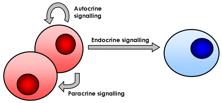
In biology, cell signaling (cell signalling in British English) or cell communication is the ability of a cell to receive, process, and transmit signals with its environment and with itself. Cell signaling is a fundamental property of all cellular life in prokaryotes and eukaryotes. Signals that originate from outside a cell (or extracellular signals) can be physical agents like mechanical pressure, voltage, temperature, light, or chemical signals (e.g., small molecules, peptides, or gas). Cell signaling can occur over short or long distances, and as a result can be classified as autocrine, juxtacrine, intracrine, paracrine, or endocrine. Signaling molecules can be synthesized from various biosynthetic pathways and released through passive or active transports, or even from cell damage. Receptors play a key role in cell signaling as they are able to detect chemical signals or physical stimuli. Receptors are generally proteins located on the cell surface or within the interio ... [...More Info...] [...Related Items...] OR: [Wikipedia] [Google] [Baidu] |


