|
Brodmann Area 6
Brodmann area 6 (BA6) is part of the frontal cortex in the human brain. Situated just anterior to the primary motor cortex ( BA4), it is composed of the premotor cortex and, medially, the supplementary motor area (SMA). This large area of the frontal cortex is believed to play a role in planning complex, coordinated movements. Brodmann area 6 is also called agranular frontal area 6 in humans because it lacks an internal granular cortical layer (layer IV). It is a subdivision of the cytoarchitecturally defined precentral region of cerebral cortex. In the human brain, it is located on the portions of the precentral gyrus that are not occupied by Brodmann area 4; furthermore, BA6 extends onto the caudal portions of the superior frontal and middle frontal gyri. It extends from the cingulate sulcus on the medial aspect of the hemisphere to the lateral sulcus on the lateral aspect. It is bounded rostrally by the granular frontal region and caudally by the gigantopyramidal area 4 (Brod ... [...More Info...] [...Related Items...] OR: [Wikipedia] [Google] [Baidu] |
Frontal Lobe
The frontal lobe is the largest of the four major lobes of the brain in mammals, and is located at the front of each cerebral hemisphere (in front of the parietal lobe and the temporal lobe). It is parted from the parietal lobe by a groove between tissues called the central sulcus and from the temporal lobe by a deeper groove called the lateral sulcus (Sylvian fissure). The most anterior rounded part of the frontal lobe (though not well-defined) is known as the frontal pole, one of the three poles of the cerebrum. The frontal lobe is covered by the frontal cortex. The frontal cortex includes the premotor cortex, and the primary motor cortex – parts of the motor cortex. The front part of the frontal cortex is covered by the prefrontal cortex. There are four principal gyri in the frontal lobe. The precentral gyrus is directly anterior to the central sulcus, running parallel to it and contains the primary motor cortex, which controls voluntary movements of specific body parts ... [...More Info...] [...Related Items...] OR: [Wikipedia] [Google] [Baidu] |
Cingulate Sulcus
The cingulate sulcus is a sulcus (brain fold) on the cingulate cortex in the medial wall of the cerebral cortex. The frontal and parietal lobes are separated from the cingulate gyrus by the cingulate sulcus. It terminates as the marginal sulcus of the cingulate sulcus. It sends a ramus to separate the paracentral lobule from the frontal gyri, the paracentral sulcus. Additional images File:Cingulate sulcus animation small.gif, Position of cingulate sulcus (shown in red). File:LobesCaptsMedial1.png, Medial surface of right cerebral hemisphere. Cingulate sulcus (labeled as sulcus cinguli) and brain lobes. File:Slide2ZEN.JPG, Medial surface of cerebral hemisphere.Medial view.Deep dissection. File:Slide3ZEN.JPG, Medial surface of cerebral hemisphere.Medial view.Deep dissection. File:Slide4ZE.JPG, Medial surface of cerebral hemisphere.Medial view.Deep dissection. External links * NIF Search - Cingulate Sulcusvia the Neuroscience Information Framework The Neuroscience Information ... [...More Info...] [...Related Items...] OR: [Wikipedia] [Google] [Baidu] |
Korbinian Brodmann
Korbinian Brodmann (17 November 1868 – 22 August 1918) was a German neurologist who became famous for mapping the cerebral cortex and defining 52 distinct regions, known as Brodmann areas, based on their cytoarchitectonic (histological) characteristics. Life and career Brodmann was born in Liggersdorf, Province of Hohenzollern. He studied medicine in Munich, Würzburg, Berlin, and Freiburg, where he received his medical diploma in 1895. Subsequently he studied at the Medical School in the University of Lausanne in Switzerland, and then worked in the University Clinic in Munich. He received a doctor of medicine degree from the University of Leipzig in 1898, with a thesis on chronic ependymal sclerosis. From 1900 to 1901, Brodmann also worked in the Psychiatric Clinic at the University of Jena, with Ludwig Binswanger, and in the Municipal Mental Asylum in Frankfurt. There, he met Alois Alzheimer, who was influential in his decision to pursue basic neuroscience research. Fol ... [...More Info...] [...Related Items...] OR: [Wikipedia] [Google] [Baidu] |
List Of Regions In The Human Brain
The human brain anatomical regions are ordered following standard neuroanatomy hierarchies. Functional, connective, and developmental regions are listed in parentheses where appropriate. Hindbrain (rhombencephalon) Myelencephalon * Medulla oblongata **Medullary pyramids **Arcuate nucleus **Olivary body ***Inferior olivary nucleus **Rostral ventrolateral medulla **Caudal ventrolateral medulla **Solitary nucleus (Nucleus of the solitary tract) **Respiratory center- Respiratory groups ***Dorsal respiratory group ***Ventral respiratory group or Apneustic centre ****Pre-Bötzinger complex ****Botzinger complex ****Retrotrapezoid nucleus ****Nucleus retrofacialis ****Nucleus retroambiguus ****Nucleus para-ambiguus **Paramedian reticular nucleus **Gigantocellular reticular nucleus **Parafacial zone **Cuneate nucleus ** Gracile nucleus ** Perihypoglossal nuclei *** Intercalated nucleus *** Prepositus nucleus *** Sublingual nucleus **Area postrema **Medullary cranial nerve nucl ... [...More Info...] [...Related Items...] OR: [Wikipedia] [Google] [Baidu] |
Brodmann Area
A Brodmann area is a region of the cerebral cortex, in the human or other primate brain, defined by its cytoarchitecture, or histological structure and organization of cells. History Brodmann areas were originally defined and numbered by the German anatomist Korbinian Brodmann based on the cytoarchitectural organization of neurons he observed in the cerebral cortex using the Nissl method of cell staining. Brodmann published his maps of cortical areas in humans, monkeys, and other species in 1909, along with many other findings and observations regarding the general cell types and laminar organization of the mammalian cortex. The same Brodmann area number in different species does not necessarily indicate homologous areas. A similar, but more detailed cortical map was published by Constantin von Economo and Georg N. Koskinas in 1925. Present importance Brodmann areas have been discussed, debated, refined, and renamed exhaustively for nearly a century and remain the most wid ... [...More Info...] [...Related Items...] OR: [Wikipedia] [Google] [Baidu] |
Granular Layer (cerebral Cortex)
The internal granular layer of the cortex, also commonly referred to as the granular layer of the cortex, is the layer IV in the subdivision of the mammalian cortex into 6 layers. The adjective internal is used in opposition to the external granular layer of the cortex, the term granular refers to the granule cells found here. This layer receives the afferent connections from the thalamus and from other cortical regions and sends connections to the other layers. The line of Gennari The line of Gennari (also called the "band" or "stria" of Gennari) is a band of myelinated axons that runs parallel to the surface of the cerebral cortex on the banks of the calcarine fissure in the occipital lobe. This formation is visible to the ... (occipital stripe) is also present in this layer. Cerebral cortex {{neuroanatomy-stub ... [...More Info...] [...Related Items...] OR: [Wikipedia] [Google] [Baidu] |
White Matter
White matter refers to areas of the central nervous system (CNS) that are mainly made up of myelinated axons, also called tracts. Long thought to be passive tissue, white matter affects learning and brain functions, modulating the distribution of action potentials, acting as a relay and coordinating communication between different brain regions. White matter is named for its relatively light appearance resulting from the lipid content of myelin. However, the tissue of the freshly cut brain appears pinkish-white to the naked eye because myelin is composed largely of lipid tissue veined with capillaries. Its white color in prepared specimens is due to its usual preservation in formaldehyde. Structure White matter White matter is composed of bundles, which connect various grey matter areas (the locations of nerve cell bodies) of the brain to each other, and carry nerve impulses between neurons. Myelin acts as an insulator, which allows electrical signals to jump, rather than c ... [...More Info...] [...Related Items...] OR: [Wikipedia] [Google] [Baidu] |
Homology (biology)
In biology, homology is similarity due to shared ancestry between a pair of structures or genes in different taxa. A common example of homologous structures is the forelimbs of vertebrates, where the wings of bats and birds, the arms of primates, the front flippers of whales and the forelegs of four-legged vertebrates like dogs and crocodiles are all derived from the same ancestral tetrapod structure. Evolutionary biology explains homologous structures adapted to different purposes as the result of descent with modification from a common ancestor. The term was first applied to biology in a non-evolutionary context by the anatomist Richard Owen in 1843. Homology was later explained by Charles Darwin's theory of evolution in 1859, but had been observed before this, from Aristotle onwards, and it was explicitly analysed by Pierre Belon in 1555. In developmental biology, organs that developed in the embryo in the same manner and from similar origins, such as from matching p ... [...More Info...] [...Related Items...] OR: [Wikipedia] [Google] [Baidu] |
Guenon
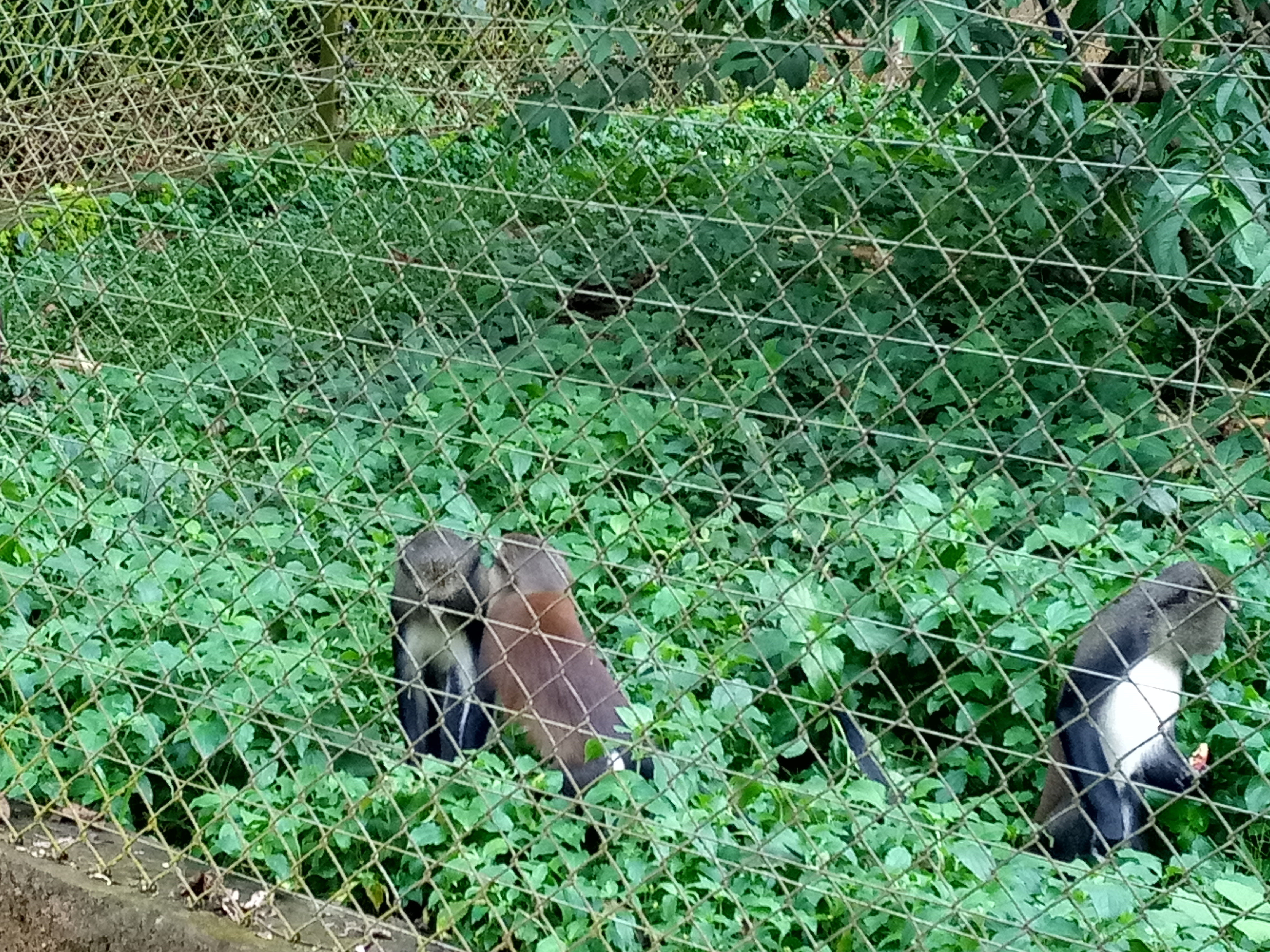
The guenons (, ) are Old World monkeys of the genus ''Cercopithecus'' (). Not all members of this genus have the word "guenon" in their common names; also, because of changes in scientific classification, some monkeys in other genera may have common names that include the word "guenon". Nonetheless, the use of the term guenon for monkeys of this genus is widely accepted. All members of the genus are endemic to sub-Saharan Africa, and most are forest monkeys. Many of the species are quite local in their ranges, and some have even more local subspecies. Many are threatened or endangered because of habitat loss. The species currently placed in the genus ''Chlorocebus'', such as vervet monkeys and green monkeys, were formerly considered as a single species in this genus, ''Cercopithecus aethiops''. In the English language, the word "guenon" is apparently of French origin. In French, ''guenon'' was the common name for all species and individuals, both males and females, from the gen ... [...More Info...] [...Related Items...] OR: [Wikipedia] [Google] [Baidu] |
Lateral Sulcus
In neuroanatomy, the lateral sulcus (also called Sylvian fissure, after Franciscus Sylvius, or lateral fissure) is one of the most prominent features of the human brain. The lateral sulcus is a deep fissure in each hemisphere that separates the frontal and parietal lobes from the temporal lobe. The insular cortex lies deep within the lateral sulcus. Anatomy The lateral sulcus divides both the frontal lobe and parietal lobe above from the temporal lobe below. It is in both hemispheres of the brain. The lateral sulcus is one of the earliest-developing sulci of the human brain. It first appears around the fourteenth gestational week. The insular cortex lies deep within the lateral sulcus. The lateral sulcus has a number of side branches. Two of the most prominent and most regularly found are the ascending (also called vertical) ramus and the horizontal ramus of the lateral fissure, which subdivide the inferior frontal gyrus. The lateral sulcus also contains the transverse tempor ... [...More Info...] [...Related Items...] OR: [Wikipedia] [Google] [Baidu] |
Middle Frontal Gyrus
The middle frontal gyrus makes up about one-third of the frontal lobe of the human brain. (A ''gyrus'' is one of the prominent "bumps" or "ridges" on the surface of the human brain.) The middle frontal gyrus, like the inferior frontal gyrus and the superior frontal gyrus, is more of a region in the frontal gyrus than a true gyrus. The borders of the middle frontal gyrus are the ''inferior frontal sulcus'' below; the ''superior frontal sulcus'' above; and the precentral sulcus The precentral sulcus is a part of the human brain that lies parallel to, and in front of, the central sulcus. (A ''sulcus'' is one of the prominent grooves on the surface of the human brain.) The precentral sulcus divides the inferior, middl ... behind. Additional images File:Middle frontal gyrus animation small.gif, Position of middle frontal gyrus (shown in red). File:Gray725 middle frontal gyrus.png, Left cerebral hemisphere seen from above. File:Gray726 middle frontal gyrus.png, Lateral ... [...More Info...] [...Related Items...] OR: [Wikipedia] [Google] [Baidu] |
Cerebral Cortex
The cerebral cortex, also known as the cerebral mantle, is the outer layer of neural tissue of the cerebrum of the brain in humans and other mammals. The cerebral cortex mostly consists of the six-layered neocortex, with just 10% consisting of allocortex. It is separated into two cortices, by the longitudinal fissure that divides the cerebrum into the left and right cerebral hemispheres. The two hemispheres are joined beneath the cortex by the corpus callosum. The cerebral cortex is the largest site of neural integration in the central nervous system. It plays a key role in attention, perception, awareness, thought, memory, language, and consciousness. The cerebral cortex is part of the brain responsible for cognition. In most mammals, apart from small mammals that have small brains, the cerebral cortex is folded, providing a greater surface area in the confined volume of the cranium. Apart from minimising brain and cranial volume, cortical folding is crucial for the brain ... [...More Info...] [...Related Items...] OR: [Wikipedia] [Google] [Baidu] |


