|
Brodmann Area
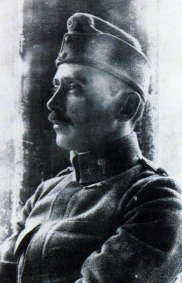
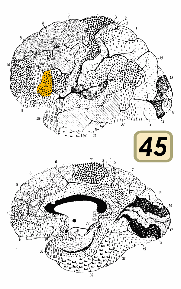
A Brodmann area is a region of the cerebral cortex, in the human or other primate brain, defined by its cytoarchitecture, or histological structure and organization of cells. History Brodmann areas were originally defined and numbered by the German anatomist Korbinian Brodmann based on the cytoarchitectural organization of neurons he observed in the cerebral cortex using the Nissl method of cell staining. Brodmann published his maps of cortical areas in humans, monkeys, and other species in 1909, along with many other findings and observations regarding the general cell types and laminar organization of the mammalian cortex. The same Brodmann area number in different species does not necessarily indicate homologous areas. A similar, but more detailed cortical map was published by Constantin von Economo and Georg N. Koskinas in 1925. Present importance Brodmann areas have been discussed, debated, refined, and renamed exhaustively for nearly a century and remain the most w ... [...More Info...] [...Related Items...] OR: [Wikipedia] [Google] [Baidu] |
Cerebrum
The cerebrum, telencephalon or endbrain is the largest part of the brain containing the cerebral cortex (of the two cerebral hemispheres), as well as several subcortical structures, including the hippocampus, basal ganglia, and olfactory bulb. In the human brain, the cerebrum is the uppermost region of the central nervous system. The cerebrum develops prenatally from the forebrain (prosencephalon). In mammals, the dorsal telencephalon, or pallium, develops into the cerebral cortex, and the ventral telencephalon, or subpallium, becomes the basal ganglia. The cerebrum is also divided into approximately symmetric left and right cerebral hemispheres. With the assistance of the cerebellum, the cerebrum controls all voluntary actions in the human body. Structure The cerebrum is the largest part of the brain. Depending upon the position of the animal it lies either in front or on top of the brainstem. In humans, the cerebrum is the largest and best-developed of the five ma ... [...More Info...] [...Related Items...] OR: [Wikipedia] [Google] [Baidu] |
Constantin Von Economo
Constantin Freiherr von Economo ( gr, Κωνσταντίνος Οικονόμου; 21 August 1876 – 21 October 1931) was an Austrian psychiatrist and neurologist of Greek descent, born in modern-day Romania (then Ottoman Empire). He is mostly known for his discovery of encephalitis lethargica and his atlas of cytoarchitectonics of the cerebral cortex. Biography Youth and schooling Constantin Economo von San Serff was born in Brăila, Romania, to Johannes and Helene Economo, a wealthy family with large holdings in Thessaly and Macedonia. The Economo (Οικονόμου, '' Oikonomou'') family originated from Edessa, in the Ottoman Sanjak of Salonica (modern Edessa, Central Macedonia, Greece) where some of Constantin's ancestors were notables, and his family included many bishops. In 1877, the family moved to Trieste, Austria-Hungary,Economo, K. (1932). ''Constantin Freiherr von Economo''. Wien: Mayer & Co. and Constantin spent his childhood and youth in Trieste. He ... [...More Info...] [...Related Items...] OR: [Wikipedia] [Google] [Baidu] |
Neocortex
The neocortex, also called the neopallium, isocortex, or the six-layered cortex, is a set of layers of the mammalian cerebral cortex involved in higher-order brain functions such as sensory perception, cognition, generation of motor commands, spatial reasoning and language. The neocortex is further subdivided into the true isocortex and the proisocortex. In the human brain, the neocortex is the largest part of the cerebral cortex (the outer layer of the cerebrum). The neocortex makes up the largest part of the cerebral cortex, with the allocortex making up the rest. The neocortex is made up of six layers, labelled from the outermost inwards, I to VI. Etymology The term is from ''cortex'', Latin, "bark" or "rind", combined with ''neo-'', Greek, "new". ''Neopallium'' is a similar hybrid, from Latin ''pallium'', "cloak". ''Isocortex'' and ''allocortex'' are hybrids with Greek ''isos'', "same", and ''allos'', "other". Anatomy The neocortex is the most developed in its organisa ... [...More Info...] [...Related Items...] OR: [Wikipedia] [Google] [Baidu] |
Neuroscientist
A neuroscientist (or neurobiologist) is a scientist who has specialised knowledge in neuroscience, a branch of biology that deals with the physiology, biochemistry, psychology, anatomy and molecular biology of neurons, neural circuits, and glial cells and especially their behavioral, biological, and psychological aspect in health and disease. Neuroscientists generally work as researchers within a college, university, government agency, or private industry setting. In research-oriented careers, neuroscientists typically spend their time designing and carrying out scientific experiments that contribute to the understanding of the nervous system and its function. They can engage in basic or applied research. Basic research seeks to add information to our current understanding of the nervous system, whereas applied research seeks to address a specific problem, such as developing a treatment for a neurological disorder. Biomedically-oriented neuroscientists typically engage in app ... [...More Info...] [...Related Items...] OR: [Wikipedia] [Google] [Baidu] |
Brodmann Areas 44 And 45
Broca's area, or the Broca area (, also , ), is a region in the frontal lobe of the dominant hemisphere, usually the left, of the brain with functions linked to speech production. Language processing has been linked to Broca's area since Pierre Paul Broca reported impairments in two patients. They had lost the ability to speak after injury to the posterior inferior frontal gyrus (pars triangularis) (BA45) of the brain. Since then, the approximate region he identified has become known as Broca's area, and the deficit in language production as Broca's aphasia, also called expressive aphasia. Broca's area is now typically defined in terms of the pars opercularis and pars triangularis of the inferior frontal gyrus, represented in Brodmann's cytoarchitectonic map as Brodmann area 44 and Brodmann area 45 of the dominant hemisphere. Functional magnetic resonance imaging (fMRI) has shown language processing to also involve the third part of the inferior frontal gyrus the pars orbita ... [...More Info...] [...Related Items...] OR: [Wikipedia] [Google] [Baidu] |
Broca's Area
Broca's area, or the Broca area (, also , ), is a region in the frontal lobe of the dominant hemisphere, usually the left, of the brain with functions linked to speech production. Language processing has been linked to Broca's area since Pierre Paul Broca reported impairments in two patients. They had lost the ability to speak after injury to the posterior inferior frontal gyrus (pars triangularis) (BA45) of the brain. Since then, the approximate region he identified has become known as Broca's area, and the deficit in language production as Broca's aphasia, also called expressive aphasia. Broca's area is now typically defined in terms of the pars opercularis and pars triangularis of the inferior frontal gyrus, represented in Brodmann's cytoarchitectonic map as Brodmann area 44 and Brodmann area 45 of the dominant hemisphere. Functional magnetic resonance imaging (fMRI) has shown language processing to also involve the third part of the inferior frontal gyrus the pars or ... [...More Info...] [...Related Items...] OR: [Wikipedia] [Google] [Baidu] |
Functional Magnetic Resonance Imaging
Functional magnetic resonance imaging or functional MRI (fMRI) measures brain activity by detecting changes associated with blood flow. This technique relies on the fact that cerebral blood flow and neuronal activation are coupled. When an area of the brain is in use, blood flow to that region also increases. The primary form of fMRI uses the blood-oxygen-level dependent (BOLD) contrast, discovered by Seiji Ogawa in 1990. This is a type of specialized brain and body scan used to map neural activity in the brain or spinal cord of humans or other animals by imaging the change in blood flow (hemodynamic response) related to energy use by brain cells. Since the early 1990s, fMRI has come to dominate brain mapping research because it does not involve the use of injections, surgery, the ingestion of substances, or exposure to ionizing radiation. This measure is frequently corrupted by noise from various sources; hence, statistical procedures are used to extract the underlying signal. ... [...More Info...] [...Related Items...] OR: [Wikipedia] [Google] [Baidu] |
Neurophysiology
Neurophysiology is a branch of physiology and neuroscience that studies nervous system function rather than nervous system architecture. This area aids in the diagnosis and monitoring of neurological diseases. Historically, it has been dominated by electrophysiology—the electrical recording of neural activity ranging from the molar (the electroencephalogram, EEG) to the cellular (intracellular recording of the properties of single neurons), such as patch clamp, voltage clamp, extracellular single-unit recording and recording of local field potentials. However, since the neurone is an electrochemical machine, it is difficult to isolate electrical events from the metabolic and molecular processes that cause them. Thus, neurophysiologists currently utilise tools from chemistry (calcium imaging), physics (functional magnetic resonance imaging, fMRI), and molecular biology (site directed mutations) to examine brain activity. The word originates from the Greek word meaning "nerve" ... [...More Info...] [...Related Items...] OR: [Wikipedia] [Google] [Baidu] |
Higher-order Thinking
Higher-order thinking, known as higher order thinking skills (HOTS), is a concept of education reform based on learning taxonomies (such as Bloom's taxonomy). The idea is that some types of learning require more cognitive processing than others, but also have more generalized benefits. In Bloom's taxonomy, for example, skills involving analysis, evaluation and synthesis (creation of new knowledge) are thought to be of a higher order than the learning of facts and concepts which requires different learning and teaching methods. Higher-order thinking involves the learning of complex judgmental skills such as critical thinking and problem solving. Higher-order thinking is more difficult to learn or teach but also more valuable because such skills are more likely to be usable in novel situations (i.e., situations other than those in which the skill was learned). Education reform It is a notion that students must master the lower level skills before they can engage in higher-order ... [...More Info...] [...Related Items...] OR: [Wikipedia] [Google] [Baidu] |
Primary Auditory Cortex
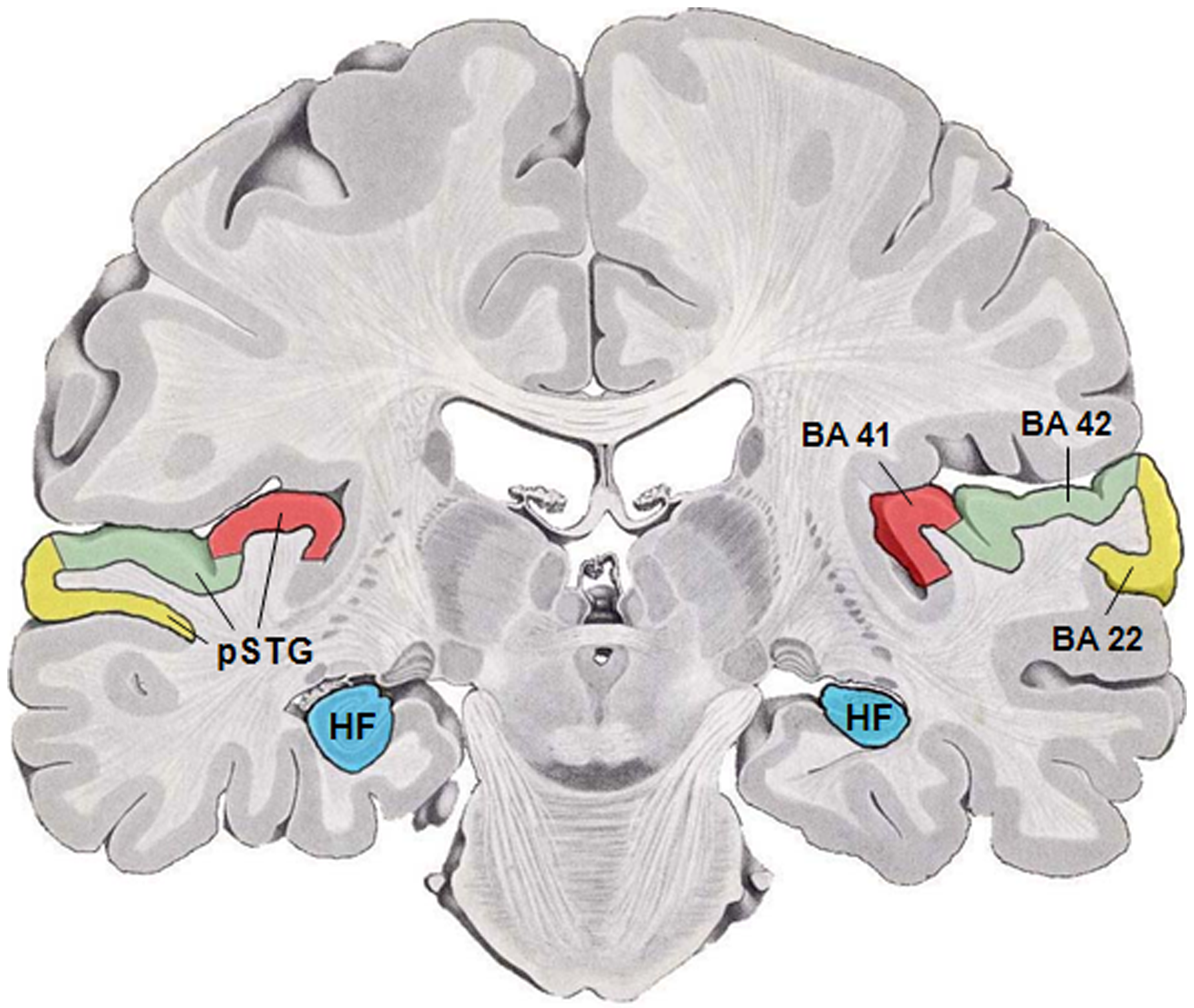
The auditory cortex is the part of the temporal lobe that processes auditory information in humans and many other vertebrates. It is a part of the auditory system, performing basic and higher functions in hearing, such as possible relations to language switching.Cf. Pickles, James O. (2012). ''An Introduction to the Physiology of Hearing'' (4th ed.). Bingley, UK: Emerald Group Publishing Limited, p. 238. It is located bilaterally, roughly at the upper sides of the temporal lobes – in humans, curving down and onto the medial surface, on the superior temporal plane, within the lateral sulcus and comprising parts of the transverse temporal gyri, and the superior temporal gyrus, including the planum polare and planum temporale (roughly Brodmann areas 41 and 42, and partially 22). The auditory cortex takes part in the spectrotemporal, meaning involving time and frequency, analysis of the inputs passed on from the ear. The cortex then filters and passes on the information to th ... [...More Info...] [...Related Items...] OR: [Wikipedia] [Google] [Baidu] |
Primary Visual Cortex
The visual cortex of the brain is the area of the cerebral cortex that processes visual information. It is located in the occipital lobe. Sensory input originating from the eyes travels through the lateral geniculate nucleus in the thalamus and then reaches the visual cortex. The area of the visual cortex that receives the sensory input from the lateral geniculate nucleus is the primary visual cortex, also known as visual area 1 ( V1), Brodmann area 17, or the striate cortex. The extrastriate areas consist of visual areas 2, 3, 4, and 5 (also known as V2, V3, V4, and V5, or Brodmann area 18 and all Brodmann area 19). Both hemispheres of the brain include a visual cortex; the visual cortex in the left hemisphere receives signals from the right visual field, and the visual cortex in the right hemisphere receives signals from the left visual field. Introduction The primary visual cortex (V1) is located in and around the calcarine fissure in the occipital lobe. Each hemisphere' ... [...More Info...] [...Related Items...] OR: [Wikipedia] [Google] [Baidu] |


