|
Frontal Lobe
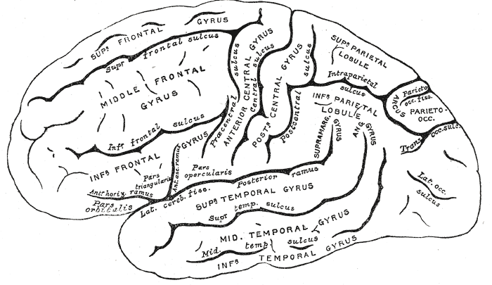
The frontal lobe is the largest of the four major lobes of the brain in mammals, and is located at the front of each cerebral hemisphere (in front of the parietal lobe and the temporal lobe). It is parted from the parietal lobe by a groove between tissues called the central sulcus and from the temporal lobe by a deeper groove called the lateral sulcus (Sylvian fissure). The most anterior rounded part of the frontal lobe (though not well-defined) is known as the frontal pole, one of the three poles of the cerebrum. The frontal lobe is covered by the frontal cortex. The frontal cortex includes the premotor cortex, and the primary motor cortex – parts of the motor cortex. The front part of the frontal cortex is covered by the prefrontal cortex. There are four principal gyri in the frontal lobe. The precentral gyrus is directly anterior to the central sulcus, running parallel to it and contains the primary motor cortex, which controls voluntary movements of specific body parts ... [...More Info...] [...Related Items...] OR: [Wikipedia] [Google] [Baidu] |
Cerebrum
The cerebrum, telencephalon or endbrain is the largest part of the brain containing the cerebral cortex (of the two cerebral hemispheres), as well as several subcortical structures, including the hippocampus, basal ganglia, and olfactory bulb. In the human brain, the cerebrum is the uppermost region of the central nervous system. The cerebrum prenatal development, develops prenatally from the forebrain (prosencephalon). In mammals, the Dorsum (biology), dorsal telencephalon, or Pallium (neuroanatomy), pallium, develops into the cerebral cortex, and the ventral telencephalon, or Pallium (neuroanatomy), subpallium, becomes the basal ganglia. The cerebrum is also divided into approximately symmetric Lateralization of brain function, left and right cerebral hemispheres. With the assistance of the cerebellum, the cerebrum controls all voluntary actions in the human body. Structure The cerebrum is the largest part of the brain. Depending upon the position of the animal it lies eithe ... [...More Info...] [...Related Items...] OR: [Wikipedia] [Google] [Baidu] |
Gyrus
In neuroanatomy, a gyrus (pl. gyri) is a ridge on the cerebral cortex. It is generally surrounded by one or more sulci (depressions or furrows; sg. ''sulcus''). Gyri and sulci create the folded appearance of the brain in humans and other mammals. Structure The gyri are part of a system of folds and ridges that create a larger surface area for the human brain and other mammalian brains. Because the brain is confined to the skull, brain size is limited. Ridges and depressions create folds allowing a larger cortical surface area, and greater cognitive function, to exist in the confines of a smaller cranium. Development The human brain undergoes gyrification during fetal and neonatal development. In embryonic development, all mammalian brains begin as smooth structures derived from the neural tube. A cerebral cortex without surface convolutions is lissencephalic, meaning 'smooth-brained'. As development continues, gyri and sulci begin to take shape on the fetal brain, with ... [...More Info...] [...Related Items...] OR: [Wikipedia] [Google] [Baidu] |
Attention
Attention is the behavioral and cognitive process of selectively concentrating on a discrete aspect of information, whether considered subjective or objective, while ignoring other perceivable information. William James (1890) wrote that "Attention is the taking possession by the mind, in clear and vivid form, of one out of what seem several simultaneously possible objects or trains of thought. Focalization, concentration, of consciousness are of its essence." Attention has also been described as the allocation of limited cognitive processing resources. Attention is manifested by an attentional bottleneck, in terms of the amount of data the brain can process each second; for example, in human vision, only less than 1% of the visual input data (at around one megabyte per second) can enter the bottleneck, leading to inattentional blindness. Attention remains a crucial area of investigation within education, psychology, neuroscience, cognitive neuroscience, and neuropsychology. ... [...More Info...] [...Related Items...] OR: [Wikipedia] [Google] [Baidu] |
Reward System
The reward system (the mesocorticolimbic circuit) is a group of neural structures responsible for incentive salience (i.e., "wanting"; desire or craving for a reward and motivation), associative learning (primarily positive reinforcement and classical conditioning), and positively-valenced emotions, particularly ones involving pleasure as a core component (e.g., joy, euphoria and ecstasy). Reward is the attractive and motivational property of a stimulus that induces appetitive behavior, also known as approach behavior, and consummatory behavior. A rewarding stimulus has been described as "any stimulus, object, event, activity, or situation that has the potential to make us approach and consume it is by definition a reward". In operant conditioning, rewarding stimuli function as positive reinforcers; however, the converse statement also holds true: positive reinforcers are rewarding. The reward system motivates animals to approach stimuli or engage in behaviour that increases ... [...More Info...] [...Related Items...] OR: [Wikipedia] [Google] [Baidu] |
Dopaminergic Pathways
Dopaminergic pathways (dopamine pathways, dopaminergic projections) in the human brain are involved in both physiological and behavioral processes including movement, cognition, executive functions, reward, motivation, and neuroendocrine control. Each pathway is a set of projection neuron, projection neurons, consisting of individual dopaminergic neurons. The four major dopaminergic pathways are the mesolimbic pathway, the mesocortical pathway, the nigrostriatal pathway, and the tuberoinfundibular pathway.The mesolimbic pathway and the mesocortical pathway form the mesocorticolimbic system. Two other dopaminergic pathways to be considered are the hypothalamospinal tract and the incertohypothalamic pathway. Parkinson's disease, attention deficit hyperactivity disorder (ADHD), substance use disorders (addiction), and restless legs syndrome (RLS) can be attributed to dysfunction in specific dopaminergic pathways. The dopamine neurons of the dopaminergic pathways synthesize and r ... [...More Info...] [...Related Items...] OR: [Wikipedia] [Google] [Baidu] |
Cerebral Cortex
The cerebral cortex, also known as the cerebral mantle, is the outer layer of neural tissue of the cerebrum of the brain in humans and other mammals. The cerebral cortex mostly consists of the six-layered neocortex, with just 10% consisting of allocortex. It is separated into two cortices, by the longitudinal fissure that divides the cerebrum into the left and right cerebral hemispheres. The two hemispheres are joined beneath the cortex by the corpus callosum. The cerebral cortex is the largest site of neural integration in the central nervous system. It plays a key role in attention, perception, awareness, thought, memory, language, and consciousness. The cerebral cortex is part of the brain responsible for cognition. In most mammals, apart from small mammals that have small brains, the cerebral cortex is folded, providing a greater surface area in the confined volume of the cranium. Apart from minimising brain and cranial volume, cortical folding is crucial for the brain ... [...More Info...] [...Related Items...] OR: [Wikipedia] [Google] [Baidu] |
Dopaminergic Pathways
Dopaminergic pathways (dopamine pathways, dopaminergic projections) in the human brain are involved in both physiological and behavioral processes including movement, cognition, executive functions, reward, motivation, and neuroendocrine control. Each pathway is a set of projection neuron, projection neurons, consisting of individual dopaminergic neurons. The four major dopaminergic pathways are the mesolimbic pathway, the mesocortical pathway, the nigrostriatal pathway, and the tuberoinfundibular pathway.The mesolimbic pathway and the mesocortical pathway form the mesocorticolimbic system. Two other dopaminergic pathways to be considered are the hypothalamospinal tract and the incertohypothalamic pathway. Parkinson's disease, attention deficit hyperactivity disorder (ADHD), substance use disorders (addiction), and restless legs syndrome (RLS) can be attributed to dysfunction in specific dopaminergic pathways. The dopamine neurons of the dopaminergic pathways synthesize and r ... [...More Info...] [...Related Items...] OR: [Wikipedia] [Google] [Baidu] |
Opercular Part Of Inferior Frontal Gyrus
The inferior frontal gyrus (IFG), (gyrus frontalis inferior), is the lowest positioned gyrus of the frontal gyri, of the frontal lobe, and is part of the prefrontal cortex. Its superior border is the inferior frontal sulcus (which divides it from the middle frontal gyrus), its inferior border is the lateral sulcus (which divides it from the superior temporal gyrus) and its posterior border is the inferior precentral sulcus. Above it is the middle frontal gyrus, behind it is the precentral gyrus. The inferior frontal gyrus contains Broca's area, which is involved in language processing and speech production. Structure The inferior frontal gyrus is highly convoluted and has three cytoarchitecturally diverse regions. The three subdivisions are an opercular part, a triangular part, and an orbital part. These divisions are marked by two rami arising from the lateral sulcus. The ascending ramus separates the opercular and triangular parts. The anterior (horizontal) ramus separates ... [...More Info...] [...Related Items...] OR: [Wikipedia] [Google] [Baidu] |
Brodmann Area 44
Brodmann area 44, or BA44, is part of the frontal cortex in the human brain. Situated just anterior to premotor cortex ( BA6) and on the lateral surface, inferior to BA9. This area is also known as pars opercularis (of the inferior frontal gyrus), and it refers to a subdivision of the cytoarchitecturally defined frontal region of cerebral cortex. In the human it corresponds approximately to the opercular part of the inferior frontal gyrus. Thus, it is bounded caudally by the inferior precentral sulcus (H) and rostrally by the anterior ascending limb of lateral sulcus (H). It surrounds the diagonal sulcus (H). In the depth of the lateral sulcus it borders on the insula. Cytoarchitectonically it is bounded caudally and dorsally by the agranular frontal area 6, dorsally by the granular frontal area 9 and rostrally by the triangular part of inferior frontal gyrus (Brodmann area 45 BA 45). Functions Together with left-hemisphere BA45, the left hemisphere BA44 comprises Broca ... [...More Info...] [...Related Items...] OR: [Wikipedia] [Google] [Baidu] |
Orbital Part Of Inferior Frontal Gyrus
The orbital part of inferior frontal gyrus also known as the pars orbitalis is the orbital part of the inferior frontal gyrus. In humans, this region is bordered by the triangular part of the inferior frontal gyrus (pars triangularis) and, surrounding the anterior horizontal limb of the lateral sulcus, a portion of the opercular part of inferior frontal gyrus (pars opercularis). Bounded caudally by the anterior ascending limb of the lateral sulcus, it borders on the insula in the depth of the lateral sulcus. It is bordered anteriorly/inferiorly by the lateral orbital sulcus. Cytoarchitectonically it is most closely represented by Brodmann area 47 Brodmann is a German surname. Notable people with the surname include: *Ines Brodmann (birth date unknown), Swiss orienteer *Korbinian Brodmann (1868–1918), German neurologist *Mario Brodmann (born 1966), Swiss former ice hockey forward *René Br ... (BA47).Brodmann, K. (1909). Vergleichende Lokalisationslehre der Grosshirnrinde in ... [...More Info...] [...Related Items...] OR: [Wikipedia] [Google] [Baidu] |
Inferior Frontal Gyrus
The inferior frontal gyrus (IFG), (gyrus frontalis inferior), is the lowest positioned gyrus of the frontal gyri, of the frontal lobe, and is part of the prefrontal cortex. Its superior border is the inferior frontal sulcus (which divides it from the middle frontal gyrus), its inferior border is the lateral sulcus (which divides it from the superior temporal gyrus) and its posterior border is the inferior precentral sulcus. Above it is the middle frontal gyrus, behind it is the precentral gyrus. The inferior frontal gyrus contains Broca's area, which is involved in language processing and speech production. Structure The inferior frontal gyrus is highly convoluted and has three cytoarchitecturally diverse regions. The three subdivisions are an opercular part, a triangular part, and an orbital part. These divisions are marked by two rami arising from the lateral sulcus. The ascending ramus separates the opercular and triangular parts. The anterior (horizontal) ramus separates ... [...More Info...] [...Related Items...] OR: [Wikipedia] [Google] [Baidu] |
Middle Frontal Gyrus
The middle frontal gyrus makes up about one-third of the frontal lobe of the human brain. (A ''gyrus'' is one of the prominent "bumps" or "ridges" on the surface of the human brain.) The middle frontal gyrus, like the inferior frontal gyrus and the superior frontal gyrus, is more of a region in the frontal gyrus than a true gyrus. The borders of the middle frontal gyrus are the ''inferior frontal sulcus'' below; the ''superior frontal sulcus'' above; and the precentral sulcus The precentral sulcus is a part of the human brain that lies parallel to, and in front of, the central sulcus. (A ''sulcus'' is one of the prominent grooves on the surface of the human brain.) The precentral sulcus divides the inferior, middl ... behind. Additional images File:Middle frontal gyrus animation small.gif, Position of middle frontal gyrus (shown in red). File:Gray725 middle frontal gyrus.png, Left cerebral hemisphere seen from above. File:Gray726 middle frontal gyrus.png, Lateral ... [...More Info...] [...Related Items...] OR: [Wikipedia] [Google] [Baidu] |


