|
Axillary Spaces
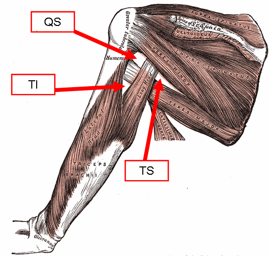
The axillary spaces are anatomic spaces. through which axillary contents leave the axilla. They consist of the quadrangular space, triangular space, and triangular interval. It is bounded by teres major, teres minor, medial border of the humerus, and long head of triceps brachii. They should not be confused with the true "axillary space" within the borders of the axilla. Structure Axilla The true axilla is a conical space with its apex at the Cervico-axillary Canal, Base at the axillary fascia and skin of the armpit. When viewed in an axillary plane (axillary cut), it is more triangle with: Medial Wall: Serratus Anterior, Anterior Wall: pectoral muscles, Posterior Wall: subscapularis muscle, where the "apex" of the triangle is the humerus Quadrangular space This space is in the posterior wall of the axilla. It is a quadrangular space bounded laterally by surgical neck of the humerus, medially by long head of triceps brachii and inferiorly by teres major. It is bounded super ... [...More Info...] [...Related Items...] OR: [Wikipedia] [Google] [Baidu] |
Spatium
{{set index article In anatomy, a spatium or anatomic space is a space (cavity or gap). Anatomic spaces are often landmarks to find other important structures. When they fill with gases (such as air) or liquids (such as blood) in pathological ways, they can suffer conditions such as pneumothorax, edema, or pericardial effusion. Many anatomic spaces are potential spaces, which means that they are potential rather than realized (with their realization being dynamic according to physiologic or pathophysiologic events). In other words, they are like an empty plastic bag that has not been opened (two walls collapsed against each other; no interior volume until opened) or a balloon that has not been inflated. Examples of anatomic spaces (or potential spaces) include: *Axillary space *Buccal space * Canine space *Cystohepatic triangle * Deep perineal space * Deep temporal space *Epidural space * Extraperitoneal space *Fascial spaces of the head and neck * Infratemporal space *Intercosta ... [...More Info...] [...Related Items...] OR: [Wikipedia] [Google] [Baidu] |
Quadrangular Space
The quadrangular space, also known as the quadrilateral space f Velpeau'' and the foramen humerotricipitale, is one of the three spaces in the axillary space. The other two spaces are: triangular space and triangular interval. Structure The quadrangular space is one of the three spaces in the axillary space. Boundaries The quadrangular space is defined by: - "Scapular Region: Quadrangular Space of Scapular Region" * ''above/superior:'' teres minor muscle. * ''below/inferior:'' teres major muscle. * ''medially:'' long head of the triceps brachii muscle (lateral margin). * ''laterally:'' surgical neck of the humerus. * ''anteriorly:'' subscapularis muscle. Contents The quadrangular space transmits the axillary nerve, and the posterior humeral circumflex artery. Clinical significance The quadrangular space is a clinically important anatomic space in the arm as it provides the anterior regions of the axilla a passageway to the posterior regions. In the quadrangular space, the axi ... [...More Info...] [...Related Items...] OR: [Wikipedia] [Google] [Baidu] |
Triangular Space
The triangular space (also known as the medial triangular space, upper triangular space, medial axillary space or foramen omotricipitale) is one of the three spaces found at the axillary space. The other two spaces are the quadrangular space and the triangular interval. Boundaries It has the following boundaries: * Inferior: the superior border of the teres major; * Lateral: the long head of the triceps; * Superior: Teres minor or Subscapularis For the superior border, some sources list the teres minor, while others list the subscapularis. Contents It contains the scapular circumflex vessels. Unlike the quadrangular space or the triangular interval, no major nerve passes through the triangular space. See also * Quadrangular space * Triangular interval The triangular interval (also known as the lateral triangular space, lower triangular space, and triceps hiatus) is a space found in the axilla. It is one of the three intermuscular spaces found in the axillary space. The ... [...More Info...] [...Related Items...] OR: [Wikipedia] [Google] [Baidu] |
Triangular Interval
The triangular interval (also known as the lateral triangular space, lower triangular space, and triceps hiatus) is a space found in the axilla. It is one of the three intermuscular spaces found in the axillary space. The other two spaces are: quadrangular space and triangular space. Borders Two of its borders are as follows: * teres major - superior * long head of the triceps brachii - medial Some sources state the lateral border is the humerus, while others define it as the lateral head of the triceps. (The effective difference is relatively minor, though.) Contents The radial nerve is visible through the triangular interval, on its way to the posterior compartment of the arm. Profunda Brachii also passes through the triangular interval from anterior to posterior. Additional images File:Gray412-spaces.png, Muscles on the dorsum of the scapula, and the Triceps brachii. Triangular Interval Syndrome Triangular Interval Syndrome (TIS) was described as a differential diagnos ... [...More Info...] [...Related Items...] OR: [Wikipedia] [Google] [Baidu] |
Teres Major
The teres major muscle is a muscle of the upper limb. It attaches to the scapula and the humerus and is one of the seven scapulohumeral muscles. It is a thick but somewhat flattened muscle. The teres major muscle (from Latin ''teres'', meaning "rounded") is positioned above the latissimus dorsi muscle and assists in the Anatomical terms of motion#Flexion and extension, extension and Anatomical terms of motion#Rotation, medial rotation of the humerus. This muscle is commonly confused as a rotator cuff muscle, but it is not because it does not attach to the capsule of the shoulder joint, unlike the teres minor muscle for example. Structure The teres major muscle originates on the dorsal surface of the Scapula, inferior angle and the lower part of the Scapula, lateral border of the scapula. The fibers of teres major insert into the medial lip of the Bicipital groove, intertubercular sulcus of the humerus. Relations The tendon, at its insertion, lies behind that of the latissimus ... [...More Info...] [...Related Items...] OR: [Wikipedia] [Google] [Baidu] |
Teres Minor
The teres minor (Latin ''teres'' meaning 'rounded') is a narrow, elongated muscle of the rotator cuff. The muscle originates from the lateral border and adjacent posterior surface of the corresponding right or left scapula and inserts at both the greater tubercle of the humerus and the posterior surface of the joint capsule. The primary function of the teres minor is to modulate the action of the deltoid, preventing the humeral head from sliding upward as the arm is abducted. It also functions to rotate the humerus laterally. The teres minor is innervated by the axillary nerve. Structure It arises from the dorsal surface of the axillary border of the scapula for the upper two-thirds of its extent, and from two aponeurotic laminae, one of which separates it from the infraspinatus muscle, the other from the teres major muscle. Its fibers run obliquely upwards and laterally; the upper ones end in a tendon which is inserted into the lowest of the three impressions on the greater tub ... [...More Info...] [...Related Items...] OR: [Wikipedia] [Google] [Baidu] |
Humerus
The humerus (; ) is a long bone in the arm that runs from the shoulder to the elbow. It connects the scapula and the two bones of the lower arm, the radius and ulna, and consists of three sections. The humeral upper extremity consists of a rounded head, a narrow neck, and two short processes (tubercles, sometimes called tuberosities). The body is cylindrical in its upper portion, and more prismatic below. The lower extremity consists of 2 epicondyles, 2 processes (trochlea & capitulum), and 3 fossae (radial fossa, coronoid fossa, and olecranon fossa). As well as its true anatomical neck, the constriction below the greater and lesser tubercles of the humerus is referred to as its surgical neck due to its tendency to fracture, thus often becoming the focus of surgeons. Etymology The word "humerus" is derived from la, humerus, umerus meaning upper arm, shoulder, and is linguistically related to Gothic ''ams'' shoulder and Greek ''ōmos''. Structure Upper extremity The upper or pr ... [...More Info...] [...Related Items...] OR: [Wikipedia] [Google] [Baidu] |
Triceps Brachii
The triceps, or triceps brachii (Latin for "three-headed muscle of the arm"), is a large muscle on the back of the upper limb of many vertebrates Vertebrates () comprise all animal taxa within the subphylum Vertebrata () ( chordates with backbones), including all mammals, birds, reptiles, amphibians, and fish. Vertebrates represent the overwhelming majority of the phylum Chordata, .... It consists of 3 parts: the medial, lateral, and long head. It is the muscle principally responsible for Extension (kinesiology), extension of the elbow joint (straightening of the arm). Structure The long head arises from the infraglenoid tubercle of the scapula. It extends distally anterior to the Teres minor muscle, teres minor and posterior to the Teres major muscle, teres major. The medial head arises proximally in the humerus, just inferior to the Radial sulcus, groove of the radial nerve; from the dorsal (back) surface of the humerus; from the Medial intermuscular septum of arm ... [...More Info...] [...Related Items...] OR: [Wikipedia] [Google] [Baidu] |
Subscapularis
The subscapularis is a large triangular muscle which fills the subscapular fossa and inserts into the lesser tubercle of the humerus and the front of the capsule of the shoulder-joint. Structure It arises from its medial two-thirds and Some fibers arise from tendinous laminae, which intersect the muscle and are attached to ridges on the bone; others from an aponeurosis, which separates the muscle from the teres major and the long head of the triceps brachii. The fibers pass laterally and coalesce into a tendon that is inserted into the lesser tubercle of the humerus and the anterior part of the shoulder-joint capsule. Tendinous fibers extend to the greater tubercle with insertions into the bicipital groove. Relations The tendon of the muscle is separated from the neck of the scapula by a large bursa, which communicates with the cavity of the shoulder-joint through an aperture in the capsule. The subscapularis is separated from the serratus anterior books.google.com/books? ... [...More Info...] [...Related Items...] OR: [Wikipedia] [Google] [Baidu] |
Axillary Nerve
The axillary nerve or the circumflex nerve is a nerve of the human body, that originates from the brachial plexus (upper trunk, posterior division, posterior cord) at the level of the axilla (armpit) and carries nerve fibers from C5 and C6. The axillary nerve travels through the quadrangular space with the posterior circumflex humeral artery and vein to innervate the deltoid and teres minor. Structure The nerve lies at first behind the axillary artery, and in front of the subscapularis, and passes downward to the lower border of that muscle. It then winds from anterior to posterior around the neck of the humerus, in company with the posterior humeral circumflex artery, through the quadrangular space (bounded above by the teres minor, below by the teres major, medially by the long head of the triceps brachii, and laterally by the surgical neck of the humerus), and divides into an anterior, a posterior, and a collateral branch to the long head of the triceps brachii branch. * The ... [...More Info...] [...Related Items...] OR: [Wikipedia] [Google] [Baidu] |
Posterior Humeral Circumflex Artery
The posterior humeral circumflex artery (posterior circumflex artery, posterior circumflex humeral artery) arises from the third part of axillary artery at the lower border of the subscapularis, and runs posteriorly with the axillary nerve through the quadrangular space. It winds around the surgical neck of the humerus and is distributed to the deltoid muscle and shoulder-joint, anastomosing with the anterior humeral circumflex and deep artery of the arm. It supplies the teres major, teres minor, deltoid, and (long head only) triceps muscles. Additional images File:Gray810.png, Suprascapular and axillary nerves of right side, seen from behind. File:Slide4SSS.JPG, Posterior humeral circumflex artery See also * Anterior humeral circumflex artery The anterior humeral circumflex artery (anterior circumflex artery, anterior circumflex humeral artery) is an artery in the arm. It is one of two circumflexing arteries that branch from the axillary artery, the other being the posteri ... [...More Info...] [...Related Items...] OR: [Wikipedia] [Google] [Baidu] |
Scapular Circumflex Artery
The circumflex scapular artery (scapular circumflex artery, dorsalis scapulae artery) is a branch of the subscapular artery and part of the scapular anastomoses. It curves around the axillary border of the scapula, traveling through the anatomical "Triangular space" made up of the Teres minor superiorly, the Teres major inferiorly, and the long head of the Triceps laterally. It enters the infraspinatous fossa under cover of the Teres minor, and anastomoses with the transverse scapular artery (suprascapular) and the descending branch of the transverse cervical (a.k.a. dorsal scapular artery). Branches In its course it gives off two branches: * one (infrascapular) enters the subscapular fossa beneath the Subscapularis, which it supplies, anastomosing with the transverse scapular artery and the descending branch of the transverse cervical. * the other is continued along the axillary border of the scapula, between the Teres major and minor, and at the dorsal surface of the infer ... [...More Info...] [...Related Items...] OR: [Wikipedia] [Google] [Baidu] |


