|
ATP7A
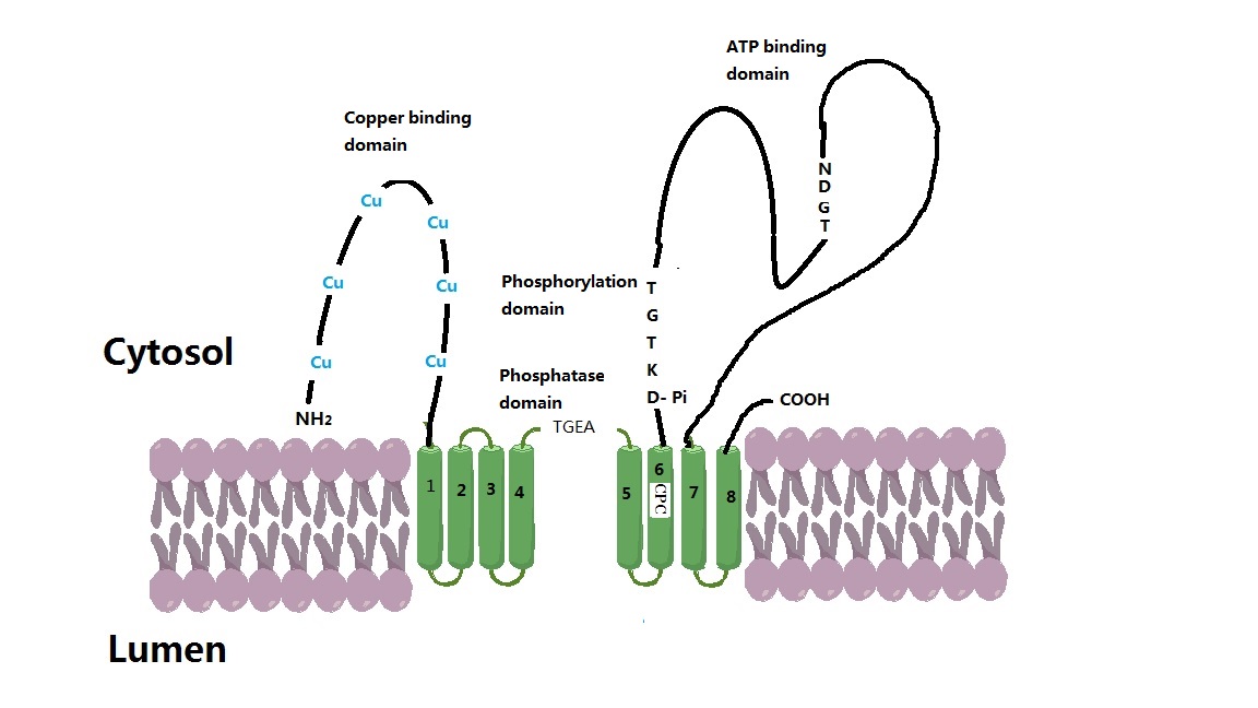
ATP7A, also known as Menkes' protein (MNK), is a copper-transporting P-type ATPase which uses the energy arising from ATP hydrolysis to transport Cu(I) across cell membranes. The ATP7A protein is a transmembrane protein and is expressed in the intestine and all tissues except liver. In the intestine, ATP7A regulates Cu(I) absorption in the human body by transporting Cu(I) from the small intestine into the blood. In other tissues, ATP7A shuttles between the Golgi apparatus and the cell membrane to maintain proper Cu(I) concentrations (since there is no free Cu(I) in the cell, Cu(I) ions are all tightly bound) in the cell and provides certain enzymes with Cu(I) (e.g. peptidyl-α-monooxygenase, tyrosinase, and lysyl oxidase). The X-linked, inherited, lethal genetic disorder of the ''ATP7A'' gene causes Menkes disease, a copper deficiency resulting in early childhood death. Gene The ''ATP7A'' gene is located on the long (q) arm of the X chromosome at band Xq21.1. The encoded ATP ... [...More Info...] [...Related Items...] OR: [Wikipedia] [Google] [Baidu] |
Menkes Disease
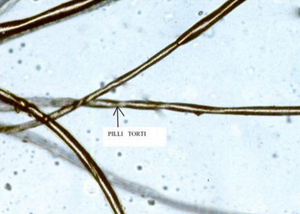
Menkes disease (MNK), also known as Menkes syndrome, is an X-linked recessive disorder caused by mutations in genes coding for the copper-transport protein ATP7A, leading to copper deficiency. Characteristic findings include kinky hair, growth failure, and nervous system deterioration. Like all X-linked recessive conditions, Menkes disease is more common in males than in females. The disorder was first described by John Hans Menkes in 1962. Onset occurs during infancy, with incidence of about 1 in 100,000 to 250,000 newborns; affected infants often do not live past the age of three years, though there are rare cases in which less severe symptoms emerge later in childhood. Signs and symptoms Affected infants may be born prematurely. Signs and symptoms appear during infancy, typically after a two- to three-month period of normal or slightly slowed development that is followed by a loss of early developmental skills and subsequent developmental delay. Patients exhibit hyp ... [...More Info...] [...Related Items...] OR: [Wikipedia] [Google] [Baidu] |
ATP7B
Wilson disease protein (WND), also known as ATP7B protein, is a copper-transporting P-type ATPase which is encoded by the ''ATP7B'' gene. The ATP7B protein is located in the trans-Golgi network of the liver and brain and balances the copper level in the body by excreting excess copper into bile and plasma. Genetic disorder of the ATP7B gene may cause Wilson's disease, a disease in which copper accumulates in tissues, leading to neurological or psychiatric issues and liver diseases. Gene Wilson disease protein is associated with ''ATP7B'' gene, approximately 80 Kb, located on human chromosome 13 and consists of 21 exons. The mRNA transcribed by ''ATP7B'' gene has a size of 7.5 Kb, and which encodes a protein of 1465 amino acids. The gene is a member of the P-type cation transport ATPase family and encodes a protein with several membrane-spanning domains, an ATPase consensus sequence, a hinge domain, a phosphorylation site, and at least two putative copper-binding sites. Thi ... [...More Info...] [...Related Items...] OR: [Wikipedia] [Google] [Baidu] |
ATOX1
ATOX1 is a copper metallochaperone protein that is encoded by the ''ATOX1'' gene in humans. In mammals, ATOX1 plays a key role in copper homeostasis as it delivers copper from the cytosol to transporters ATP7A and ATP7B. Homologous proteins are found in a wide variety of eukaryotes, including ''Saccharomyces cerevisiae'' as ATX1, and all contain a conserved metal binding domain. Function ATOX1 is an abbreviation of the full name Antioxidant Protein 1. The nomenclature stems from initial characterization that showed that ATOX1 protected cells from reactive oxygen species. Since then, the primary role of ATOX1 has been established as a copper metallochaperone protein found in the cytoplasm of eukaryotes. A metallochaperone is an important protein that has metal trafficking and sequestration roles. As a metal sequestration protein, ATOX1 is capable of binding free metals ''in vivo'', in order to protect cells from generation of reactive oxygen species and mismetallation of met ... [...More Info...] [...Related Items...] OR: [Wikipedia] [Google] [Baidu] |
P-type ATPase
The P-type ATPases, also known as E1-E2 ATPases, are a large group of evolutionarily related ion pump, ion and lipid pumps that are found in bacteria, archaea, and eukaryotes. P-type ATPases are α-helical bundle active transport, primary transporters named based upon their ability to catalyze auto- (or self-) phosphorylation (hence P) of a key conserved aspartate residue within the pump and their energy source, adenosine triphosphate (ATP). In addition, they all appear to interconvert between at least two different conformations, denoted by E1 and E2. P-type ATPases fall under the P-type ATPase (P-ATPase) SuperfamilyTC# 3.A.3 which, as of early 2016, includes 20 different protein families. Most members of this transporter superfamily catalyze cation uptake and/or efflux, however one subfamily, the flippases,TC# 3.A.3.8 is involved in flipping phospholipids to maintain the asymmetric nature of the biomembrane. In humans, P-type ATPases serve as a basis for Action potential, nerve ... [...More Info...] [...Related Items...] OR: [Wikipedia] [Google] [Baidu] |
GLRX
Glutaredoxin-1 is a protein that in humans is encoded by the ''GLRX'' gene. Interactions GLRX has been shown to interact with Wilson disease protein and ATP7A ATP7A, also known as Menkes' protein (MNK), is a copper-transporting P-type ATPase which uses the energy arising from ATP hydrolysis to transport Cu(I) across cell membranes. The ATP7A protein is a transmembrane protein and is expressed in the i .... References Further reading * * * * * * * * * * * * * * * * * * * {{gene-5-stub ... [...More Info...] [...Related Items...] OR: [Wikipedia] [Google] [Baidu] |
Phosphorylation
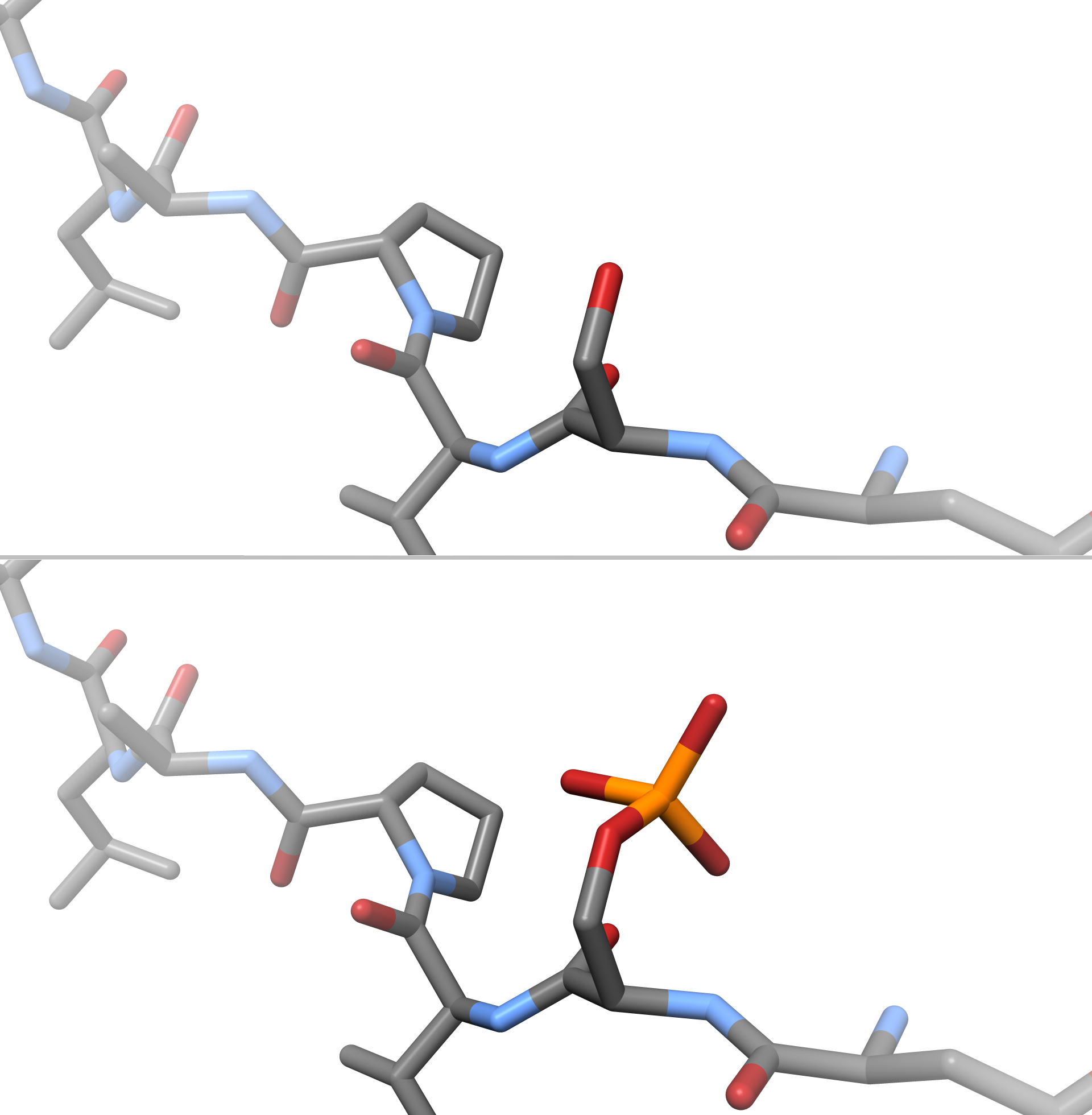
In chemistry, phosphorylation is the attachment of a phosphate group to a molecule or an ion. This process and its inverse, dephosphorylation, are common in biology and could be driven by natural selection. Text was copied from this source, which is available under a Creative Commons Attribution 4.0 International License. Protein phosphorylation often activates (or deactivates) many enzymes. Glucose Phosphorylation of sugars is often the first stage in their catabolism. Phosphorylation allows cells to accumulate sugars because the phosphate group prevents the molecules from diffusing back across their transporter. Phosphorylation of glucose is a key reaction in sugar metabolism. The chemical equation for the conversion of D-glucose to D-glucose-6-phosphate in the first step of glycolysis is given by :D-glucose + ATP → D-glucose-6-phosphate + ADP :ΔG° = −16.7 kJ/mol (° indicates measurement at standard condition) Hepatic cells are freely permeable to glucose, an ... [...More Info...] [...Related Items...] OR: [Wikipedia] [Google] [Baidu] |
S-glutathionylation
S-Glutathionylation is the posttranslational modification of protein cysteine residues by the addition of glutathione, the most abundant and important low-molecular-mass thiol within most cell types. Protein S-glutathionylation is involved in * oxidative stress * nitrosative stress * preventing irreversible oxidation of protein thiols * control of cell-signalling pathways by modulating protein Proteins are large biomolecules and macromolecules that comprise one or more long chains of amino acid residues. Proteins perform a vast array of functions within organisms, including catalysing metabolic reactions, DNA replication, respon ... function References {{Reflist Post-translational modification ... [...More Info...] [...Related Items...] OR: [Wikipedia] [Google] [Baidu] |
Endothelium
The endothelium is a single layer of squamous endothelial cells that line the interior surface of blood vessels and lymphatic vessels. The endothelium forms an interface between circulating blood or lymph in the lumen and the rest of the vessel wall. Endothelial cells form the barrier between vessels and tissue and control the flow of substances and fluid into and out of a tissue. Endothelial cells in direct contact with blood are called vascular endothelial cells whereas those in direct contact with lymph are known as lymphatic endothelial cells. Vascular endothelial cells line the entire circulatory system, from the heart to the smallest capillaries. These cells have unique functions that include fluid filtration, such as in the glomerulus of the kidney, blood vessel tone, hemostasis, neutrophil recruitment, and hormone trafficking. Endothelium of the interior surfaces of the heart chambers is called endocardium. An impaired function can lead to serious health i ... [...More Info...] [...Related Items...] OR: [Wikipedia] [Google] [Baidu] |
Syncytiotrophoblast
Syncytiotrophoblast (from the Greek 'syn'- "together"; 'cytio'- "of cells"; 'tropho'- "nutrition"; 'blast'- "bud") is the epithelial covering of the highly vascular embryonic placental villi, which invades the wall of the uterus to establish nutrient circulation between the embryo and the mother. It is a multi-nucleate, terminally differentiated syncytium, extending to 13cm. Function It is the outer layer of the trophoblasts and actively invades the uterine wall, during implantation, rupturing maternal capillaries and thus establishing an interface between maternal blood and embryonic extracellular fluid, facilitating passive exchange of material between the mother and the embryo. The syncytial property is important since the mother's immune system includes white blood cells that are able to migrate into tissues by "squeezing" in between cells. If they were to reach the fetal side of the placenta many foreign proteins would be recognised, triggering an immune reaction. H ... [...More Info...] [...Related Items...] OR: [Wikipedia] [Google] [Baidu] |
Cytotrophoblast
"Cytotrophoblast" is the name given to both the inner layer of the trophoblast (also called layer of Langhans) or the cells that live there. It is interior to the syncytiotrophoblast and external to the wall of the blastocyst in a developing embryo. The cytotrophoblast is considered to be the trophoblastic stem cell because the layer surrounding the blastocyst remains while daughter cells differentiate and proliferate to function in multiple roles. There are two lineages that cytotrophoblastic cells may differentiate through: fusion and invasive. The fusion lineage yields syncytiotrophoblast and the invasive lineage yields interstitial cytotrophoblast cells. Cytotrophoblastic cells play an important role in the implantation of an embryo in the uterus. Fusion lineage The formation of all syncytiotrophoblast is from the fusion of two or more cytotrophoblasts via this fusion pathway. This pathway is important because the syncytiotrophoblast plays an important role in fetal-maternal ... [...More Info...] [...Related Items...] OR: [Wikipedia] [Google] [Baidu] |
Parenchyma
Parenchyma () is the bulk of functional substance in an animal organ or structure such as a tumour. In zoology it is the name for the tissue that fills the interior of flatworms. Etymology The term ''parenchyma'' is New Latin from the word παρέγχυμα ''parenchyma'' meaning 'visceral flesh', and from παρεγχεῖν ''parenchyma'' meaning 'to pour in' from παρα- ''para-'' 'beside' + ἐν ''en-'' 'in' + χεῖν ''chyma'' 'to pour'. Originally, Erasistratus and other anatomists used it to refer to certain human tissues. Later, it was also applied to plant tissues by Nehemiah Grew. Structure The parenchyma is the ''functional'' parts of an organ, or of a structure such as a tumour in the body. This is in contrast to the stroma, which refers to the ''structural'' tissue of organs or of structures, namely, the connective tissues. Brain The brain parenchyma refers to the functional tissue in the brain that is made up of the two types of brain cell, neurons an ... [...More Info...] [...Related Items...] OR: [Wikipedia] [Google] [Baidu] |
Renal Tubules
The nephron is the minute or microscopic structural and functional unit of the kidney. It is composed of a renal corpuscle and a Nephron#Renal tubule, renal tubule. The renal corpuscle consists of a tuft of capillary, capillaries called a glomerulus (kidney), glomerulus and a cup-shaped structure called Bowman's capsule. The renal tubule extends from the capsule. The capsule and tubule are connected and are composed of epithelial cells with a Lumen (anatomy), lumen. A healthy adult has 1 to 1.5 million nephrons in each kidney. Blood is filtered as it passes through three layers: the endothelial cells of the capillary wall, its basement membrane, and between the foot processes of the podocytes of the lining of the capsule. The tubule has adjacent peritubular capillaries that run between the descending and ascending portions of the tubule. As the fluid from the capsule flows down into the tubule, it is processed by the epithelial cells lining the tubule: water is reabsorbed and subst ... [...More Info...] [...Related Items...] OR: [Wikipedia] [Google] [Baidu] |


