|
Infratemporal Space
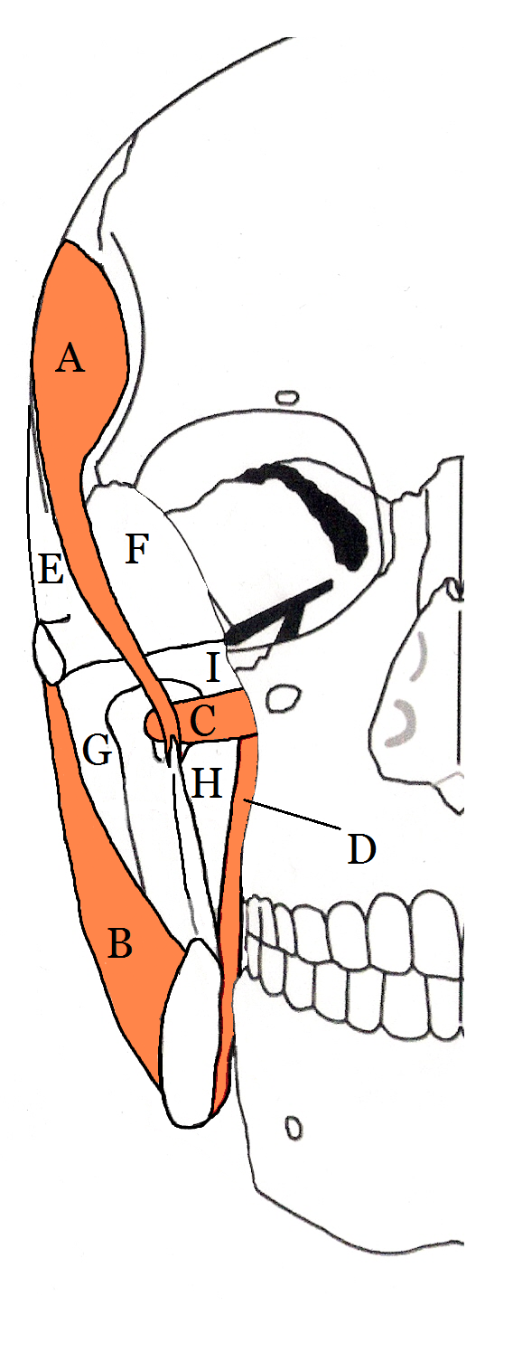
The Infratemporal space (also termed the infra-temporal space or the infra-temporal portion of the deep temporal space) is a fascial space of the head and neck (sometimes also termed fascial spaces or tissue spaces). It is a potential space in the side of the head, and is paired on either side. It is located posterior to the maxilla, between the lateral pterygoid plate of the sphenoid bone medially and by the base of skull superiorly. The term is derived from '' infra-'' meaning below and ''temporal'' which refers to the temporalis muscle. The infratemporal space is the inferior portion of the deep temporal space, which is one of the four compartments of the masticator space, along with the pterygomandibular space, the submasseteric space and the superficial temporal space. The deep temporal space is separated from the pterygomandibular space by the lateral pterygoid muscle inferiorly and from the superficial temporal space by the temporalis muscle laterally. The deep tempora ... [...More Info...] [...Related Items...] OR: [Wikipedia] [Google] [Baidu] |
Fascial Spaces Of The Head And Neck
Fascial spaces (also termed fascial tissue spaces or tissue spaces) are potential spaces that exist between the fasciae and underlying organs and other tissues. In health, these spaces do not exist; they are only created by pathology, e.g. the spread of pus or cellulitis in an infection. The fascial spaces can also be opened during the dissection of a cadaver. The fascial spaces are different from the fasciae themselves, which are bands of connective tissue that surround structures, e.g. muscles. The opening of fascial spaces may be facilitated by pathogenic bacterial release of enzymes which cause tissue lysis (e.g. hyaluronidase and collagenase). The spaces filled with loose areolar connective tissue may also be termed clefts. Other contents such as salivary glands, blood vessels, nerves and lymph nodes are dependent upon the location of the space. Those containing neurovascular tissue (nerves and blood vessels) may also be termed compartments. Generally, the spread of infection ... [...More Info...] [...Related Items...] OR: [Wikipedia] [Google] [Baidu] |
Masticator Space
Fascial spaces (also termed fascial tissue spaces or tissue spaces) are potential spaces that exist between the fasciae and underlying organs and other tissues. In health, these spaces do not exist; they are only created by pathology, e.g. the spread of pus or cellulitis in an infection. The fascial spaces can also be opened during the dissection of a cadaver. The fascial spaces are different from the fasciae themselves, which are bands of connective tissue that surround structures, e.g. muscles. The opening of fascial spaces may be facilitated by pathogenic bacterial release of enzymes which cause tissue lysis (e.g. hyaluronidase and collagenase). The spaces filled with loose areolar connective tissue may also be termed clefts. Other contents such as salivary glands, blood vessels, nerves and lymph nodes are dependent upon the location of the space. Those containing neurovascular tissue (nerves and blood vessels) may also be termed compartments. Generally, the spread of infection ... [...More Info...] [...Related Items...] OR: [Wikipedia] [Google] [Baidu] |
Otorhinolaryngology
Otorhinolaryngology ( , abbreviated ORL and also known as otolaryngology, otolaryngology–head and neck surgery (ORL–H&N or OHNS), or ear, nose, and throat (ENT)) is a surgical subspeciality within medicine that deals with the surgical and medical management of conditions of the head and neck. Doctors who specialize in this area are called otorhinolaryngologists, otolaryngologists, head and neck surgeons, or ENT surgeons or physicians. Patients seek treatment from an otorhinolaryngologist for diseases of the ear, nose, throat, base of the skull, head, and neck. These commonly include functional diseases that affect the senses and activities of eating, drinking, speaking, breathing, swallowing, and hearing. In addition, ENT surgery encompasses the surgical management of cancers and benign tumors and reconstruction of the head and neck as well as plastic surgery of the face and neck. Etymology The term is a combination of New Latin combining forms ('' oto-'' + ''rhino-'' + ... [...More Info...] [...Related Items...] OR: [Wikipedia] [Google] [Baidu] |
Mouth
In animal anatomy, the mouth, also known as the oral cavity, or in Latin cavum oris, is the opening through which many animals take in food and issue vocal sounds. It is also the cavity lying at the upper end of the alimentary canal, bounded on the outside by the lips and inside by the pharynx. In tetrapods, it contains the tongue and, except for some like birds, teeth. This cavity is also known as the buccal cavity, from the Latin ''bucca'' ("cheek"). Some animal phyla, including arthropods, molluscs and chordates, have a complete digestive system, with a mouth at one end and an anus at the other. Which end forms first in ontogeny is a criterion used to classify bilaterian animals into protostomes and deuterostomes. Development In the first multicellular animals, there was probably no mouth or gut and food particles were engulfed by the cells on the exterior surface by a process known as endocytosis. The particles became enclosed in vacuoles into which enzymes were secr ... [...More Info...] [...Related Items...] OR: [Wikipedia] [Google] [Baidu] |
Wisdom Tooth
A third molar, commonly called wisdom tooth, is one of the three molars per quadrant of the human dentition. It is the most posterior of the three. The age at which wisdom teeth come through ( erupt) is variable, but this generally occurs between late teens and early twenties. Most adults have four wisdom teeth, one in each of the four quadrants, but it is possible to have none, fewer, or more, in which case the extras are called supernumerary teeth. Wisdom teeth may get stuck ( impacted) against other teeth if there is not enough space for them to come through normally. Impacted wisdom teeth are still sometimes removed for orthodontic treatment, believing that they move the other teeth and cause crowding, though this is not held anymore as true. Impacted wisdom teeth may suffer from tooth decay if oral hygiene becomes more difficult. Wisdom teeth which are partially erupted through the gum may also cause inflammation and infection in the surrounding gum tissues, termed pericoron ... [...More Info...] [...Related Items...] OR: [Wikipedia] [Google] [Baidu] |
Odontogenic Infection
An odontogenic infection is an infection that originates within a tooth or in the closely surrounding tissues. The term is derived from '' odonto-'' (Ancient Greek: , – 'tooth') and '' -genic'' (Ancient Greek: , ; – 'birth'). The most common causes for odontogenic infection to be established are dental caries, deep fillings, failed root canal treatments, periodontal disease, and pericoronitis. Odontogenic infection starts as localised infection and may remain localised to the region where it started, or spread into adjacent or distant areas. It is estimated that 90-95% of all orofacial infections originate from the teeth or their supporting structures and are the most common infections in the oral and maxilofacial region. Odontogenic infections can be severe if not treated and are associated with mortality rate of 10 to 40%. Furthermore, about 70% of odontogenic infections occur as periapical inflammation, i.e. acute periapical periodontitis or a periapical abscess. The next m ... [...More Info...] [...Related Items...] OR: [Wikipedia] [Google] [Baidu] |
Zygomatic Bone
In the human skull, the zygomatic bone (from grc, ζῠγόν, zugón, yoke), also called cheekbone or malar bone, is a paired irregular bone which articulates with the maxilla, the temporal bone, the sphenoid bone and the frontal bone. It is situated at the upper and lateral part of the face and forms the prominence of the cheek, part of the lateral wall and floor of the orbit, and parts of the temporal fossa and the infratemporal fossa. It presents a malar and a temporal surface; four processes (the frontosphenoidal, orbital, maxillary, and temporal), and four borders. Etymology The term ''zygomatic'' derives from the Ancient Greek , ''zygoma'', meaning "yoke". The zygomatic bone is occasionally referred to as the zygoma, but this term may also refer to the zygomatic arch. Structure Surfaces The ''malar surface'' is convex and perforated near its center by a small aperture, the zygomaticofacial foramen, for the passage of the zygomaticofacial nerve and vessels; below ... [...More Info...] [...Related Items...] OR: [Wikipedia] [Google] [Baidu] |


