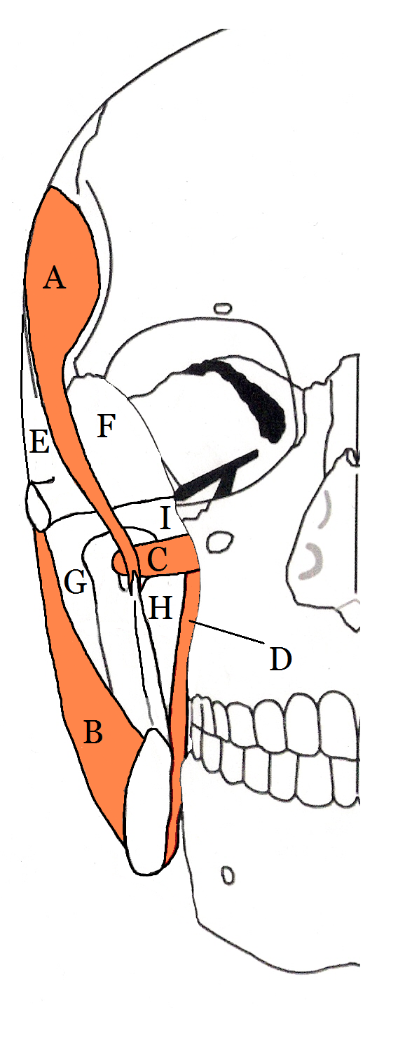
Masticator Space on:
[Wikipedia]
[Google]
[Amazon]
Fascial spaces (also termed fascial tissue spaces or tissue spaces) are
 This term is sometimes used, and is a collective name for the submasseteric (masseteric), pterygomandibular, superficial temporal and deep temporal spaces. The infratemporal space is the inferior portion of the deep temporal space. The superficial temporal and the deep temporal spaces are sometimes together called the temporal spaces. The masticator spaces are paired structures on either side of the head. The muscles of mastication are enclosed in a layer of fascia, formed by cervical fascia ascending from the neck which divides at the inferior border of the mandible to envelope the area. Each masticator space also contains the sections of the
potential space
In anatomy, a potential space is a space between two adjacent structures that are normally pressed together (directly apposed). Many anatomic spaces are potential spaces, which means that they are potential rather than realized (with their realiz ...
s that exist between the fasciae and underlying organs and other tissues. In health, these spaces do not exist; they are only created by pathology, e.g. the spread of pus
Pus is an exudate, typically white-yellow, yellow, or yellow-brown, formed at the site of inflammation during bacterial or fungal infection. An accumulation of pus in an enclosed tissue space is known as an abscess, whereas a visible collection ...
or cellulitis
Cellulitis is usually a bacterial infection involving the inner layers of the skin. It specifically affects the dermis and subcutaneous fat. Signs and symptoms include an area of redness which increases in size over a few days. The borders of t ...
in an infection
An infection is the invasion of tissues by pathogens, their multiplication, and the reaction of host tissues to the infectious agent and the toxins they produce. An infectious disease, also known as a transmissible disease or communicable dise ...
. The fascial spaces can also be opened during the dissection
Dissection (from Latin ' "to cut to pieces"; also called anatomization) is the dismembering of the body of a deceased animal or plant to study its anatomical structure. Autopsy is used in pathology and forensic medicine to determine the cause o ...
of a cadaver
A cadaver or corpse is a dead human body that is used by medical students, physicians and other scientists to study anatomy, identify disease sites, determine causes of death, and provide tissue to repair a defect in a living human being. Stud ...
. The fascial spaces are different from the fasciae themselves, which are bands of connective tissue
Connective tissue is one of the four primary types of animal tissue, along with epithelial tissue, muscle tissue, and nervous tissue. It develops from the mesenchyme derived from the mesoderm the middle embryonic germ layer. Connective tiss ...
that surround structures, e.g. muscle
Skeletal muscles (commonly referred to as muscles) are organs of the vertebrate muscular system and typically are attached by tendons to bones of a skeleton. The muscle cells of skeletal muscles are much longer than in the other types of muscl ...
s. The opening of fascial spaces may be facilitated by pathogenic
In biology, a pathogen ( el, πάθος, "suffering", "passion" and , "producer of") in the oldest and broadest sense, is any organism or agent that can produce disease. A pathogen may also be referred to as an infectious agent, or simply a germ ...
bacteria
Bacteria (; singular: bacterium) are ubiquitous, mostly free-living organisms often consisting of one biological cell. They constitute a large domain of prokaryotic microorganisms. Typically a few micrometres in length, bacteria were among ...
l release of enzyme
Enzymes () are proteins that act as biological catalysts by accelerating chemical reactions. The molecules upon which enzymes may act are called substrates, and the enzyme converts the substrates into different molecules known as products. A ...
s which cause tissue lysis
Lysis ( ) is the breaking down of the membrane of a cell, often by viral, enzymic, or osmotic (that is, "lytic" ) mechanisms that compromise its integrity. A fluid containing the contents of lysed cells is called a ''lysate''. In molecular bio ...
(e.g. hyaluronidase
Hyaluronidases are a family of enzymes that catalyse the degradation of hyaluronic acid (HA). Karl Meyer classified these enzymes in 1971, into three distinct groups, a scheme based on the enzyme reaction products. The three main types of hyal ...
and collagenase
Collagenases are enzymes that break the peptide bonds in collagen. They assist in destroying extracellular structures in the pathogenesis of bacteria such as ''Clostridium''. They are considered a virulence factor, facilitating the spread of ...
). The spaces filled with loose areolar connective tissue
Loose connective tissue, sometimes called areolar tissue, is a cellular connective tissue with thin and relatively sparse collagen fibers. Its ground substance occupies more volume than the fibers do. It has a viscous to gel-like consistenc ...
may also be termed clefts. Other contents such as salivary gland
The salivary glands in mammals are exocrine glands that produce saliva through a system of ducts. Humans have three paired major salivary glands (parotid, submandibular, and sublingual), as well as hundreds of minor salivary glands. Salivary gla ...
s, blood vessel
The blood vessels are the components of the circulatory system that transport blood throughout the human body. These vessels transport blood cells, nutrients, and oxygen to the tissues of the body. They also take waste and carbon dioxide away ...
s, nerve
A nerve is an enclosed, cable-like bundle of nerve fibers (called axons) in the peripheral nervous system.
A nerve transmits electrical impulses. It is the basic unit of the peripheral nervous system. A nerve provides a common pathway for the e ...
s and lymph node
A lymph node, or lymph gland, is a kidney-shaped organ of the lymphatic system and the adaptive immune system. A large number of lymph nodes are linked throughout the body by the lymphatic vessels. They are major sites of lymphocytes that inclu ...
s are dependent upon the location of the space. Those containing neurovascular tissue (nerves and blood vessels) may also be termed compartment
Compartment may refer to:
Biology
* Compartment (anatomy), a space of connective tissue between muscles
* Compartment (chemistry), in which different parts of the same protein serves different functions
* Compartment (development), fields of cells ...
s.
Generally, the spread of infection is determined by barriers such as muscle, bone and fasciae. Pus moves by the path of least resistance, e.g. the fluid will more readily dissect apart loosely connected tissue planes, such the fascial spaces, than erode through bone
A bone is a Stiffness, rigid Organ (biology), organ that constitutes part of the skeleton in most vertebrate animals. Bones protect the various other organs of the body, produce red blood cell, red and white blood cells, store minerals, provid ...
or muscles. In the head and neck, potential spaces are primarily defined by the complex attachment of muscles, especially mylohyoid, buccinator, masseter, medial pterygoid, superior constrictor and orbicularis oris.
Infections involving fascial spaces of the head and neck may give varying signs and symptoms depending upon the spaces involved. Trismus
Trismus, commonly called ''lockjaw'' as associated with tetanus, is a condition of limited jaw mobility. It may be caused by spasm of the muscles of mastication or a variety of other causes. Temporary trismus occurs much more frequently than perma ...
(difficulty opening the mouth) is a sign that the muscles of mastication
There are four classical muscles of mastication. During mastication, three muscles of mastication (''musculi masticatorii'') are responsible for adduction of the jaw, and one (the lateral pterygoid) helps to abduct it. All four move the jaw late ...
(the muscles that move the jaw) are involved. Dysphagia
Dysphagia is difficulty in swallowing. Although classified under "symptoms and signs" in ICD-10, in some contexts it is classified as a disease#Terminology, condition in its own right.
It may be a sensation that suggests difficulty in the passag ...
(difficulty swallowing) and dyspnoea
Shortness of breath (SOB), also medically known as dyspnea (in AmE) or dyspnoea (in BrE), is an uncomfortable feeling of not being able to breathe well enough. The American Thoracic Society defines it as "a subjective experience of breathing disc ...
(difficulty breathing) may be a sign that the airway is being compressed by the swelling.
Classification
Different classifications are used. One method distinguishes four anatomic groups: * The mandible and below ** The buccal vestibule ** The body of the mandible ** The mental space ** The submental space ** The sublingual space ** The submandibular space * The cheek and lateral face **The buccal vestibule of the maxilla ** The buccal space ** The submasseteric space ** The temporal space * The pharyngeal and cervical areas **The pterygomandibular space ** The parapharyngeal spaces ** The cervical spaces * The midface ** The palate ** The base of the upper lip ** The canine spaces (infraorbital spaces) ** The periorbital spaces Since thehyoid bone
The hyoid bone (lingual bone or tongue-bone) () is a horseshoe-shaped bone situated in the anterior midline of the neck between the chin and the thyroid cartilage. At rest, it lies between the base of the mandible and the third cervical vertebr ...
is the most important anatomic structure in the neck that limits the spread of infection, the spaces can be classified according to their relation to the hyoid bone:
* Suprahyoid (above the hyoid)
* Infrahyoid (below the hyoid)
* Fascial spaces traversing the length of the neck
In oral and maxillofacial surgery, the fascial spaces are almost always of relevance due to the spread of odontogenic infection
An odontogenic infection is an infection that originates within a tooth or in the closely surrounding tissues. The term is derived from '' odonto-'' (Ancient Greek: , – 'tooth') and '' -genic'' (Ancient Greek: , ; – 'birth'). The most common ...
s. As such, the spaces can also be classified according to their relation to the upper and lower teeth, and whether infection may directly spread into the space (primary space), or must spread via another space (secondary space):
* Primary maxillary spaces
** Canine space
** Buccal space
** Infratemporal space
* Primary mandibular spaces
** Submental space
** Buccal space
** Submandibular space
** Sublingual space
** Submasseteric space
* Cervical spaces
Perimandibular spaces
The submaxillary space is a historical term for the combination of the submandibular, submental and sublingual spaces, which in modern practice are referred to separately or collectively termed the perimandibular spaces. The term submaxillary may be confusing to modern students and clinicians since these spaces are located below the mandible, but historically the maxilla and mandible together were termed "maxillae", and sometimes the mandible was termed the "inferior maxilla". Sometimes the term submaxillary space is used synonymously with submandibular space. Confusion exists, as some sources describe the sublingual and the submandibular spaces as compartments of the "submandibular space".Submandibular space
Submental space
Sublingual space
Mental space
Buccal space
Canine space (infra-orbital space)
Masticator space
 This term is sometimes used, and is a collective name for the submasseteric (masseteric), pterygomandibular, superficial temporal and deep temporal spaces. The infratemporal space is the inferior portion of the deep temporal space. The superficial temporal and the deep temporal spaces are sometimes together called the temporal spaces. The masticator spaces are paired structures on either side of the head. The muscles of mastication are enclosed in a layer of fascia, formed by cervical fascia ascending from the neck which divides at the inferior border of the mandible to envelope the area. Each masticator space also contains the sections of the
 mandibular division of the trigeminal nerve
In neuroanatomy, the mandibular nerve (V) is the largest of the three divisions of the trigeminal nerve, the fifth cranial nerve (CN V). Unlike the other divisions of the trigeminal nerve (ophthalmic nerve, maxillary nerve) which contain only aff ...
and the internal maxillary artery
The maxillary artery supplies deep structures of the face. It branches from the external carotid artery just deep to the neck of the mandible.
Structure
The maxillary artery, the larger of the two terminal branches of the external carotid artery, ...
.
The masticator space could therefore be described as a potential space with four separate compartments. Infections usually only occupy one of these compartments, but severe or long standing infections can spread to involve the entire masticator space. The compartments of the masticator space are located on either side of the mandibular ramus and on either side of the temporalis muscle
In anatomy, the temporalis muscle, also known as the temporal muscle, is one of the muscles of mastication (chewing). It is a broad, fan-shaped convergent muscle on each side of the head that fills the temporal fossa, superior to the zygomati ...
.
Submasseteric space
This is also referred to as the masseter space or the superifical masticator space. The submasseteric space is logically located under (deep to) the masseter muscle, created by the insertions of masseter onto the lateral surface of the mandibular ramus. Submasseteric abscesses are rare and are associated with marked trismus.Pterygomandibular space
The pterygomandibular space lies between the medial side of the ramus of the mandible and the lateral surface of the medial pterygoid muscle.Deep temporal space (infra-temporal space)
The infra-temporal space is the inferior portion of the deep temporal space.Superficial temporal space
History
Modern understanding of the fascial spaces of the head and neck developed from the landmark research of Grodinsky and Holyoke in the 1930s. They injected a dye into cadavers to simulate pus. Theirhypothesis
A hypothesis (plural hypotheses) is a proposed explanation for a phenomenon. For a hypothesis to be a scientific hypothesis, the scientific method requires that one can test it. Scientists generally base scientific hypotheses on previous obse ...
was that infection in the head and neck mainly spread by hydrostatic pressure. This is now accepted to be true for most infections in the head and neck, with the exception of actinomycosis
Actinomycosis is a rare infectious bacterial disease caused by ''Actinomyces'' species. The name refers to ray-like appearance of the organisms in the granules. About 70% of infections are due to either ''Actinomyces israelii'' or '' A. gerencseria ...
which tends to burrow into the skin, and mycotuberculoid infections which tend to spread via the lymphatics.
References
{{reflist, 30em Human anatomy