|
Three-dimensional X-ray Diffraction
Three-dimensional X-ray diffraction (3DXRD) is a microscopy technique using hard X-rays (with energy in the 30-100 keV range) to investigate the internal structure of polycrystalline materials in three dimensions. For a given sample, 3DXRD returns the shape, juxtaposition, and orientation of the crystallites (''"grains"'') it is made of. 3DXRD allows investigating micrometer- to millimetre-sized samples with resolution ranging from hundreds of nanometers to micrometers. Other techniques employing X-rays to investigate the internal structure of polycrystalline materials include X-ray diffraction contrast tomography (DCT) and high energy X-ray diffraction (HEDM). Compared with destructive techniques, e.g. three-dimensional electron backscatter diffraction (3D EBSD), with which the sample is serially sectioned and imaged, 3DXRD and similar X-ray nondestructive techniques have the following advantages: * They require less sample preparation, thus limiting the introduction of new struc ... [...More Info...] [...Related Items...] OR: [Wikipedia] [Google] [Baidu] |
X-rays
An X-ray (also known in many languages as Röntgen radiation) is a form of high-energy electromagnetic radiation with a wavelength shorter than those of ultraviolet rays and longer than those of gamma rays. Roughly, X-rays have a wavelength ranging from 10 Nanometre, nanometers to 10 Picometre, picometers, corresponding to frequency, frequencies in the range of 30 Hertz, petahertz to 30 Hertz, exahertz ( to ) and photon energies in the range of 100 electronvolt, eV to 100 keV, respectively. X-rays were discovered in 1895 in science, 1895 by the German scientist Wilhelm Röntgen, Wilhelm Conrad Röntgen, who named it ''X-radiation'' to signify an unknown type of radiation.Novelline, Robert (1997). ''Squire's Fundamentals of Radiology''. Harvard University Press. 5th edition. . X-rays can penetrate many solid substances such as construction materials and living tissue, so X-ray radiography is widely used in medical diagnostics (e.g., checking for Bo ... [...More Info...] [...Related Items...] OR: [Wikipedia] [Google] [Baidu] |
Crystallites
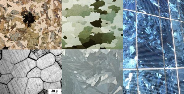
A crystallite is a small or even microscopic crystal which forms, for example, during the cooling of many materials. Crystallites are also referred to as grains. Bacillite is a type of crystallite. It is rodlike with parallel longulites. Structure The orientation of crystallites can be random with no preferred direction, called random texture, or directed, possibly due to growth and processing conditions. While the structure of a single crystal is highly ordered and its lattice is continuous and unbroken, amorphous materials, such as glass and many polymers, are non-crystalline and do not display any structures, as their constituents are not arranged in an ordered manner. Polycrystalline structures and paracrystalline phases are in between these two extremes. Polycrystalline materials, or polycrystals, are solids that are composed of many crystallites of varying size and orientation. Most materials are polycrystalline, made of a large number crystallites held together by th ... [...More Info...] [...Related Items...] OR: [Wikipedia] [Google] [Baidu] |
Bragg Condition
In many areas of science, Bragg's law — also known as Georg Wulff, Wulff–Bragg's condition or Laue–Bragg interference — is a special case of Laue diffraction that gives the angles for coherent scattering of waves from a large crystal lattice. It describes how the superposition of wave fronts scattered by lattice planes leads to a strict relation between the wavelength and scattering angle. This law was initially formulated for X-rays, but it also applies to all types of Matter wave, matter waves including neutron and electron waves if there are a large number of atoms, as well as to visible light with artificial periodic microscale lattices. History Bragg diffraction (also referred to as the Bragg formulation of X-ray diffraction) was first proposed by Lawrence Bragg and his father, William Henry Bragg, in 1913 after their discovery that crystalline solids produced surprising patterns of reflected X-rays (in contrast to those produced with, for instance, a liquid). They ... [...More Info...] [...Related Items...] OR: [Wikipedia] [Google] [Baidu] |
European Synchrotron Radiation Facility
The European Synchrotron (ESRF) is a joint research facility situated in Grenoble, France, supported by 19 countries (13 member countries: Belgium, Denmark, Finland, France, Germany, Italy, the Netherlands, Norway, Russia, Spain, Sweden, Switzerland, and the UK; and 6 associate countries: Austria, the Czech Republic, Israel, Poland, Portugal and South Africa). Some 10,000 scientists visit this particle accelerator each year, conducting upwards of 2,000 experiments and producing around 1,800 scientific publications. History Inaugurated in September 1994, it has an annual operating budget of around 100 million euros, employs around 700 people and is host to more than 10,000 visiting scientists each year. The ESRF was the world's first third generation synchrotron when it opened for user operation in 1994. In 2009, the ESRF began a major refurbishment programme that, at term, has seen its performances increase by 100-fold. In 2015, the facility built an 8000 m2 extension to ... [...More Info...] [...Related Items...] OR: [Wikipedia] [Google] [Baidu] |
Algebraic Reconstruction Technique
frame, Animated sequence of reconstruction steps, one iteration. The algebraic reconstruction technique (ART) is an iterative reconstruction technique used in computed tomography. It reconstructs an image from a series of angular projections (a sinogram). Gordon, Bender and Herman first showed its use in image reconstruction; whereas the method is known as Kaczmarz method in numerical linear algebra. An advantage of ART over other reconstruction methods (such as filtered backprojection) is that it is relatively easy to incorporate prior knowledge into the reconstruction process. ART can be considered as an iterative solver of a system of linear equations A x = b , where: : A is a sparse m \times n matrix whose values represent the relative contribution of each output pixel to different points in the sinogram ( m being the number of individual values in the sinogram, and n being the number of output pixels); : x represents the pixels in the generated (output) imag ... [...More Info...] [...Related Items...] OR: [Wikipedia] [Google] [Baidu] |
Monte Carlo Method
Monte Carlo methods, or Monte Carlo experiments, are a broad class of computational algorithms that rely on repeated random sampling to obtain numerical results. The underlying concept is to use randomness to solve problems that might be deterministic in principle. The name comes from the Monte Carlo Casino in Monaco, where the primary developer of the method, mathematician Stanisław Ulam, was inspired by his uncle's gambling habits. Monte Carlo methods are mainly used in three distinct problem classes: optimization, numerical integration, and generating draws from a probability distribution. They can also be used to model phenomena with significant uncertainty in inputs, such as calculating the risk of a nuclear power plant failure. Monte Carlo methods are often implemented using computer simulations, and they can provide approximate solutions to problems that are otherwise intractable or too complex to analyze mathematically. Monte Carlo methods are widely used in va ... [...More Info...] [...Related Items...] OR: [Wikipedia] [Google] [Baidu] |
Electrons
The electron (, or in nuclear reactions) is a subatomic particle with a negative one elementary charge, elementary electric charge. It is a fundamental particle that comprises the ordinary matter that makes up the universe, along with up quark, up and down quark, down quarks. Electrons are extremely lightweight particles that orbit the positively charged atomic nucleus, nucleus of atoms. Their negative charge is balanced by the positive charge of protons in the nucleus, giving atoms their overall electric charge#Charge neutrality, neutral charge. Ordinary matter is composed of atoms, each consisting of a positively charged nucleus surrounded by a number of orbiting electrons equal to the number of protons. The configuration and energy levels of these orbiting electrons determine the chemical properties of an atom. Electrons are bound to the nucleus to different degrees. The outermost or valence electron, valence electrons are the least tightly bound and are responsible for th ... [...More Info...] [...Related Items...] OR: [Wikipedia] [Google] [Baidu] |
Neutrons
The neutron is a subatomic particle, symbol or , that has no electric charge, and a mass slightly greater than that of a proton. The neutron was discovered by James Chadwick in 1932, leading to the discovery of nuclear fission in 1938, the first self-sustaining nuclear reactor (Chicago Pile-1, 1942) and the first nuclear weapon (Trinity, 1945). Neutrons are found, together with a similar number of protons in the nuclei of atoms. Atoms of a chemical element that differ only in neutron number are called isotopes. Free neutrons are produced copiously in nuclear fission and fusion. They are a primary contributor to the nucleosynthesis of chemical elements within stars through fission, fusion, and neutron capture processes. Neutron stars, formed from massive collapsing stars, consist of neutrons at the density of atomic nuclei but a total mass more than the Sun. Neutron properties and interactions are described by nuclear physics. Neutrons are not elementary particles; each is ... [...More Info...] [...Related Items...] OR: [Wikipedia] [Google] [Baidu] |
Dark-field X-ray Microscopy
Dark-field X-ray microscopy (DFXM or DFXRM) is an imaging technique used for multiscale Characterization (materials science), structural characterisation. It is capable of mapping deeply embedded structural elements with nm-resolution using synchrotron X-ray diffraction-based imaging. The technique works by using scattered X-rays to create a high degree of Diffraction contrast tomography, contrast, and by measuring the intensity and spatial distribution of the diffracted beams, it is possible to obtain a three-dimensional map of the sample's Crystal structure, structure, Orientation (geometry), orientation, and local Strain (mechanics), strain. History The first experimental demonstration of dark-field X-ray microscopy was reported in 2006 by a group at the European Synchrotron Radiation Facility in Grenoble, France. Since then, the technique has been rapidly evolving and has shown great promise in multiscale structural characterization. Its development is largely due to advance ... [...More Info...] [...Related Items...] OR: [Wikipedia] [Google] [Baidu] |
Diffraction Contrast Tomography
Diffraction is the deviation of waves from straight-line propagation without any change in their energy due to an obstacle or through an aperture. The diffracting object or aperture effectively becomes a secondary source of the propagating wave. Diffraction is the same physical effect as interference, but interference is typically applied to superposition of a few waves and the term diffraction is used when many waves are superposed. Italian scientist Francesco Maria Grimaldi coined the word ''diffraction'' and was the first to record accurate observations of the phenomenon in 1660. In classical physics, the diffraction phenomenon is described by the Huygens–Fresnel principle that treats each point in a propagating wavefront as a collection of individual spherical wavelets. The characteristic pattern is most pronounced when a wave from a coherent source (such as a laser) encounters a slit/aperture that is comparable in size to its wavelength, as shown in the inserted image. ... [...More Info...] [...Related Items...] OR: [Wikipedia] [Google] [Baidu] |
X-ray Diffraction Computed Tomography
X-ray diffraction computed tomography is an experimental technique that combines X-ray diffraction with the computed tomography data acquisition approach. X-ray diffraction (XRD) computed tomography (CT) was first introduced in 1987 by Harding et al. using a laboratory diffractometer and a monochromatic X-ray pencil beam. The first implementation of the technique at synchrotron facilities was performed in 1998 by Kleuker ''et al.'' X-ray diffraction computed tomography can be divided into two main categories depending on how the XRD data are being treated, specifically the XRD data can be treated either as powder diffraction or single crystal diffraction data and this depends on the sample properties. If the sample contains small and randomly oriented crystals, then it generates smooth powder diffraction "rings" when using a 2D area detector. If the sample contains large crystals, then it generates "spotty" 2D diffraction patterns. The latter can be performed using also a letterbo ... [...More Info...] [...Related Items...] OR: [Wikipedia] [Google] [Baidu] |


