|
Supraorbital Nerve
The supraorbital nerve is one of two terminal branches - the other being the supratrochlear nerve - of the frontal nerve (itself a branch of the ophthalmic nerve (CN V1)). It exits the orbit via the supraorbital foramen/notch before splitting into a medial branch and a lateral branch. It innervates the skin of the forehead, upper eyelid, and the root of the nose. Structure Origin The supraorbital nerve branches from the frontal nerve midway between the base and apex of the orbit. Course It travels anteriorly superior to the levator palpebrae superioris muscle. It exits the orbit through the supraorbital foramen/notch in the superior margin orbit, exiting it lateral to the supratrochlear nerve. It then ascends onto the forehead deep to the corrugator supercilii muscle and frontalis muscles. Fate It divides into a medial branch and lateral branch - usually after emerging from the orbit, but sometimes already within the orbit. Distribution The supraorbital nerv ... [...More Info...] [...Related Items...] OR: [Wikipedia] [Google] [Baidu] |
Conjunctiva
In the anatomy of the eye, the conjunctiva (: conjunctivae) is a thin mucous membrane that lines the inside of the eyelids and covers the sclera (the white of the eye). It is composed of non-keratinized, stratified squamous epithelium with goblet cells, stratified columnar epithelium and stratified cuboidal epithelium (depending on the zone). The conjunctiva is highly Angiogenesis, vascularised, with many microvessels easily accessible for imaging studies. Structure The conjunctiva is typically divided into three parts: Blood supply Blood to the bulbar conjunctiva is primarily derived from the ophthalmic artery. The blood supply to the palpebral conjunctiva (the eyelid) is derived from the external carotid artery. However, the circulations of the bulbar conjunctiva and palpebral conjunctiva are linked, so both bulbar conjunctival and palpebral conjunctival vessels are supplied by both the ophthalmic artery and the external carotid artery, to varying extents. Nerve supply Se ... [...More Info...] [...Related Items...] OR: [Wikipedia] [Google] [Baidu] |
Frontal Sinus
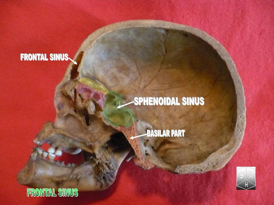
The frontal sinuses are one of the four pairs of paranasal sinuses that are situated behind the brow ridges. Sinuses are mucosa-lined airspaces within the bones of the face and skull. Each opens into the anterior part of the corresponding middle nasal meatus of the nose through the frontonasal duct which traverses the anterior part of the labyrinth of the ethmoid. These structures then open into the semilunar hiatus in the middle meatus. Structure Each frontal sinus is situated between the external and internal plates of the frontal bone. Their average measurements are as follows: height 28 mm, breadth 24 mm, depth 20 mm, creating a space of 6-7 ml. Each frontal sinus extends into the squamous part of the frontal bone superiorly, and into the orbital part of frontal bone posteriorly to come to occupy the medial part of the roof of the orbit. Each sinus drains through an opening in its inferomedial part into the frontonasal duct. Vasculature The mucous me ... [...More Info...] [...Related Items...] OR: [Wikipedia] [Google] [Baidu] |
Frontal Nerve
The frontal nerve is the largest branch of the ophthalmic nerve (V1), itself a branch of the trigeminal nerve (CN V). It supplies sensation to the skin of the forehead, the mucosa of the frontal sinus, and the skin of the upper eyelid. It may be affected by schwannoma. Structure The frontal nerve is a branch of the ophthalmic nerve (V1), itself a branch of the trigeminal nerve (CN V). The frontal nerve branches immediately before entering the superior orbital fissure. In then travels superolateral to the annulus of Zinn between the lacrimal nerve and inferior ophthalmic vein. After entering the orbit it travels anteriorly between the roof periosteum and the levator palpebrae superioris. Midway between the apex and base of the orbit it divides into two branches, the supratrochlear nerve and supraorbital nerve. Functions The two branches of the frontal nerve provide sensory innervation to the skin of the forehead, mucosa of the frontal sinus (an air sinus), and the skin of th ... [...More Info...] [...Related Items...] OR: [Wikipedia] [Google] [Baidu] |
Supratrochlear Nerve
The supratrochlear nerve is a branch of the frontal nerve, itself a branch of the ophthalmic nerve (CN V1) from the trigeminal nerve (CN V). It provides sensory innervation to the skin of the forehead and the upper eyelid. Structure Origin The supratrochlear nerve is the smaller of the two terminal branches of the frontal nerve (the other being the supraorbital nerve). It arises midway between the base and apex of the orbit where the frontal nerve splits into said terminal branches. Course The supratrochlear nerve passes medially above the trochlea of the superior oblique muscle. It then travels anteriorly above the levator palpebrae superioris muscle. It exits the orbit through the supratrochlear notch or foramen. It then ascends onto the forehead beneath the corrugator supercilii muscle and frontalis muscle. It finally divides into sensory branches. The supratrochlear nerve travels with the supratrochlear artery, a branch of the ophthalmic artery. Branches Be ... [...More Info...] [...Related Items...] OR: [Wikipedia] [Google] [Baidu] |
Frontal Nerve
The frontal nerve is the largest branch of the ophthalmic nerve (V1), itself a branch of the trigeminal nerve (CN V). It supplies sensation to the skin of the forehead, the mucosa of the frontal sinus, and the skin of the upper eyelid. It may be affected by schwannoma. Structure The frontal nerve is a branch of the ophthalmic nerve (V1), itself a branch of the trigeminal nerve (CN V). The frontal nerve branches immediately before entering the superior orbital fissure. In then travels superolateral to the annulus of Zinn between the lacrimal nerve and inferior ophthalmic vein. After entering the orbit it travels anteriorly between the roof periosteum and the levator palpebrae superioris. Midway between the apex and base of the orbit it divides into two branches, the supratrochlear nerve and supraorbital nerve. Functions The two branches of the frontal nerve provide sensory innervation to the skin of the forehead, mucosa of the frontal sinus (an air sinus), and the skin of th ... [...More Info...] [...Related Items...] OR: [Wikipedia] [Google] [Baidu] |
Ophthalmic Nerve
The ophthalmic nerve (CN V1) is a sensory nerve of the head. It is one of three divisions of the trigeminal nerve (CN V), a cranial nerve. It has three major branches which provide sensory innervation to the eye, and the skin of the upper face and anterior scalp, as well as other structures of the head. Structure Origin The ophthalmic nerve is the first branch of the trigeminal nerve (CN V), the first and smallest of its three divisions. It arises from the superior part of the trigeminal ganglion. Course It passes anterior-ward along the lateral wall of the cavernous sinus inferior to the oculomotor nerve (CN III) and trochlear nerve (N IV). It exits the skull into the orbit through the superior orbital fissure. Branches Within the skull, the ophthalmic nerve produces: * meningeal branch (tentorial nerve) The ophthalmic nerve divides into three major branches which pass through the superior orbital fissure: * frontal nerve ** supraorbital nerve ** supratrochlea ... [...More Info...] [...Related Items...] OR: [Wikipedia] [Google] [Baidu] |
Supraorbital Foramen
The supraorbital foramen, is a bony elongated opening located above the orbit (eye socket) and under the forehead. It is part of the frontal bone of the skull. The supraorbital foramen lies directly under the eyebrow. In some people this foramen is incomplete and is then known as the supraorbital notch. Structure The supraorbital foramen is a small groove at superior and medial margin of the orbit in the frontal bone. It is part of the frontal bone of the skull. It arches transversely below the superciliary arches and is the upper part of the brow ridge. It is thin and prominent in its lateral two-thirds, but rounded in its medial third. Between these two parts, the supraorbital nerve, the supraorbital artery, and the supraorbital vein pass. The supraorbital nerve divides into superficial and deep branches after it has left the supraorbital foramen. Additional images File:Gray135.png, Frontal bone. Inner surface. File:Gray1193.svg, Side view of head, showing surface relations ... [...More Info...] [...Related Items...] OR: [Wikipedia] [Google] [Baidu] |
Levator Palpebrae Superioris Muscle
The levator palpebrae superioris () is the muscle in the orbit that elevates the upper eyelid. Structure The levator palpebrae superioris originates from inferior surface of the lesser wing of the sphenoid bone, just above the optic foramen. It broadens and decreases in thickness (becomes thinner) and becomes the levator aponeurosis. This portion inserts on the skin of the upper eyelid, as well as the superior tarsal plate. It is a skeletal muscle. The superior tarsal muscle, a smooth muscle, is attached to the levator palpebrae superioris, and inserts on the superior tarsal plate as well. Blood supply The levator palebrae superioris receives its blood supply from branches of the ophthalmic artery, specifically, muscular branches and the supraorbital artery. Blood is drained into the superior ophthalmic vein. Nerve supply The levator palpebrae superioris receives motor innervation from the superior division of the oculomotor nerve. The smooth muscle that originates from ... [...More Info...] [...Related Items...] OR: [Wikipedia] [Google] [Baidu] |
Corrugator Supercilii
The corrugator supercilii muscle is a small, narrow, pyramidal muscle of the face. It arises from the medial end of the superciliary arch; it inserts into the deep surface of the skin of the eyebrow. It draws the eyebrow downward and medially, producing the vertical "frowning" wrinkles of the forehead. It may be thought as the principal muscle in the facial expression of suffering. It also shields the eyes from strong sunlight. Structure The corrugator supercilii muscle is located at the medial end of the eyebrow. Its fibers pass laterally and somewhat superiorly from its origin to its insertion. Origin It arises from bone at the medial extremity of the superciliary arch. Insertion It inserts between the palpebral and orbital portions of the orbicularis oculi muscle. It inserts into the deep surface of the skin of the eyebrow, above the middle of the orbital arch. Innervation Motor innervation is provided by the temporal branches of facial nerve (CN VII). Vasculatu ... [...More Info...] [...Related Items...] OR: [Wikipedia] [Google] [Baidu] |
Frontalis Muscles
The frontalis muscle () is a muscle which covers parts of the forehead of the skull. Some sources consider the frontalis muscle to be a distinct muscle. However, Terminologia Anatomica currently classifies it as part of the occipitofrontalis muscle along with the occipitalis muscle. In humans, the frontalis muscle only serves for facial expressions. The frontalis muscle is supplied by the facial nerve and receives blood from the supraorbital and supratrochlear arteries. Structure The frontalis muscle is thin, of a quadrilateral form, and intimately adherent to the superficial fascia. It is broader than the occipitalis and its fibers are longer and paler in color. It is located on the front of the head. The muscle has no bony attachments. Its medial fibers are continuous with those of the procerus; its intermediate fibers blend with the corrugator and orbicularis oculi muscles, thus attached to the skin of the eyebrows; and its lateral fibers are also blended with the latter m ... [...More Info...] [...Related Items...] OR: [Wikipedia] [Google] [Baidu] |
Upper Eyelid
An eyelid ( ) is a thin fold of skin that covers and protects an eye. The levator palpebrae superioris muscle retracts the eyelid, exposing the cornea to the outside, giving vision. This can be either voluntarily or involuntarily. "Palpebral" (and "blepharal") means relating to the eyelids. Its key function is to regularly spread the tears and other secretions on the eye surface to keep it moist, since the cornea must be continuously moist. They keep the eyes from drying out when asleep. Moreover, the blink reflex protects the eye from foreign bodies. A set of specialized hairs known as lashes grow from the upper and lower eyelid margins to further protect the eye from dust and debris. The appearance of the human upper eyelid often varies between different populations. The prevalence of an epicanthic fold covering the inner corner of the eye account for the majority of East Asian and Southeast Asian populations, and is also found in varying degrees among other population ... [...More Info...] [...Related Items...] OR: [Wikipedia] [Google] [Baidu] |

