|
Stapes Human Ear
The ''stapes'' or stirrup is a bone in the middle ear of humans and other tetrapods which is involved in the conduction of sound vibrations to the inner ear. This bone is connected to the oval window by its annular ligament, which allows the footplate (or base) to transmit sound energy through the oval window into the inner ear. The ''stapes'' is the smallest and lightest bone in the human body, and is so-called because of its resemblance to a stirrup (). Structure The ''stapes'' is the third bone of the three ossicles in the middle ear and the smallest in the human body. It measures roughly , greater along the head-base span. It rests on the oval window, to which it is connected by an annular ligament and articulates with the ''incus'', or anvil through the incudostapedial joint. They are connected by anterior and posterior limbs (). Development The ''stapes'' develops from the second pharyngeal arch during the sixth to eighth week of embryological life. The central cav ... [...More Info...] [...Related Items...] OR: [Wikipedia] [Google] [Baidu] |
Branchial Arch
Branchial arches or gill arches are a series of paired bony/ cartilaginous "loops" behind the throat ( pharyngeal cavity) of fish, which support the fish gills. As chordates, all vertebrate embryos develop pharyngeal arches, though the eventual fate of these arches varies between taxa. In all jawed fish (gnathostomes), the first arch pair (mandibular arches) develops into the jaw, the second gill arches (the hyoid arches) develop into the hyomandibular complex (which supports the back of the jaw and the front of the gill series), and the remaining posterior arches (simply called branchial arches) support the gills. In tetrapods, a mostly terrestrial clade evolved from lobe-finned fish, many pharyngeal arch elements are lost, including the gill arches. In amphibians and reptiles, only the oral jaws and a hyoid apparatus remains, and in mammals and birds the hyoid is simplified further to support the tongue and floor of the mouth. In mammals, the first and second pharyng ... [...More Info...] [...Related Items...] OR: [Wikipedia] [Google] [Baidu] |
Pharyngeal Arch
The pharyngeal arches, also known as visceral arches'','' are transient structures seen in the Animal embryonic development, embryonic development of humans and other vertebrates, that are recognisable precursors for many structures. In fish, the arches support the Fish gill, gills and are known as the branchial arches, or gill arches. In the human embryo, the arches are first seen during the fourth week of human embryonic development, development. They appear as a series of outpouchings of mesoderm on both sides of the developing pharynx. The vasculature of the pharyngeal arches are the aortic arches that arise from the aortic sac. Structure In humans and other vertebrates, the pharyngeal arches are derived from all three germ layers (the primary layers of cells that form during embryonic development). Neural crest cells enter these arches where they contribute to features of the skull and facial skeleton such as bone and cartilage. However, the existence of pharyngeal structu ... [...More Info...] [...Related Items...] OR: [Wikipedia] [Google] [Baidu] |
Otosclerosis
Otosclerosis is a condition of the middle ear, middle and inner ear where portions of the dense enchondral layer of the bony labyrinth Tissue remodeling, remodel into one or more lesions of irregularly-laid spongy bone. As the lesions reach the stapes the bone is Bone resorption, resorbed, then hardened (Sclerosis (medicine), sclerotized), which limits its movement and results in hearing loss, tinnitus, vertigo or a combination of these. The term otosclerosis is something of a misnomer: much of the clinical course is characterized by lucent rather than sclerotic bony changes, so the disease is also known as otospongiosis. Etymology The word ''otosclerosis'' derives from Greek Language, Greek ὠτός (''ōtos''), genitive of οὖς (''oûs'') "ear" + σκλήρωσις (''sklērōsis''), "hardening". Presentation The primary form of hearing loss in otosclerosis is conductive hearing loss (CHL) whereby sounds reach the ear drum but are incompletely transferred via the ossicular ... [...More Info...] [...Related Items...] OR: [Wikipedia] [Google] [Baidu] |
Facial Nerve
The facial nerve, also known as the seventh cranial nerve, cranial nerve VII, or simply CN VII, is a cranial nerve that emerges from the pons of the brainstem, controls the muscles of facial expression, and functions in the conveyance of taste sensations from the anterior two-thirds of the tongue. The nerve typically travels from the pons through the facial canal in the temporal bone and exits the skull at the stylomastoid foramen. It arises from the brainstem from an area posterior to the cranial nerve VI (abducens nerve) and anterior to cranial nerve VIII (vestibulocochlear nerve). The facial nerve also supplies preganglionic parasympathetic fibers to several head and neck ganglia. The facial and intermediate nerves can be collectively referred to as the nervus intermediofacialis. The path of the facial nerve can be divided into six segments: # intracranial (cisternal) segment (from brainstem pons to internal auditory canal) # meatal (canalicular) segment (with ... [...More Info...] [...Related Items...] OR: [Wikipedia] [Google] [Baidu] |
Stapedius
The stapedius is the smallest skeletal muscle in the human body. At just over one millimeter in length, its purpose is to stabilize the smallest bone in the body, the stapes or stirrup bone of the middle ear. Structure The stapedius emerges from a pinpoint foramen or opening in the apex of the pyramidal eminence (a hollow, cone-shaped prominence in the posterior wall of the tympanic cavity), and inserts into the neck of the stapes. Nerve supply The stapedius is supplied by the nerve to stapedius, a branch of the facial nerve. Function The stapedius dampens the vibrations of the stapes by pulling on the neck of that bone. As one of the muscles involved in the acoustic reflex it prevents excess movement of the stapes, helping to control the amplitude of sound, sound waves from the general external environment to the inner ear. Clinical significance Paralysis of the stapedius allows wider oscillation of the stapes, resulting in heightened reaction of the Auditory Ossicles, audit ... [...More Info...] [...Related Items...] OR: [Wikipedia] [Google] [Baidu] |
Amphibian
Amphibians are ectothermic, anamniote, anamniotic, tetrapod, four-limbed vertebrate animals that constitute the class (biology), class Amphibia. In its broadest sense, it is a paraphyletic group encompassing all Tetrapod, tetrapods, but excluding the amniotes (tetrapods with an amniotic membrane, such as modern reptiles, birds and mammals). All extant taxon, extant (living) amphibians belong to the monophyletic subclass (biology), subclass Lissamphibia, with three living order (biology), orders: Anura (frogs and toads), Urodela (salamanders), and Gymnophiona (caecilians). Evolved to be mostly semiaquatic, amphibians have adapted to inhabit a wide variety of habitats, with most species living in freshwater ecosystem, freshwater, wetland or terrestrial ecosystems (such as riparian woodland, fossorial and even arboreal habitats). Their biological life cycle, life cycle typically starts out as aquatic animal, aquatic larvae with gills known as tadpoles, but some species have devel ... [...More Info...] [...Related Items...] OR: [Wikipedia] [Google] [Baidu] |
Spiracle (vertebrates)
Spiracles () are openings on the surface of some animals, which usually lead to respiratory systems. The spiracle is a small hole behind each eye that opens to the mouth in some fish. In the Agnatha, jawless fish, the first gill opening immediately behind the mouth is essentially similar to the other gill openings. With the evolution of the jaw in the early gnathostomes, jawed vertebrates, this gill slit was caught between the forward gill-rod (now functioning as the jaw) and the next rod, the Hyomandibula, hyomandibular bone, supporting the jaw hinge and anchoring the jaw to the skull proper. The gill opening was closed off from below, the remaining opening was small and hole-like, and is termed a spiracle. In many species of Shark, sharks and all Batoidea, rays the spiracle is responsible for the intake of water into the buccal space before being expelled from the gills. The spiracle is often located towards the top of the animal allowing breathing even while the animal is most ... [...More Info...] [...Related Items...] OR: [Wikipedia] [Google] [Baidu] |
Gill Arch
Branchial arches or gill arches are a series of paired bony/ cartilaginous "loops" behind the throat ( pharyngeal cavity) of fish, which support the fish gills. As chordates, all vertebrate embryos develop pharyngeal arches, though the eventual fate of these arches varies between taxa. In all jawed fish (gnathostomes), the first arch pair (mandibular arches) develops into the jaw, the second gill arches (the hyoid arches) develop into the hyomandibular complex (which supports the back of the jaw and the front of the gill series), and the remaining posterior arches (simply called branchial arches) support the gills. In tetrapods, a mostly terrestrial clade evolved from lobe-finned fish, many pharyngeal arch elements are lost, including the gill arches. In amphibians and reptiles, only the oral jaws and a hyoid apparatus remains, and in mammals and birds the hyoid is simplified further to support the tongue and floor of the mouth. In mammals, the first and second pharyngea ... [...More Info...] [...Related Items...] OR: [Wikipedia] [Google] [Baidu] |
Hyomandibula
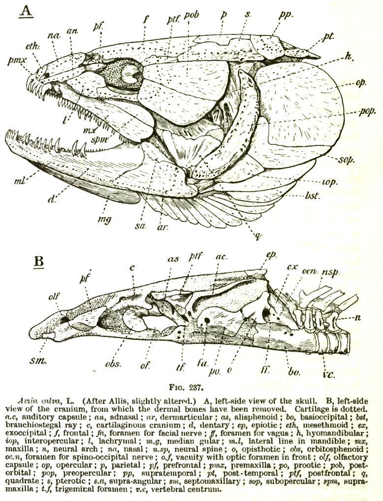
The hyomandibula, commonly referred to as hyomandibular one(, from , "upsilon-shaped" (υ), and Latin: mandibula, "jawbone"), is a set of bones that is found in the hyoid region in most fishes. It usually plays a role in suspending the jaws and/or operculum ( teleostomi only). It is commonly suggested that in tetrapods (land animals), the hyomandibula evolved into the columella ( stapes). Evolutionary context In jawless fishes, a series of gills opened behind the mouth, and these gills became supported by cartilaginous elements. The first set of these elements surrounded the mouth to form the jaw. There is ample evidence For example: (1) both sets of bones are made from neural crest cells (rather than mesodermal tissue like most other bones); (2) both structures form the upper and lower bars that bend forward and are hinged in the middle; and (3) the musculature of the jaw seem homologous to the gill arches of jawless fishes. (Gilbert 2000) that vertebrate jaws are ... [...More Info...] [...Related Items...] OR: [Wikipedia] [Google] [Baidu] |
Reptile
Reptiles, as commonly defined, are a group of tetrapods with an ectothermic metabolism and Amniotic egg, amniotic development. Living traditional reptiles comprise four Order (biology), orders: Testudines, Crocodilia, Squamata, and Rhynchocephalia. About 12,000 living species of reptiles are listed in the Reptile Database. The study of the traditional reptile orders, customarily in combination with the study of modern amphibians, is called herpetology. Reptiles have been subject to several conflicting Taxonomy, taxonomic definitions. In Linnaean taxonomy, reptiles are gathered together under the Class (biology), class Reptilia ( ), which corresponds to common usage. Modern Cladistics, cladistic taxonomy regards that group as Paraphyly, paraphyletic, since Genetics, genetic and Paleontology, paleontological evidence has determined that birds (class Aves), as members of Dinosauria, are more closely related to living crocodilians than to other reptiles, and are thus nested among re ... [...More Info...] [...Related Items...] OR: [Wikipedia] [Google] [Baidu] |
Columella (auditory System)
In the auditory system, the columella contributes to hearing in amphibians, reptiles and birds. The columella form thin, bony structures in the interior of the skull and serve the purpose of transmitting sounds from the eardrum. It is an evolutionary Homology (biology), homolog of the stapes, one of the Evolution of mammalian auditory ossicles, auditory ossicles in mammals. In many species, the extracolumella is a cartilaginous structure that grows in association with the columella. During development, the columella is derived from the dorsal end of the Pharyngeal arch, hyoid arch. Evolution The evolution of the columella is closely related to the evolution of the Temporomandibular joint, jaw joint. It is an ancestral homolog of the stapes, and is derived from the Hyomandibula, hyomandibular bone of fishes. As the columella is derived from the hyomandibula, many of its functional relationships remain the same. The columella resides in the air-filled tympanic cavity of the middl ... [...More Info...] [...Related Items...] OR: [Wikipedia] [Google] [Baidu] |
Homology (biology)
In biology, homology is similarity in anatomical structures or genes between organisms of different taxa due to shared ancestry, ''regardless'' of current functional differences. Evolutionary biology explains homologous structures as retained heredity from a common descent, common ancestor after having been subjected to adaptation (biology), adaptive modifications for different purposes as the result of natural selection. The term was first applied to biology in a non-evolutionary context by the anatomist Richard Owen in 1843. Homology was later explained by Charles Darwin's theory of evolution in 1859, but had been observed before this from Aristotle's biology onwards, and it was explicitly analysed by Pierre Belon in 1555. A common example of homologous structures is the forelimbs of vertebrates, where the bat wing development, wings of bats and origin of avian flight, birds, the arms of primates, the front flipper (anatomy), flippers of whales, and the forelegs of quadrupedalis ... [...More Info...] [...Related Items...] OR: [Wikipedia] [Google] [Baidu] |


