|
Perirhinal Cortex
The perirhinal cortex is a brain cortex, cortical region in the medial temporal lobe that is made up of Brodmann areas Brodmann area 35, 35 and Brodmann area 36, 36. It receives highly processed sensory information from all sensory regions, and is generally accepted to be an important region for memory. It is bordered caudally by postrhinal cortex or parahippocampal gyrus, parahippocampal cortex (homologous regions in rodents and primates, respectively) and ventrally and Anatomical terms of location#Left and right (lateral), and medial, medially by entorhinal cortex. Structure The perirhinal cortex is composed of two regions: areas 36 and 35. Area 36 is sometimes divided into three subdivisions: 36d is the most rostral and dorsal, 36r ventral and caudal, and 36c the most caudal. Area 35 can be divided in the same manner, into 35d and 35v (for dorsal and ventral, respectively). Area 36 is six-layered, dysgranular, meaning that its layer IV is relatively sparse. Area 35 is agran ... [...More Info...] [...Related Items...] OR: [Wikipedia] [Google] [Baidu] |
Cerebral Cortex
The cerebral cortex, also known as the cerebral mantle, is the outer layer of neural tissue of the cerebrum of the brain in humans and other mammals. It is the largest site of Neuron, neural integration in the central nervous system, and plays a key role in attention, perception, awareness, thought, memory, language, and consciousness. The six-layered neocortex makes up approximately 90% of the Cortex (anatomy), cortex, with the allocortex making up the remainder. The cortex is divided into left and right parts by the longitudinal fissure, which separates the two cerebral hemispheres that are joined beneath the cortex by the corpus callosum and other commissural fibers. In most mammals, apart from small mammals that have small brains, the cerebral cortex is folded, providing a greater surface area in the confined volume of the neurocranium, cranium. Apart from minimising brain and cranial volume, gyrification, cortical folding is crucial for the Neural circuit, brain circuitry ... [...More Info...] [...Related Items...] OR: [Wikipedia] [Google] [Baidu] |
Visual Cortex
The visual cortex of the brain is the area of the cerebral cortex that processes visual information. It is located in the occipital lobe. Sensory input originating from the eyes travels through the lateral geniculate nucleus in the thalamus and then reaches the visual cortex. The area of the visual cortex that receives the sensory input from the lateral geniculate nucleus is the primary visual cortex, also known as visual area 1 ( V1), Brodmann area 17, or the striate cortex. The extrastriate areas consist of visual areas 2, 3, 4, and 5 (also known as V2, V3, V4, and V5, or Brodmann area 18 and all Brodmann area 19). Both hemispheres of the brain include a visual cortex; the visual cortex in the left hemisphere receives signals from the right visual field, and the visual cortex in the right hemisphere receives signals from the left visual field. Introduction The primary visual cortex (V1) is located in and around the calcarine fissure in the occipital lobe. Each h ... [...More Info...] [...Related Items...] OR: [Wikipedia] [Google] [Baidu] |
Subiculum
The subiculum (Latin for "support") also known as the subicular complex, or subicular cortex, is the most inferior component of the hippocampal formation. It lies between the entorhinal cortex and the CA1 hippocampal subfield. The subicular complex comprises a set of four related structures including the prosubiculum, presubiculum, postsubiculum and parasubiculum. Name The subiculum got its name from Karl Friedrich Burdach in his three-volume work ''Vom Bau und Leben des Gehirns'' (Vol. 2, §199). He originally named it subiculum cornu ammonis and so associated it with the rest of the hippocampal subfields. Structure The subicular complex receives input from CA1 and entorhinal cortical layer III pyramidal neurons and is the main output of the hippocampus proper. The pyramidal neurons send projections to the nucleus accumbens, septal nuclei, prefrontal cortex, lateral hypothalamus, nucleus reuniens, mammillary nuclei, entorhinal cortex and amygdala. The pyramidal neuro ... [...More Info...] [...Related Items...] OR: [Wikipedia] [Google] [Baidu] |
Hippocampal Subfields
The hippocampal subfields are four subfields CA1, CA2, CA3, and CA4 that make up the structure of the hippocampus. Regions described in the hippocampus are the head, body, and tail, and other hippocampal subfields include the dentate gyrus, the presubiculum, and the subiculum. The CA subfields use the initials of cornu ammonis, an earlier name of the hippocampus. Structure There are four hippocampal subfields, in the hippocampus proper which form a neural circuit called the trisynaptic circuit. CA1 CA1 is the first region in the hippocampal circuit, from which a significant output pathway goes to layer V of the entorhinal cortex. The main output of CA1 is to the subiculum. CA2 CA2 is a small region located between CA1 and CA3. It receives some input from layer II of the entorhinal cortex via the perforant path. Its pyramidal cells are more like those in CA3 than those in CA1. It is often ignored due to its small size. CA3 CA3 receives input from the mossy fibers of the g ... [...More Info...] [...Related Items...] OR: [Wikipedia] [Google] [Baidu] |
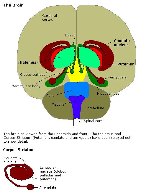
Hippocampus
The hippocampus (: hippocampi; via Latin from Ancient Greek, Greek , 'seahorse'), also hippocampus proper, is a major component of the brain of humans and many other vertebrates. In the human brain the hippocampus, the dentate gyrus, and the subiculum are components of the hippocampal formation located in the limbic system. The hippocampus plays important roles in the Memory consolidation, consolidation of information from short-term memory to long-term memory, and in spatial memory that enables Navigation#Navigation in spatial cognition, navigation. In humans, and other primates the hippocampus is located in the archicortex, one of the three regions of allocortex, in each cerebral hemisphere, hemisphere with direct neural projections to, and reciprocal indirect projections from the neocortex. The hippocampus, as the medial pallium, is a structure found in all vertebrates. In Alzheimer's disease (and other forms of dementia), the hippocampus is one of the first regions of th ... [...More Info...] [...Related Items...] OR: [Wikipedia] [Google] [Baidu] |
Amygdala
The amygdala (; : amygdalae or amygdalas; also '; Latin from Greek language, Greek, , ', 'almond', 'tonsil') is a paired nucleus (neuroanatomy), nuclear complex present in the Cerebral hemisphere, cerebral hemispheres of vertebrates. It is considered part of the limbic system. In Primate, primates, it is located lateral and medial, medially within the temporal lobes. It consists of many nuclei, each made up of further subnuclei. The subdivision most commonly made is into the Basolateral amygdala, basolateral, Central nucleus of the amygdala, central, cortical, and medial nuclei together with the intercalated cells of the amygdala, intercalated cell clusters. The amygdala has a primary role in the processing of memory, decision making, decision-making, and emotions, emotional responses (including fear, anxiety, and aggression). The amygdala was first identified and named by Karl Friedrich Burdach in 1822. Structure Thirteen Nucleus (neuroanatomy), nuclei have been identif ... [...More Info...] [...Related Items...] OR: [Wikipedia] [Google] [Baidu] |
Basal Forebrain
Part of the human brain, the basal forebrain structures are located in the forebrain to the front of and below the striatum. They include the ventral basal ganglia (including nucleus accumbens and ventral pallidum), nucleus basalis, diagonal band of Broca, substantia innominata, and the medial septal nucleus. These structures are important in the production of acetylcholine, which is then distributed widely throughout the brain. The basal forebrain is considered to be the major cholinergic output of the central nervous system (CNS) centred on the output of the nucleus basalis. The presence of non-cholinergic neurons projecting to the cortex have been found to act with the cholinergic neurons to dynamically modulate activity in the cortex. Function Acetylcholine is known to promote wakefulness in the basal forebrain. Stimulating the basal forebrain gives rise to acetylcholine release, which induces wakefulness and REM sleep, whereas inhibition of acetylcholine release in the ba ... [...More Info...] [...Related Items...] OR: [Wikipedia] [Google] [Baidu] |
Thalamus
The thalamus (: thalami; from Greek language, Greek Wikt:θάλαμος, θάλαμος, "chamber") is a large mass of gray matter on the lateral wall of the third ventricle forming the wikt:dorsal, dorsal part of the diencephalon (a division of the forebrain). Nerve fibers project out of the thalamus to the cerebral cortex in all directions, known as the thalamocortical radiations, allowing hub (network science), hub-like exchanges of information. It has several functions, such as the relaying of sensory neuron, sensory and motor neuron, motor signals to the cerebral cortex and the regulation of consciousness, sleep, and alertness. Anatomically, the thalami are paramedian symmetrical structures (left and right), within the vertebrate brain, situated between the cerebral cortex and the midbrain. It forms during embryonic development as the main product of the diencephalon, as first recognized by the Swiss embryologist and anatomist Wilhelm His Sr. in 1893. Anatomy The thalami ar ... [...More Info...] [...Related Items...] OR: [Wikipedia] [Google] [Baidu] |
Basal Ganglia
The basal ganglia (BG) or basal nuclei are a group of subcortical Nucleus (neuroanatomy), nuclei found in the brains of vertebrates. In humans and other primates, differences exist, primarily in the division of the globus pallidus into external and internal regions, and in the division of the striatum. Positioned at the base of the forebrain and the top of the midbrain, they have strong connections with the cerebral cortex, thalamus, brainstem and other brain areas. The basal ganglia are associated with a variety of functions, including regulating voluntary motor control, motor movements, procedural memory, procedural learning, habituation, habit formation, conditional learning, eye movements, cognition, and emotion. The main functional components of the basal ganglia include the striatum, consisting of both the dorsal striatum (caudate nucleus and putamen) and the ventral striatum (nucleus accumbens and olfactory tubercle), the globus pallidus, the ventral pallidum, the substa ... [...More Info...] [...Related Items...] OR: [Wikipedia] [Google] [Baidu] |
Infralimbic Cortex
The infralimbic cortex (IL) is a cortical region in the ventromedial prefrontal cortex which is important in tonic inhibition of subcortical structures and emotional responses, such as fear. Structure Connectivity Primates GABAergic neurons within the amygdala, known as intercalated (ITC) cells, receive a strong projection from the IL medial prefrontal cortex (mPFC) in primates. ITC cells are thought to play a role as the 'off switch' for the amygdala, inhibiting the amygdala's central nucleus output neurons and its basolateral nucleus neurons.Quirk, G.J. & Mueller, D. (2007). Neural mechanisms of extinction learning and retrieval. Neuropsychopharmacology Reviews, 1-17. Further, it has been shown that electrical stimulation of IL reduces conditioned fear and strengthens extinction memory, explaining cortical control over extinction processes, one of the simplest forms of emotional regulation. Rodents Amygdala ITC cells receive strong projection from the IL mPFC in rodents as ... [...More Info...] [...Related Items...] OR: [Wikipedia] [Google] [Baidu] |
Prelimbic Cortex
The dorsomedial prefrontal cortex (dmPFC or DMPFC is a section of the prefrontal cortex in some species' brain anatomy. It includes portions of Brodmann areas BA8, BA9, BA10, BA24 and BA32, although some authors identify it specifically with BA8 and BA9. Some notable sub-components include the dorsal anterior cingulate cortex (BA24 and BA32), the prelimbic cortex, and the infralimbic cortex. Functions Evidence shows that the dmPFC plays several roles in humans. The dmPFC is identified to play roles in processing a sense of self, integrating social impressions, theory of mind, morality judgments, empathy, decision making, altruism, fear and anxiety information processing, and top-down motor cortex inhibition. The dmPFC also modulates or regulates emotional responses and heart rate in situations of fear or stress and plays a role in long-term memory . Some argue that the dmPFC is made up of several smaller subregions that are more task-specific. The dmPFC is attribute ... [...More Info...] [...Related Items...] OR: [Wikipedia] [Google] [Baidu] |
Prefrontal Cortex
In mammalian brain anatomy, the prefrontal cortex (PFC) covers the front part of the frontal lobe of the cerebral cortex. It is the association cortex in the frontal lobe. The PFC contains the Brodmann areas BA8, BA9, BA10, BA11, BA12, BA13, BA14, BA24, BA25, BA32, BA44, BA45, BA46, and BA47. This brain region is involved in a wide range of higher-order cognitive functions, including speech formation (Broca's area), gaze ( frontal eye fields), working memory ( dorsolateral prefrontal cortex), and risk processing (e.g. ventromedial prefrontal cortex). The basic activity of this brain region is considered to be orchestration of thoughts and actions in accordance with internal goals. Many authors have indicated an integral link between a person's will to live, personality, and the functions of the prefrontal cortex. This brain region has been implicated in executive functions, such as planning, decision making, working memory, personality expression, moderating ... [...More Info...] [...Related Items...] OR: [Wikipedia] [Google] [Baidu] |


