|
Filamin
Filamins are a class of proteins that hold two actin filaments at large angles. Filamin protein in mammals is made up of an actin-binding domain at its N-terminus that is followed by 24 immunoglobulin-like repeat modules of roughly 95 amino acids Amino acids are organic compounds that contain both amino and carboxylic acid functional groups. Although over 500 amino acids exist in nature, by far the most important are the Proteinogenic amino acid, 22 α-amino acids incorporated into p .... There are two hinge regions; between repeats 15-16 and 23-24. Filamin gets cleaved at these hinge regions to generate smaller fragments of the protein. Filamin has two actin-binding sites with a V-linkage between them, so that it cross-links actin filaments into a network with the filaments orientated almost at right angles to one another. Filamin proteins include: * FLNA * FLNB * FLNC (gene), FLNC Over-expression of FLNA stops the regeneration of bladder carcinoma (BC) cells, by inhibitin ... [...More Info...] [...Related Items...] OR: [Wikipedia] [Google] [Baidu] |
FLNC (gene)
Filamin-C (FLN-C) also known as actin-binding-like protein (ABPL) or filamin-2 (FLN2) is a protein that in humans is encoded by the ''FLNC'' gene. Filamin-C is mainly expressed in cardiac and skeletal muscles, and functions at Z-discs and in subsarcolemmal regions. Structure Filamin-C is a 290.8 kDa protein composed of 2725 amino acids. Filamin-C, like the ubiquitously-expressed isoform Filamin-A, have an N-terminal filamentous actin-binding domain, followed by a lengthy C-terminal self-association domain containing a series of immunoglobulin-like domains, and a membrane glycoprotein-binding domain. Filamin-C interacts with γ-sarcoglycan and δ-sarcoglycan at the sarcolemma; myotilin and FATZ/calsarcin/myozenin at Z-lines, as well as LL5β. Filamin-C has also been shown to interact with INPPL1, KCND2, and MAP2K4. Function The family of Filamin proteins crosslink actin filaments into orthogonal networks in cortical cytoplasm and participate in the anchoring of membr ... [...More Info...] [...Related Items...] OR: [Wikipedia] [Google] [Baidu] |
FLNA
Filamin A, alpha (FLNA) is a protein that in humans is encoded by the ''FLNA'' gene. Structure The structure of Filamin A, alpha includes an actin binding N terminal domain, 24 internal repeats and 2 hinge regions. Function Actin-binding protein, or filamin, is a 280-kD protein that crosslinks actin filaments into orthogonal networks in cortical cytoplasm and participates in the anchoring of membrane proteins for the actin cytoskeleton. Remodeling of the cytoskeleton is central to the modulation of cell shape and migration. Filamin A, encoded by the FLNA gene, is a widely expressed filamin that regulates the reorganization of the actin cytoskeleton by interacting with integrins, transmembrane receptor complexes, and secondary messengers. At least 31 disease-causing mutations in this gene have been discovered. DNA repair Interaction of FLNA with the BRCA1 protein is required for efficient regulation of early stages of DNA repair processes. FLNA is implicated in the cont ... [...More Info...] [...Related Items...] OR: [Wikipedia] [Google] [Baidu] |
FLNB
Filamin B, beta (FLNB), also known as Filamin B, beta (truncated actin binding protein 278 homolog), is a cytoplasmic protein which in humans is encoded by the ''FLNB'' gene. FLNB regulates intracellular communication and signalling by cross-linking the protein actin to allow direct communication between the cell membrane and cytoskeletal network, to control and guide proper skeletal development. Mutations in the FLNB gene are involved in several lethal bone dysplasias, including boomerang dysplasia and atelosteogenesis type I. Interactions FLNB has been shown to interact with GP1BA, Filamin, FBLIM1, PSEN1, CD29 and PSEN2. See also * Larsen syndrome Larsen syndrome (LS) is a congenital disorder discovered in 1950 by Larsen and associates when they observed dislocation of the large joints and face anomalies in six of their patients.Mitra, N., Kannan, N., Kumar, V.S., Kavita, G. "Larsen Syndrom ... References External links GeneReview/NIH/UW entry on FLNB-Related Disor ... [...More Info...] [...Related Items...] OR: [Wikipedia] [Google] [Baidu] |
Actin Filament
Microfilaments, also called actin filaments, are protein filaments in the cytoplasm of eukaryotic cells that form part of the cytoskeleton. They are primarily composed of polymers of actin, but are modified by and interact with numerous other proteins in the cell. Microfilaments are usually about 7 nm in diameter and made up of two strands of actin. Microfilament functions include cytokinesis, amoeboid movement, cell motility, changes in cell shape, endocytosis and exocytosis, cell contractility, and mechanical stability. Microfilaments are flexible and relatively strong, resisting buckling by multi-piconewton compressive forces and filament fracture by nanonewton tensile forces. In inducing cell motility, one end of the actin filament elongates while the other end contracts, presumably by myosin II molecular motors. Additionally, they function as part of actomyosin-driven contractile molecular motors, wherein the thin filaments serve as tensile platforms for myosin's ... [...More Info...] [...Related Items...] OR: [Wikipedia] [Google] [Baidu] |
Platelet
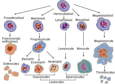
Platelets or thrombocytes () are a part of blood whose function (along with the coagulation#Coagulation factors, coagulation factors) is to react to bleeding from blood vessel injury by clumping to form a thrombus, blood clot. Platelets have no cell nucleus; they are fragments of cytoplasm from megakaryocytes which reside in bone marrow or Lung, lung tissue, and then enter the circulation. Platelets are found only in mammals, whereas in other vertebrates (e.g. birds, amphibians), thrombocytes circulate as intact agranulocyte, mononuclear cells. One major function of platelets is to contribute to hemostasis: the process of stopping bleeding at the site where the lining of vessels (endothelium) has been interrupted. Platelets gather at the site and, unless the interruption is physically too large, they plug the hole. First, platelets attach to substances outside the interrupted endothelium: ''adhesion (medicine), adhesion''. Second, they change shape, turn on receptors and secret ... [...More Info...] [...Related Items...] OR: [Wikipedia] [Google] [Baidu] |
Restrictive Cardiomyopathy
Restrictive cardiomyopathy (RCM) is a form of cardiomyopathy in which the walls of the heart are rigid (but not thickened). Thus the heart is restricted from stretching and filling with blood properly. It is the least common of the three original subtypes of cardiomyopathy: hypertrophic, dilated, and restrictive. It should not be confused with constrictive pericarditis, a disease which presents similarly but is very different in treatment and prognosis. Signs and symptoms Untreated hearts with RCM often develop the following characteristics: * M or W configuration in an invasive hemodynamic pressure tracing of the RA * Square root sign of part of the invasive hemodynamic pressure tracing Of The LV * Biatrial enlargement * Thickened LV walls (with normal chamber size) * Thickened RV free wall (with normal chamber size) * Elevated right atrial pressure (>12mmHg), * Moderate pulmonary hypertension, * Normal systolic function, * Poor diastolic function, typically Grade III - IV Dias ... [...More Info...] [...Related Items...] OR: [Wikipedia] [Google] [Baidu] |
Hypertrophic Cardiomyopathy
Hypertrophic cardiomyopathy (HCM, or HOCM when obstructive) is a condition in which muscle tissues of the heart become thickened without an obvious cause. The parts of the heart most commonly affected are the interventricular septum and the ventricles. This results in the heart being less able to pump blood effectively and also may cause electrical conduction problems. Specifically, within the bundle branches that conduct impulses through the interventricular septum and into the Purkinje fibers, as these are responsible for the depolarization of contractile cells of both ventricles. People who have HCM may have a range of symptoms. People may be asymptomatic, or may have fatigue, leg swelling, and shortness of breath. It may also result in chest pain or fainting. Symptoms may be worse when the person is dehydrated. Complications may include heart failure, an irregular heartbeat, and sudden cardiac death. HCM is most commonly inherited in an autosomal dominant pattern. I ... [...More Info...] [...Related Items...] OR: [Wikipedia] [Google] [Baidu] |
American College Of Cardiology
The American College of Cardiology (ACC), based in Washington, D.C., is a nonprofit medical association established in 1949. It bestows credentials upon cardiovascular specialists who meet its qualifications. Education is a core component of the college, which is also active in the formulation of health policy and the support of cardiovascular research. History The American College of Cardiology was chartered and incorporated as a teaching institution in 1949, and established its headquarters, called Heart House, in Bethesda, Maryland, in 1977. In 2006, the college relocated to Washington, D.C.'s West End neighborhood. Past papers for the institution are held at the National Library of Medicine in Bethesda, Maryland. Leadership The college is governed by its officers, including the president, president-elect, vice president, secretary, treasurer, chief executive officer and board of trustees (BOT). The current ACC Board of Trustees consists of 14 college members. The president ... [...More Info...] [...Related Items...] OR: [Wikipedia] [Google] [Baidu] |
Megakaryocyte
A megakaryocyte () is a large bone marrow cell with a lobation, lobated nucleus that produces blood platelets (thrombocytes), which are necessary for normal blood coagulation, clotting. In humans, megakaryocytes usually account for 1 out of 10,000 bone marrow cells, but can increase in number nearly 10-fold during the course of certain diseases. Owing to variations in neoclassical compound, combining forms and spelling, synonyms include megalokaryocyte and megacaryocyte. Structure In general, megakaryocytes are 10 to 15 times larger than a typical red blood cell, averaging 50–100 μm in diameter. During its maturation, the megakaryocyte grows in size and replicates its DNA without cytokinesis in a process called mitosis#Errors and other variations, endomitosis. As a result, the nucleus of the megakaryocyte can become very large and lobulated, which, under a light microscope, can give the false impression that there are several nuclei. In some cases, the nucleus may contain up to ... [...More Info...] [...Related Items...] OR: [Wikipedia] [Google] [Baidu] |
Motility
Motility is the ability of an organism to move independently using metabolism, metabolic energy. This biological concept encompasses movement at various levels, from whole organisms to cells and subcellular components. Motility is observed in animals, microorganisms, and even some plant structures, playing crucial roles in activities such as foraging, reproduction, and cellular functions. It is genetically determined but can be influenced by environmental factors. In multicellular organisms, motility is facilitated by systems like the Nervous system, nervous and Human musculoskeletal system, musculoskeletal systems, while at the cellular level, it involves mechanisms such as amoeboid movement and flagellar propulsion. These cellular movements can be directed by external stimuli, a phenomenon known as taxis. Examples include chemotaxis (movement along chemical gradients) and phototaxis (movement in response to light). Motility also includes physiological processes like gastroi ... [...More Info...] [...Related Items...] OR: [Wikipedia] [Google] [Baidu] |
American Society Of Hematology
The American Society of Hematology (ASH) is a professional organization representing hematologists, founded in 1958. Its annual meeting is held in December of every year and has attracted more than 30,000 attendees. The society publishes the medical journal ''Blood'', the most cited peer-reviewed publication in the field, and ''Blood Advances'', an online, peer-reviewed open-access journal. The first official ASH meeting was held in Atlantic City, New Jersey, in April 1958. More than 300 hematologists met together to discuss the key research and clinical issues related to blood and blood diseases. Since the first gathering, ASH has been an important member in the development of hematology as a discipline. For more than six decades, ASH has sponsored its annual meeting. Today, ASH has more than 17,000 members, many of whom have made major advancements in understanding and treating blood diseases. Annual meeting Held each year in December, the annual meeting brings together hema ... [...More Info...] [...Related Items...] OR: [Wikipedia] [Google] [Baidu] |


