|
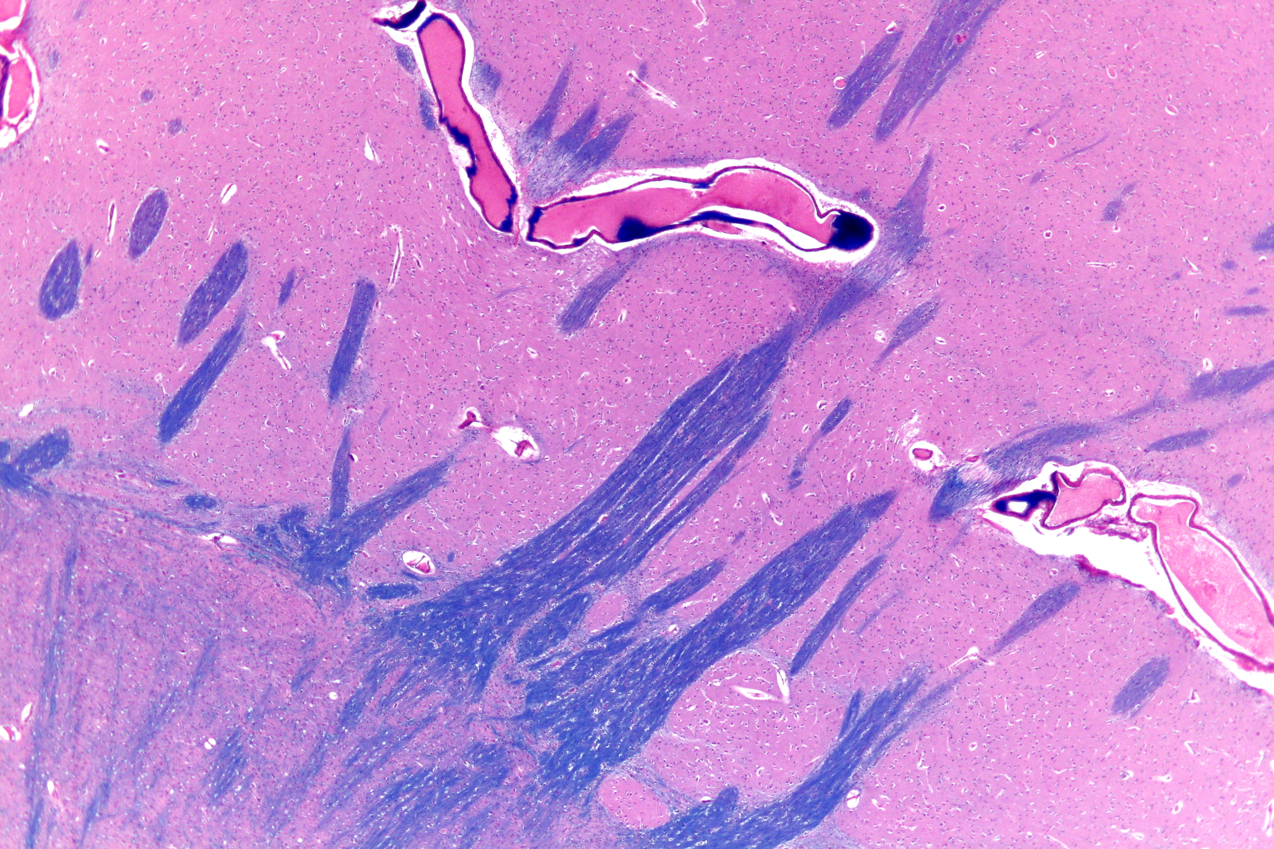
External Capsule
The external capsule is a series of white matter fiber tracts in the brain. These fibers run between the most lateral (toward the side of the head) segment of the lentiform nucleus (more specifically the putamen) and the claustrum. The white matter of the external capsule contains fibers known as corticocortical association fibers. These fibers are responsible for connecting the cerebral cortex to another cortical area. The capsule itself appears as a thin white sheet of white matter. The external capsule is a route for cholinergic fibers from the basal forebrain to the cerebral cortex. The putamen separates the external capsule from the internal capsule medially and the claustrum separates it from the extreme capsule laterally. But the external capsule eventually joins the internal capsule around the lentiform nucleus. Additional images File:Gray682.png, Superficial dissection of brain-stem. Lateral view. File:Gray689.png, Superficial dissection of brain-stem. Ventral view ... [...More Info...] [...Related Items...] OR: [Wikipedia] [Google] [Baidu] |
Horizontal Section
Horizontal may refer to: *Horizontal plane, in astronomy, geography, geometry and other sciences and contexts *Horizontal coordinate system, in astronomy *Horizontalism, in monetary circuit theory *Horizontalidad, Horizontalism, in sociology *Horizontal market, in microeconomics *Horizontal (album), ''Horizontal'' (album), a 1968 album by the Bee Gees **Horizontal (song), "Horizontal" (song)" is a 1968 song by the Bee Gees See also *Horizontal and vertical *Horizontal and vertical (other) *Horizontal fissure (other), anatomical features *Horizontal bar, an apparatus used by male gymnasts in artistic gymnastics *Vertical (other) * {{disambiguation ... [...More Info...] [...Related Items...] OR: [Wikipedia] [Google] [Baidu] |
Cerebral Hemisphere
The vertebrate cerebrum (brain) is formed by two cerebral hemispheres that are separated by a groove, the longitudinal fissure. The brain can thus be described as being divided into left and right cerebral hemispheres. Each of these hemispheres has an outer layer of grey matter, the cerebral cortex, that is supported by an inner layer of white matter. In eutherian (placental) mammals, the hemispheres are linked by the corpus callosum, a very large bundle of axon, nerve fibers. Smaller commissures, including the anterior commissure, the posterior commissure and the fornix (neuroanatomy), fornix, also join the hemispheres and these are also present in other vertebrates. These commissures transfer information between the two hemispheres to coordinate localized functions. There are three known poles of the cerebral hemispheres: the ''occipital lobe, occipital pole'', the ''frontal lobe, frontal pole'', and the ''temporal lobe, temporal pole''. The central sulcus is a prominent fissu ... [...More Info...] [...Related Items...] OR: [Wikipedia] [Google] [Baidu] |
White Matter
White matter refers to areas of the central nervous system that are mainly made up of myelinated axons, also called Nerve tract, tracts. Long thought to be passive tissue, white matter affects learning and brain functions, modulating the distribution of action potentials, acting as a relay and coordinating communication between different brain regions. White matter is named for its relatively light appearance resulting from the lipid content of myelin. Its white color in prepared specimens is due to its usual preservation in formaldehyde. It appears pinkish-white to the naked eye otherwise, because myelin is composed largely of lipid tissue veined with capillaries. Structure White matter White matter is composed of bundles, which connect various grey matter areas (the locations of nerve cell bodies) of the brain to each other, and carry nerve impulses between neurons. Myelin acts as an insulator, which allows saltatory conduction, electrical signals to jump, rather than cour ... [...More Info...] [...Related Items...] OR: [Wikipedia] [Google] [Baidu] |
Brain
The brain is an organ (biology), organ that serves as the center of the nervous system in all vertebrate and most invertebrate animals. It consists of nervous tissue and is typically located in the head (cephalization), usually near organs for special senses such as visual perception, vision, hearing, and olfaction. Being the most specialized organ, it is responsible for receiving information from the sensory nervous system, processing that information (thought, cognition, and intelligence) and the coordination of motor control (muscle activity and endocrine system). While invertebrate brains arise from paired segmental ganglia (each of which is only responsible for the respective segmentation (biology), body segment) of the ventral nerve cord, vertebrate brains develop axially from the midline dorsal nerve cord as a brain vesicle, vesicular enlargement at the rostral (anatomical term), rostral end of the neural tube, with centralized control over all body segments. All vertebr ... [...More Info...] [...Related Items...] OR: [Wikipedia] [Google] [Baidu] |
Lentiform Nucleus
The lentiform nucleus (or lentiform complex, lenticular nucleus, or lenticular complex) are the putamen (laterally) and the globus pallidus (medially), collectively. Due to their proximity, these two structures were formerly considered one, however, the two are separated by a thin layer of white matter—the external medullary lamina—and are functionally and connectionally distinct. The lentiform nucleus is a large, lens-shaped mass of gray matter just lateral to the internal capsule. It forms part of the basal ganglia. With the caudate nucleus, it forms the dorsal striatum. Structure When divided horizontally, it exhibits, to some extent, the appearance of a biconvex lens, while a coronal section of its central part presents a somewhat triangular outline. It is shorter than the caudate nucleus and does not extend as far forward. Relations It is deep/medial to the insular cortex, with which it is coextensive; the two are separated by intervening structures. It is lateral to ... [...More Info...] [...Related Items...] OR: [Wikipedia] [Google] [Baidu] |
Putamen
The putamen (; from Latin, meaning "nutshell") is a subcortical nucleus (neuroanatomy), nucleus with a rounded structure, in the basal ganglia nuclear group. It is located at the base of the forebrain and above the midbrain. The putamen and caudate nucleus together form the dorsal striatum. Through various pathways, the putamen is connected to the substantia nigra, the globus pallidus, the claustrum, and the thalamus, in addition to many regions of the cerebral cortex. A primary function of the putamen is to regulate movements at various stages such as in preparation and execution; and to influence various types of learning. It employs GABA, acetylcholine, and enkephalin to perform its functions. The putamen also plays a role in neurodegenerative diseases, such as Parkinson's disease. History The word "putamen" is from Latin, referring to that which "falls off in pruning", from "putare", meaning "to prune, to think, or to consider". Most MRI research was focused broadly on th ... [...More Info...] [...Related Items...] OR: [Wikipedia] [Google] [Baidu] |
Claustrum
The claustrum (Latin, meaning "to close" or "to shut") is a thin sheet of neurons and supporting glial cells in the brain, that connects to the cerebral cortex and subcortical regions including the amygdala, hippocampus and thalamus. It is located between the insular cortex laterally and the putamen medially, encased by the extreme and external capsules respectively. Blood to the claustrum is supplied by the middle cerebral artery. It is considered to be the most densely connected structure in the brain, and thus hypothesized to allow for the integration of various cortical inputs such as vision, sound and touch, into one experience. Other hypotheses suggest that the claustrum plays a role in salience processing, to direct attention towards the most behaviorally relevant stimuli amongst the background noise. The claustrum is difficult to study given the limited number of individuals with claustral lesions and the poor resolution of neuroimaging. The claustrum is made up of var ... [...More Info...] [...Related Items...] OR: [Wikipedia] [Google] [Baidu] |
Association Fibers
Association fibers are axons (nerve fibers) that connect cortical areas within the same cerebral hemisphere. In human neuroanatomy, axons within the brain, can be categorized on the basis of their course and connections as association fibers, projection fibers, and commissural fibers. Bundles of fibers are known as nerve tracts, and consist of association tracts, commissural tracts, and projection tracts. The association fibers unite different parts of the same cerebral hemisphere, and are of two kinds: (1) short association fibers that connect adjacent gyri; (2) long association fibers that make connections between more distant parts. Short association fibers Many of the short association fibers (also called arcuate or "U"-fibers) lie in the superficial white matter immediately beneath the gray matter of the cerebral cortex, and connect together adjacent gyri. Some pass from one wall of the sulcus to the other. Long association fibers The long association fibers connect ... [...More Info...] [...Related Items...] OR: [Wikipedia] [Google] [Baidu] |
Cerebral Cortex
The cerebral cortex, also known as the cerebral mantle, is the outer layer of neural tissue of the cerebrum of the brain in humans and other mammals. It is the largest site of Neuron, neural integration in the central nervous system, and plays a key role in attention, perception, awareness, thought, memory, language, and consciousness. The six-layered neocortex makes up approximately 90% of the Cortex (anatomy), cortex, with the allocortex making up the remainder. The cortex is divided into left and right parts by the longitudinal fissure, which separates the two cerebral hemispheres that are joined beneath the cortex by the corpus callosum and other commissural fibers. In most mammals, apart from small mammals that have small brains, the cerebral cortex is folded, providing a greater surface area in the confined volume of the neurocranium, cranium. Apart from minimising brain and cranial volume, gyrification, cortical folding is crucial for the Neural circuit, brain circuitry ... [...More Info...] [...Related Items...] OR: [Wikipedia] [Google] [Baidu] |
Cholinergic
Cholinergic agents are compounds which mimic the action of acetylcholine and/or butyrylcholine. In general, the word " choline" describes the various quaternary ammonium salts containing the ''N'',''N'',''N''-trimethylethanolammonium cation. Found in most animal tissues, choline is a primary component of the neurotransmitter acetylcholine and functions with inositol as a basic constituent of lecithin. Choline also prevents fat deposits in the liver and facilitates the movement of fats into cells. The parasympathetic nervous system, which uses acetylcholine almost exclusively to send its messages, is said to be almost entirely cholinergic. Neuromuscular junctions, preganglionic neurons of the sympathetic nervous system, the basal forebrain, and brain stem complexes are also cholinergic, as are the receptor for the merocrine sweat glands. In neuroscience and related fields, the term cholinergic is used in these related contexts: * A substance (or ligand) is cholinergic if ... [...More Info...] [...Related Items...] OR: [Wikipedia] [Google] [Baidu] |
Basal Forebrain
Part of the human brain, the basal forebrain structures are located in the forebrain to the front of and below the striatum. They include the ventral basal ganglia (including nucleus accumbens and ventral pallidum), nucleus basalis, diagonal band of Broca, substantia innominata, and the medial septal nucleus. These structures are important in the production of acetylcholine, which is then distributed widely throughout the brain. The basal forebrain is considered to be the major cholinergic output of the central nervous system (CNS) centred on the output of the nucleus basalis. The presence of non-cholinergic neurons projecting to the cortex have been found to act with the cholinergic neurons to dynamically modulate activity in the cortex. Function Acetylcholine is known to promote wakefulness in the basal forebrain. Stimulating the basal forebrain gives rise to acetylcholine release, which induces wakefulness and REM sleep, whereas inhibition of acetylcholine release in the ba ... [...More Info...] [...Related Items...] OR: [Wikipedia] [Google] [Baidu] |
Internal Capsule
The internal capsule is a paired white matter structure, as a two-way nerve tract, tract, carrying afferent nerve fiber, ascending and efferent nerve fiber, descending axon, fibers, to and from the cerebral cortex. The internal capsule is situated in the Anatomical terms of location#Medial and lateral, inferomedial part of each cerebral hemisphere of the brain. It carries information past the subcortical basal ganglia. As it courses it separates the caudate nucleus and the thalamus from the putamen and the globus pallidus. It also separates the caudate nucleus and the putamen in the dorsal striatum, a brain region involved in motor and reward pathways. The internal capsule is V-shaped in transection forming an anterior and posterior limb, with the angle between them called the genu. The corticospinal tract constitutes a large part of the internal capsule, carrying motor information from the primary motor cortex to the lower motor neurons in the spinal cord. Above the basal gangli ... [...More Info...] [...Related Items...] OR: [Wikipedia] [Google] [Baidu] |


