|
Common Hepatic Duct
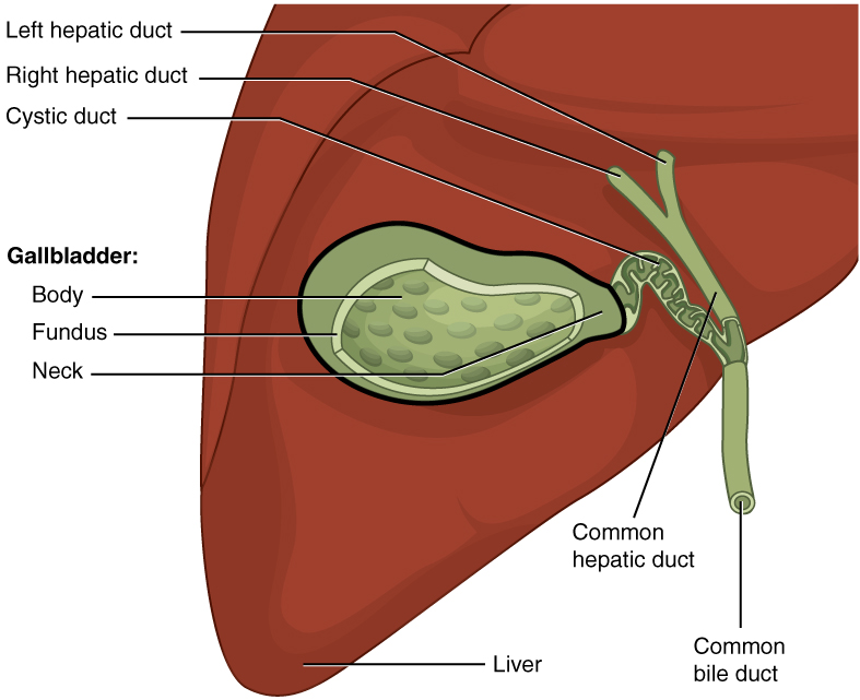
The common hepatic duct is the first part of the biliary tract. It joins the cystic duct coming from the gallbladder to form the common bile duct. Structure The common hepatic duct is the first part of the biliary tract. It is formed by the union of the right hepatic duct (which drains bile from the right functional lobe of the liver) and the left hepatic duct (which drains bile from the left functional lobe of the liver). The duct is about 3 cm long. The common hepatic duct is about 6 mm in diameter in adults, with some variation.Gray's Anatomy, 39th ed, p. 1228 Termination The common hepatic duct typically unites with the cystic duct some 1–2 cm superior to the duodenum and anterior to the right hepatic artery, with the cystic duct approaching the common hepatic duct from the right. Relations The right branch of the hepatic artery proper usually passes posterior to the duct, but may rarely pass anterior to it instead. Histology The inner surface ... [...More Info...] [...Related Items...] OR: [Wikipedia] [Google] [Baidu] |
Right Lobe Of Liver
In human anatomy, the liver is divided grossly into four parts or lobes: the right lobe, the left lobe, the caudate lobe, and the quadrate lobe. Seen from the front – the diaphragmatic surface – the liver is divided into two lobes: the right lobe and the left lobe. Viewed from the underside – the visceral surface – the other two smaller lobes, the caudate lobe and the quadrate lobe, are also visible. The two smaller lobes, the caudate lobe and the quadrate lobe, are known as superficial or accessory lobes, and both are located on the underside of the right lobe. The falciform ligament, visible on the front of the liver, makes a superficial division of the right and left lobes of the liver. From the underside, the two additional lobes are located on the right lobe. A line can be imagined running from the left of the vena cava and all the way forward to divide the liver and gallbladder into two halves. This line is called Cantlie's line and is used to mark the division b ... [...More Info...] [...Related Items...] OR: [Wikipedia] [Google] [Baidu] |
Gallbladder
In vertebrates, the gallbladder, also known as the cholecyst, is a small hollow Organ (anatomy), organ where bile is stored and concentrated before it is released into the small intestine. In humans, the pear-shaped gallbladder lies beneath the liver, although the structure and position of the gallbladder can vary significantly among animal species. It receives bile, produced by the liver, via the common hepatic duct, and stores it. The bile is then released via the common bile duct into the duodenum, where the bile helps in the digestion of fats. The gallbladder can be affected by gallstones, formed by material that cannot be dissolved – usually cholesterol or bilirubin, a product of hemoglobin breakdown. These may cause significant pain, particularly in the upper-right corner of the abdomen, and are often treated with removal of the gallbladder (called a cholecystectomy). Cholecystitis, inflammation of the gallbladder, has a wide range of causes, including result from the ... [...More Info...] [...Related Items...] OR: [Wikipedia] [Google] [Baidu] |
Gallstone
A gallstone is a stone formed within the gallbladder from precipitated bile components. The term cholelithiasis may refer to the presence of gallstones or to any disease caused by gallstones, and choledocholithiasis refers to the presence of migrated gallstones within bile ducts. Most people with gallstones (about 80%) are asymptomatic. However, when a gallstone obstructs the bile duct and causes acute cholestasis, a reflexive smooth muscle spasm often occurs, resulting in an intense cramp-like visceral pain in the right upper part of the abdomen known as a biliary colic (or "gallbladder attack"). This happens in 1–4% of those with gallstones each year. Complications from gallstones may include inflammation of the gallbladder (cholecystitis), inflammation of the pancreas (pancreatitis), obstructive jaundice, and infection in bile ducts ( cholangitis). Symptoms of these complications may include pain that lasts longer than five hours, fever, yellowish skin, vomiting, da ... [...More Info...] [...Related Items...] OR: [Wikipedia] [Google] [Baidu] |
Mirizzi's Syndrome
Mirizzi's syndrome is a rare complication in which a gallstone becomes impacted in the cystic duct or neck of the gallbladder causing compression of the common hepatic duct, resulting in obstruction and jaundice. The obstructive jaundice can be caused by direct extrinsic compression by the stone or from fibrosis caused by chronic cholecystitis (inflammation). A cholecystocholedochal fistula can occur.Vitale M. Mirizzi Syndrome Type IV: An Atypical Presentation That Is Difficult to Diagnose Preoperatively. 2009. Society for Surgery of the Alimentary Tract.http://www.ssat.com/cgi-bin/abstracts/09ddw/P7.cgi Presentation Mirizzi's syndrome has no consistent or unique clinical features that distinguish it from other more common forms of obstructive jaundice. Symptoms of recurrent cholangitis, jaundice, right upper quadrant pain, and elevated bilirubin and alkaline phosphatase may or may not be present. Acute presentations of the syndrome include symptoms consistent with cholecystitis. ... [...More Info...] [...Related Items...] OR: [Wikipedia] [Google] [Baidu] |
Cholestasis
Cholestasis is a condition where the flow of bile from the liver to the duodenum is impaired. The two basic distinctions are: * obstructive type of cholestasis, where there is a mechanical blockage in the duct system that can occur from a gallstone or malignancy, and * metabolic type of cholestasis, in which there are disturbances in bile formation that can occur because of genetic defects or acquired as a side effect of many medications. Classification is further divided into acute or chronic and extrahepatic or intrahepatic. Signs and symptoms The signs and symptoms of cholestasis vary according to the cause. In case of sudden onset, the disease is likely to be acute, while the gradual appearance of symptoms suggests chronic pathology. In many cases, patients may experience pain in the abdominal area. Localization of pain to the upper right quadrant can be indicative of cholecystitis or choledocholithiasis, which can progress to cholestasis. Pruritus or itching is often pr ... [...More Info...] [...Related Items...] OR: [Wikipedia] [Google] [Baidu] |
Cystic Artery
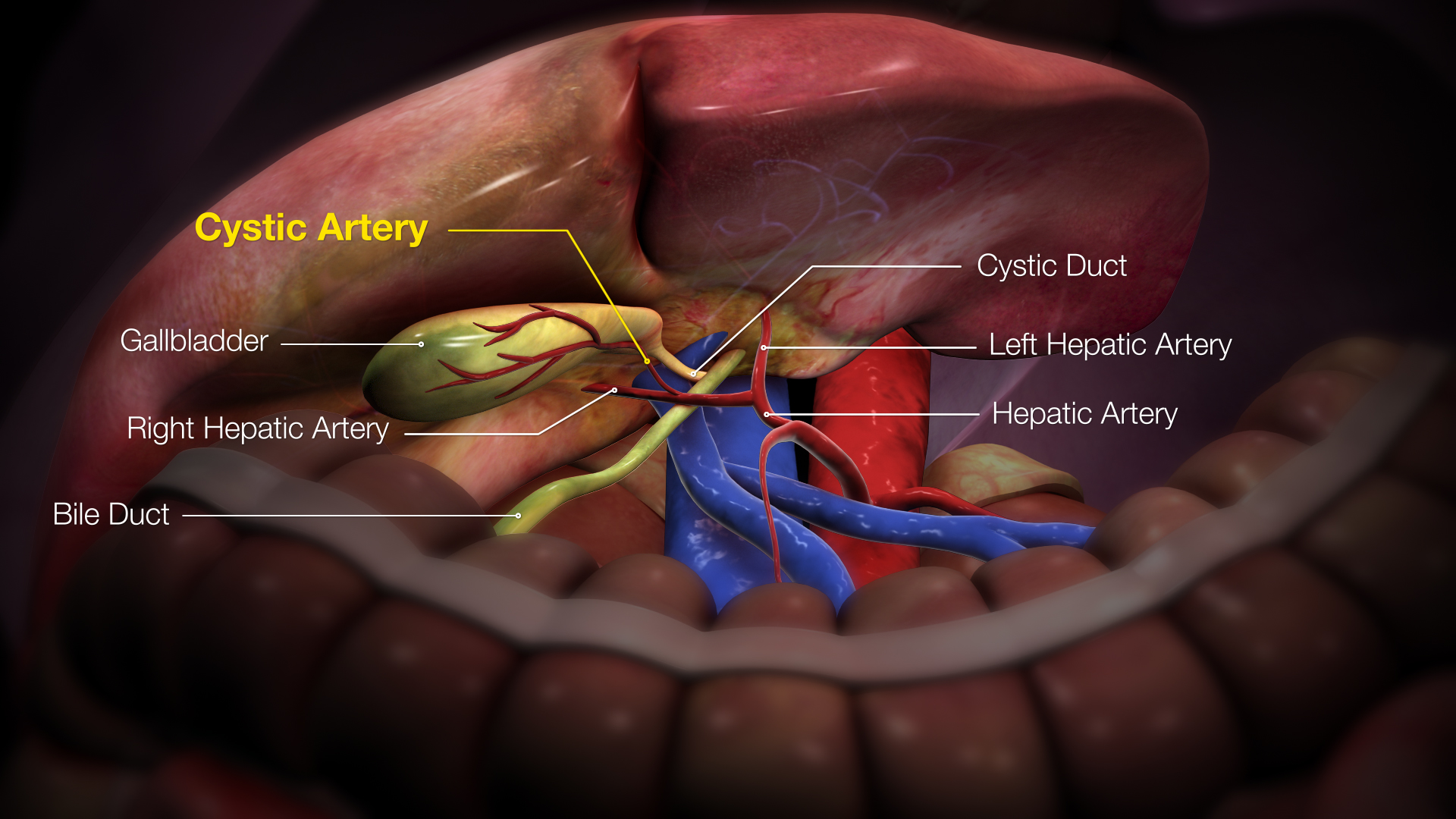
The cystic artery (also known as bachelor artery) is (usually) a branch of the right hepatic artery that provides arterial supply to the gallbladder and contributes arterial supply to the extrahepatic bile ducts. Anatomy The cystic artery usually has a diameter of less than 3mm. Origin The cystic artery arises from the right hepatic artery in about 80% of cases. Course It usually passes posterior to the common hepatic duct within the cystohepatic triangle. Within the triangle, it is usually superior to the cystic duct (if it does not pass superior to the cystic duct, it may be situated outside the triangle). Branches Upon reaching the superior aspect of the neck of the gallbladder, it splits into superficial and deep branches. These branches then form an anastomotic network over the surface of the body and fundus of the gallbladder. It produces 2 to 4 minor branches (known as ''Calot’s arteries'') that supply part of the cystic duct and cervix of the gallbladder before ... [...More Info...] [...Related Items...] OR: [Wikipedia] [Google] [Baidu] |
Cystohepatic Triangle
The cystohepatic triangle (or hepatobiliary triangle or Calot's triangle) is an anatomic space bordered by the cystic duct laterally, the common hepatic duct medially, and the inferior surface of the liver superiorly. The cystic artery lies within the hepatobiliary triangle. The triangle is used to locate the cystic artery during a laparoscopic cholecystectomy. Structure The hepatobiliary triangle is the area bounded by the: * cystic duct inferiorly * common hepatic duct medially * inferior margin of the liver superiorlySchwartz's Manual of Surgery BRUNICARDI C.F 10th edition It is covered in peritoneum both anteriorly and posteriorly. Contents The triangle contains: adipose and connective tissue, lymphatic vessels and the cystic lymph node, autonomic nerves, (usually) cystic artery, and (sometimes) an accessory cystic duct. The right hepatic artery may also pass through the hepatobiliary triangle. Clinical significance The anatomy and variant anatomy of this region is ... [...More Info...] [...Related Items...] OR: [Wikipedia] [Google] [Baidu] |
Cholecystectomy
Cholecystectomy is the surgical removal of the gallbladder. Cholecystectomy is a common treatment of symptomatic gallstones and other gallbladder conditions. In 2011, cholecystectomy was the eighth most common operating room procedure performed in hospitals in the United States. Cholecystectomy can be performed either Laparoscopy, laparoscopically, or via an Minimally invasive procedures#Open surgery, open surgical technique. The surgery is usually successful in relieving symptoms, but up to 10 percent of people may continue to experience similar symptoms after cholecystectomy, a condition called postcholecystectomy syndrome. Complications of cholecystectomy include Biliary injury, bile duct injury, wound infection, bleeding, vasculobiliary injury, retained gallstones, liver abscess formation and stenosis (narrowing) of the bile duct. Medical use Pain and complications caused by gallstones are the most common reasons for removal of the gallbladder. The gallbladder can also be r ... [...More Info...] [...Related Items...] OR: [Wikipedia] [Google] [Baidu] |
Intestine
The gastrointestinal tract (GI tract, digestive tract, alimentary canal) is the tract or passageway of the digestive system that leads from the mouth to the anus. The tract is the largest of the body's systems, after the cardiovascular system. The GI tract contains all the major organs of the digestive system, in humans and other animals, including the esophagus, stomach, and intestines. Food taken in through the mouth is digested to extract nutrients and absorb energy, and the waste expelled at the anus as feces. ''Gastrointestinal'' is an adjective meaning of or pertaining to the stomach and intestines. Most animals have a "through-gut" or complete digestive tract. Exceptions are more primitive ones: sponges have small pores ( ostia) throughout their body for digestion and a larger dorsal pore ( osculum) for excretion, comb jellies have both a ventral mouth and dorsal anal pores, while cnidarians and acoels have a single pore for both digestion and excretion. The human gas ... [...More Info...] [...Related Items...] OR: [Wikipedia] [Google] [Baidu] |
Simple Columnar Epithelium
Simple columnar epithelium is a single layer of columnar epithelial cells which are tall and slender with oval-shaped nuclei located in the basal region, attached to the basement membrane. In humans, simple columnar epithelium lines most organs of the digestive tract including the stomach, and intestines. Simple columnar epithelium also lines the uterus. Structure Simple columnar epithelium is further divided into two categories: ciliated and non-ciliated (glandular). The ciliated part of the simple columnar epithelium has tiny hairs which help move mucus and other substances up the respiratory tract. The shape of the simple columnar epithelium cells are tall and narrow giving a column like appearance. the apical surfaces of the tissue face the lumen of organs while the basal side faces the basement membrane. The nuclei are located closer along the basal side of the cell. Absorptive columnar epithelium is characterized as having a striated border on its apical side, this bord ... [...More Info...] [...Related Items...] OR: [Wikipedia] [Google] [Baidu] |
Hepatic Artery Proper
The hepatic artery proper (also proper hepatic artery) is the artery that supplies the liver and gallbladder. It raises from the common hepatic artery, a branch of the celiac artery. Structure The hepatic artery proper arises from the common hepatic artery and runs alongside the portal vein and the common bile duct to form the portal triad. A branch of the common hepatic arterythe gastroduodenal artery gives off the small supraduodenal artery to the duodenal bulb. Then the right gastric artery comes off and runs to the left along the lesser curvature of the stomach to meet the left gastric artery, which is a branch of the celiac trunk. It subsequently bifurcates into the right and left hepatic arteries. Variant anatomy Of note, the right and left hepatic arteries may demonstrate variant anatomy. A misplaced right hepatic artery may arise from the superior mesenteric artery (SMA) and a misplaced left hepatic artery may arise from the left gastric artery. The cystic artery g ... [...More Info...] [...Related Items...] OR: [Wikipedia] [Google] [Baidu] |
Common Bile Duct
The common bile duct (also bile duct) is a part of the biliary tract. It is formed by the union of the common hepatic duct and cystic duct. It ends by uniting with the pancreatic duct to form the ampulla of Vater (hepatopancreatic ampulla). Its sphincter the sphincter of Oddi, enables the regulation of bile flow. Anatomy The bile duct is some 6–8 cm long, and normally up to 8 mm in diameter. Its proximal supraduodenal part is situated within the free edge of the lesser omentum. Its middle retroduodenal part is oriented inferiorly and right-ward, and is situated posterior to the first part of the duodenum, and anterior to the inferior vena cava. Its distal paraduodenal part is oriented still more right-ward, is accommodated by a groove upon (sometimes a channel within) the posterior aspect of the head of the pancreas, and is situated anterior to the right renal vein. The bile duct terminates by uniting with the pancreatic duct (at an angle of about 60°) t ... [...More Info...] [...Related Items...] OR: [Wikipedia] [Google] [Baidu] |


