|
Biceps Femoris Muscle
The biceps femoris () is a muscle of the thigh located to the posterior, or back. As its name implies, it consists of two heads; the long head is considered part of the hamstring muscle group, while the short head is sometimes excluded from this characterization, as it only causes knee flexion (but not hip extension) and is activated by a separate nerve (the peroneal, as opposed to the tibial branch of the sciatic nerve). Structure It has two heads of origin: *the ''long head'' arises from the lower and inner impression on the posterior part of the tuberosity of the ischium. This is a common tendon origin with the semitendinosus muscle, and from the lower part of the sacrotuberous ligament. *the ''short head'', arises from the lateral lip of the linea aspera, between the adductor magnus and vastus lateralis extending up almost as high as the insertion of the gluteus maximus, from the lateral prolongation of the linea aspera to within 5 cm. of the lateral condyle; and fr ... [...More Info...] [...Related Items...] OR: [Wikipedia] [Google] [Baidu] |
Tuberosity Of The Ischium
The ischial tuberosity (or tuberosity of the ischium, tuber ischiadicum), also known colloquially as the sit bones or sitz bones, or as a pair the sitting bones, is a large posterior (anatomy), posterior bone, bony protuberance on the superior ramus of the ischium, superior ramus of the ischium. It marks the lateral boundary of the pelvic outlet. When sitting, the weight is frequently placed upon the ischial tuberosity. The Gluteus maximus muscle, gluteus maximus provides cover in the upright posture, but leaves it free in the seated position.Platzer (2004), p 236 The distance between a cyclist's ischial tuberosities is one of the factors in the choice of a bicycle saddle. Divisions The tuberosity is divided into two portions: a lower, rough, somewhat triangular part, and an upper, smooth, quadrilateral portion. * The ''lower portion'' is subdivided by a prominent longitudinal ridge, passing from base to apex, into two parts: ** The outer gives attachment to the adductor magnus ... [...More Info...] [...Related Items...] OR: [Wikipedia] [Google] [Baidu] |
Sciatic Nerve
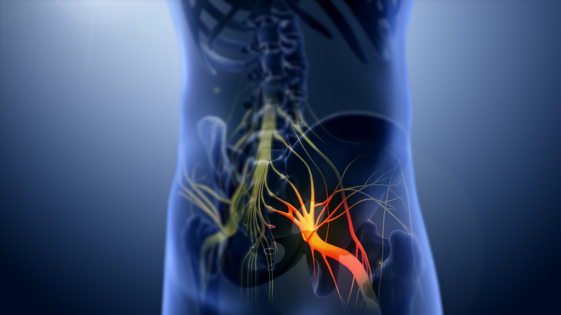
The sciatic nerve, also called the ischiadic nerve, is a large nerve in humans and other vertebrate animals. It is the largest branch of the sacral plexus and runs alongside the hip joint and down the right lower limb. It is the longest and widest single nerve in the human body, going from the top of the leg to the foot on the posterior aspect. The sciatic nerve has no cutaneous branches for the thigh. This nerve provides the connection to the nervous system for the skin of the lateral leg and the whole foot, the muscles of the back of the thigh, and those of the leg and foot. It is derived from Spinal nerve, spinal nerves Lumbar spinal nerve 4, L4 to Sacral spinal nerve 3, S3. It contains Axon, fibres from both the anterior and posterior divisions of the lumbosacral plexus. Structure In humans, the sciatic nerve is formed from the L4 to S3 segments of the sacral plexus, a collection of nerve fibres that emerge from the Sacrum, sacral part of the spinal cord. The lumbosacral trunk ... [...More Info...] [...Related Items...] OR: [Wikipedia] [Google] [Baidu] |
Fibular Collateral Ligament
The lateral collateral ligament (LCL, long external lateral ligament or fibular collateral ligament) is an extrinsic ligament of the knee located on the lateral side of the knee. Its superior attachment is at the lateral epicondyle of the femur (superoposterior to the popliteal groove); its inferior attachment is at the lateral aspect of the head of fibula (anterior to the apex). The LCL is not fused with the joint capsule. Inferiorly, the LCL splits the tendon of insertion of the biceps femoris muscle. Structure The LCL measures some 5 cm in length. It is rounded, and is more narrow and less broad compared to the medial collateral ligament. It extends obliquely inferoposteriorly from its superior attachment to its inferior attachment. In contrast to the medial collateral ligament, it is not fused with either the capsular ligament nor the lateral meniscus. Because of this, the LCL is more flexible than its medial counterpart, and is therefore less susceptible to injury. ... [...More Info...] [...Related Items...] OR: [Wikipedia] [Google] [Baidu] |
Tibia
The tibia (; : tibiae or tibias), also known as the shinbone or shankbone, is the larger, stronger, and anterior (frontal) of the two Leg bones, bones in the leg below the knee in vertebrates (the other being the fibula, behind and to the outside of the tibia); it connects the knee with the ankle bones, ankle. The tibia is found on the anatomical terms of location#Medial, medial side of the leg next to the fibula and closer to the median plane. The tibia is connected to the fibula by the interosseous membrane of leg, forming a type of fibrous joint called a syndesmosis with very little movement. The tibia is named for the flute ''aulos, tibia''. It is the second largest bone in the human body, after the femur. The leg bones are the strongest long bones as they support the rest of the body. Structure In human anatomy, the tibia is the second largest bone next to the femur. As in other vertebrates the tibia is one of two bones in the lower leg, the other being the fibula, and is a ... [...More Info...] [...Related Items...] OR: [Wikipedia] [Google] [Baidu] |
Fibula
The fibula (: fibulae or fibulas) or calf bone is a leg bone on the lateral side of the tibia, to which it is connected above and below. It is the smaller of the two bones and, in proportion to its length, the most slender of all the long bones. Its upper extremity is small, placed toward the back of the head of the tibia, below the knee joint and excluded from the formation of this joint. Its lower extremity inclines a little forward, so as to be on a plane anterior to that of the upper end; it projects below the tibia and forms the lateral part of the ankle joint. Structure The bone has the following components: * Lateral malleolus * Interosseous membrane connecting the fibula to the tibia, forming a syndesmosis joint * The superior tibiofibular articulation is an arthrodial joint between the lateral condyle of the tibia and the head of the fibula. * The inferior tibiofibular articulation (tibiofibular syndesmosis) is formed by the rough, convex surface of the medial si ... [...More Info...] [...Related Items...] OR: [Wikipedia] [Google] [Baidu] |
Aponeurosis
An aponeurosis (; : aponeuroses) is a flattened tendon by which muscle attaches to bone or fascia. Aponeuroses exhibit an ordered arrangement of collagen fibres, thus attaining high tensile strength in a particular direction while being vulnerable to tensional or shear forces in other directions. They have a shiny, whitish-silvery color, are histologically similar to tendons, and are very sparingly supplied with blood vessels and nerves. When dissected, aponeuroses are papery and peel off by sections. The primary regions with thick aponeuroses are in the ventral abdominal region, the dorsal lumbar region, the ventriculus in birds, and the palmar (palms) and plantar (soles) regions. Anatomy Anterior abdominal aponeuroses The anterior abdominal aponeuroses are located just superficial to the rectus abdominis muscle. It has for its borders the external oblique, pectoralis muscles, and the latissimus dorsi. Posterior lumbar aponeuroses The posterior lumbar aponeuroses are sit ... [...More Info...] [...Related Items...] OR: [Wikipedia] [Google] [Baidu] |
Fusiform
Fusiform (from Latin ''fusus'' ‘spindle’) means having a spindle (textiles), spindle-like shape that is wide in the middle and tapers at both ends. It is similar to the lemon (geometry), lemon-shape, but often implies a focal broadening of a structure that continues from one or both ends, such as an aneurysm on a blood vessel. Examples * Fusiform, a body shape common to many aquatic animals, characterized by being tapered at both the head and the tail * Fusiform, a classification of aneurysm * Fusiform bacteria (spindled rods, that is, fusiform bacilli), such as the Fusobacteriota * Fusiform cell (biology) * Fusiform face area, a part of the human visual system which seems to specialize in facial recognition * Fusiform gyrus, part of the temporal lobe of the brain * Fusiform muscle, where the fibres run parallel along the length of the muscle * Fusiform neuron, a spindle-shaped neuron References {{Reflist Geometric shapes See also * Streamliner, a fusiform hydro- ... [...More Info...] [...Related Items...] OR: [Wikipedia] [Google] [Baidu] |
Lateral Intermuscular Septum Of Thigh
The lateral intermuscular septum of thigh is a fold of deep fascia Deep fascia (or investing fascia) is a fascia, a layer of dense connective tissue that can surround individual muscles and groups of muscles to separate into fascial compartments. This fibrous connective tissue interpenetrates and surrounds the m ... in the thigh. It is between the vastus lateralis and biceps femoris. It separates the anterior compartment of the thigh from the posterior compartment of the thigh. See also * Medial intermuscular septum of thigh * Anterior compartment of thigh * Posterior compartment of thigh References External links Topographical Anatomy of the Lower Limb - Listed Alphabeticallyfrom UAMS Department of Neurobiology and Developmental Sciences from anatomy.med.umich.edu Lower limb anatomy {{musculoskeletal-stub ... [...More Info...] [...Related Items...] OR: [Wikipedia] [Google] [Baidu] |
Lateral Condyle Of Femur
The lateral condyle is one of the two projections on the lower extremity of the femur. The other one is the medial condyle. The lateral condyle is the more prominent and is broader both in its front-to-back and transverse diameters. Clinical significance The most common injury to the lateral femoral condyle is an osteochondral fracture combined with a patellar dislocation. The osteochondral fracture occurs on the weight-bearing portion of the lateral condyle. Typically, the condyle will fracture (and the patella may dislocate) as a result of severe impaction from activities such as downhill skiing and parachuting. Open reduction and internal fixation surgery is typically used to repair an osteochondral fracture. For a AO Type B1 partial articular fracture of the lateral condyle, interfragmentary lag screws are used to secure the bone back together. Supplementation of buttress screws or a buttress plate is used if the fracture extends to the proximal metaphysis or distal diaphysi ... [...More Info...] [...Related Items...] OR: [Wikipedia] [Google] [Baidu] |
Gluteus Maximus Muscle
The gluteus maximus is the main extensor muscle of the hip in humans. It is the largest and outermost of the three gluteal muscles and makes up a large part of the shape and appearance of each side of the hips. It is the single largest muscle in the human body. Its thick fleshy mass, in a quadrilateral shape, forms the prominence of the buttocks. The other gluteal muscles are the medius and minimus, and sometimes informally these are collectively referred to as the glutes. Its large size is one of the most characteristic features of the muscular system in humans,Norman Eizenberg et al., ''General Anatomy: Principles and Applications'' (2008), p. 17. connected as it is with the power of maintaining the trunk in the erect posture. Other primates have much flatter hips and cannot sustain standing erectly. The muscle is made up of muscle fascicles lying parallel with one another, and are collected together into larger bundles separated by fibrous septa. Structure The gluteus ma ... [...More Info...] [...Related Items...] OR: [Wikipedia] [Google] [Baidu] |
Vastus Lateralis Muscle
The vastus lateralis (), also called the vastus externus, is the largest and most powerful part of the quadriceps femoris, a muscle in the thigh. Together with other muscles of the quadriceps group, it serves to extend the knee joint, moving the lower leg forward. It arises from a series of flat, broad tendons attached to the femur, and attaches to the outer border of the patella. It ultimately joins with the other muscles that make up the quadriceps in the quadriceps tendon, which travels over the knee to connect to the tibia. The vastus lateralis is the recommended site for intramuscular injection in infants less than 7 months old and those unable to walk, with loss of muscular tone.Mann, E. (2016). ''Injection (Intramuscular): Clinician Information.'' The Johanna Briggs Institute. Structure The vastus lateralis muscle arises from several areas of the femur, including the upper part of the intertrochanteric line; the lower, anterior borders of the greater trochanter, to t ... [...More Info...] [...Related Items...] OR: [Wikipedia] [Google] [Baidu] |
Adductor Magnus Muscle
The adductor magnus is a large triangular muscle, situated on the medial side of the thigh. It consists of two parts. The portion which arises from the ischiopubic ramus (a small part of the inferior ramus of the pubis, and the inferior ramus of the ischium) is called the pubofemoral portion, adductor portion, or adductor minimus, and the portion arising from the tuberosity of the ischium is called the ischiocondylar portion, extensor portion, or "hamstring portion". Due to its common embryonic origin, innervation, and action the ischiocondylar portion (or hamstring portion) is often considered part of the hamstring group of muscles. The ischiocondylar portion of the adductor magnus is considered a muscle of the posterior compartment of the thigh while the pubofemoral portion of the adductor magnus is considered a muscle of the medial compartment. Structure Pubofemoral (adductor) portion Those fibers which arise from the ramus of the pubis are short, horizontal in directio ... [...More Info...] [...Related Items...] OR: [Wikipedia] [Google] [Baidu] |


