|
ŇĀucja Frey
ŇĀucja Frey or ŇĀucja Frey-Gottesman (November 3, 1889, in Lw√≥w ‚Äď 1942?) was a Polish-Jewish physician and neurologist, known for describing the syndrome later Frey's syndrome, named after her. She was one of the first female academic neurologists in Europe. Frey perished during the Holocaust in 1942 in Lw√≥w ghetto aged 53. Life ŇĀucja Frey was born on November 3, 1889, in Lw√≥w, then part of the Austro-Hungarian empire, as the daughter of the building contractor Szymon Symcha Frey and his wife, Dina (n√©e Weinreb)Mirjam Moltrecht: Dr. med. Lucja Frey. Eine √Ąrztin aus Lwow 1889-1942. Rekonstruktion eines Lebens. Hartung Gorre Verlag Konstanz, 2004PDF Frey and her family were Judaism, Jewish. She attended a Christian elementary school between 1896 and 1900. She graduated from Franciszek-J√≥zef secondary school as an extern pupil in 1907. After graduation she studied mathematics and philosophy under professor Marian Smoluchowski (1872-1917). She was a student of the faculty of ... [...More Info...] [...Related Items...] OR: [Wikipedia] [Google] [Baidu] |
Retrosplenial Region
The retrosplenial cortex (RSC) is a cortical area in the brain comprising Brodmann areas 29 and 30. It is secondary association cortex, making connections with numerous other brain regions. The region's name refers to its anatomical location immediately behind the splenium of the corpus callosum in primates, although in rodents it is located more towards the brain surface and is relatively larger. Its function is currently not well understood, but its location close to visual areas and also to the hippocampal spatial/memory system suggest it may have a role in mediating between perceptual and memory functions, particularly in the spatial domain. However, its exact contribution to either space or memory processing has been hard to pin down. Anatomy There is large variation in the region's size across different species. In humans it comprises roughly 0.3% of the entire cortical surface whereas in rabbits it is at least 10% and in rats it extends for more than half the cerebrum d ... [...More Info...] [...Related Items...] OR: [Wikipedia] [Google] [Baidu] |
Frontal Lobe
The frontal lobe is the largest of the four major lobes of the brain in mammals, and is located at the front of each cerebral hemisphere (in front of the parietal lobe and the temporal lobe). It is parted from the parietal lobe by a Sulcus (neuroanatomy), groove between tissues called the central sulcus and from the temporal lobe by a deeper groove called the lateral sulcus (Sylvian fissure). The most anterior rounded part of the frontal lobe (though not well-defined) is known as the frontal pole, one of the three Cerebral hemisphere#Poles, poles of the cerebrum. The frontal lobe is covered by the frontal cortex. The frontal cortex includes the premotor cortex and the primary motor cortex ‚Äď parts of the motor cortex. The front part of the frontal cortex is covered by the prefrontal cortex. The nonprimary motor cortex is a functionally defined portion of the frontal lobe. There are four principal Gyrus, gyri in the frontal lobe. The precentral gyrus is directly anterior to the ... [...More Info...] [...Related Items...] OR: [Wikipedia] [Google] [Baidu] |
Clivus (anatomy)
The clivus (, Latin for "slope") or Blumenbach clivus is a part of the occipital bone at the base of the skull, extending anteriorly from the foramen magnum. It is related to the pons and the abducens nerve (CN VI). The term is also used for the clivus ocularis, an unrelated feature of the retina. Structure The clivus is a shallow depression behind the dorsum sellae of the sphenoid bone, extending inferiorly to the foramen magnum. It slopes gradually to the anterior part of the basilar occipital bone at its junction with the sphenoid bone. Synchondrosis of these two bones forms the clivus. On axial planes, it sits just posterior to the sphenoid sinuses. It is medial to the foramen lacerum and proximal to the anastomosis of the internal carotid artery with the Circle of Willis. (The artery reaches the middle cranial fossa above the foramen lacerum). It is anterior to the basilar artery. On sagittal plane, it can be divided into two surfaces, the pharyngeal (inferior) surf ... [...More Info...] [...Related Items...] OR: [Wikipedia] [Google] [Baidu] |
Charcot Joints
Neuropathic arthropathy (also known as Charcot neuroarthropathy or diabetic arthropathy) refers to a progressive fragmentation of bones and joints in the presence of neuropathy. It can occur in any joint where denervation is present, although it most frequently presents in the foot and ankle. It follows an episodic pattern of early inflammation followed by periarticular destruction, bony coalescence, and finally bony remodeling.  This can lead to considerable deformity and morbidity, including limb instability, ulceration, infection, and amputation. The diagnosis of Charcot neuroarthropathy is made clinically and should be considered whenever a patient presents with warmth and swelling around a joint in the presence of neuropathy. Although counterintuitive, pain is present in many cases despite the neuropathy. Some sort of trauma or microtrauma is thought to initiate the cycle but often patients will not remember because of numbness. Misdiagnosis is common. The goal of treatm ... [...More Info...] [...Related Items...] OR: [Wikipedia] [Google] [Baidu] |
Amyotrophic Lateral Sclerosis
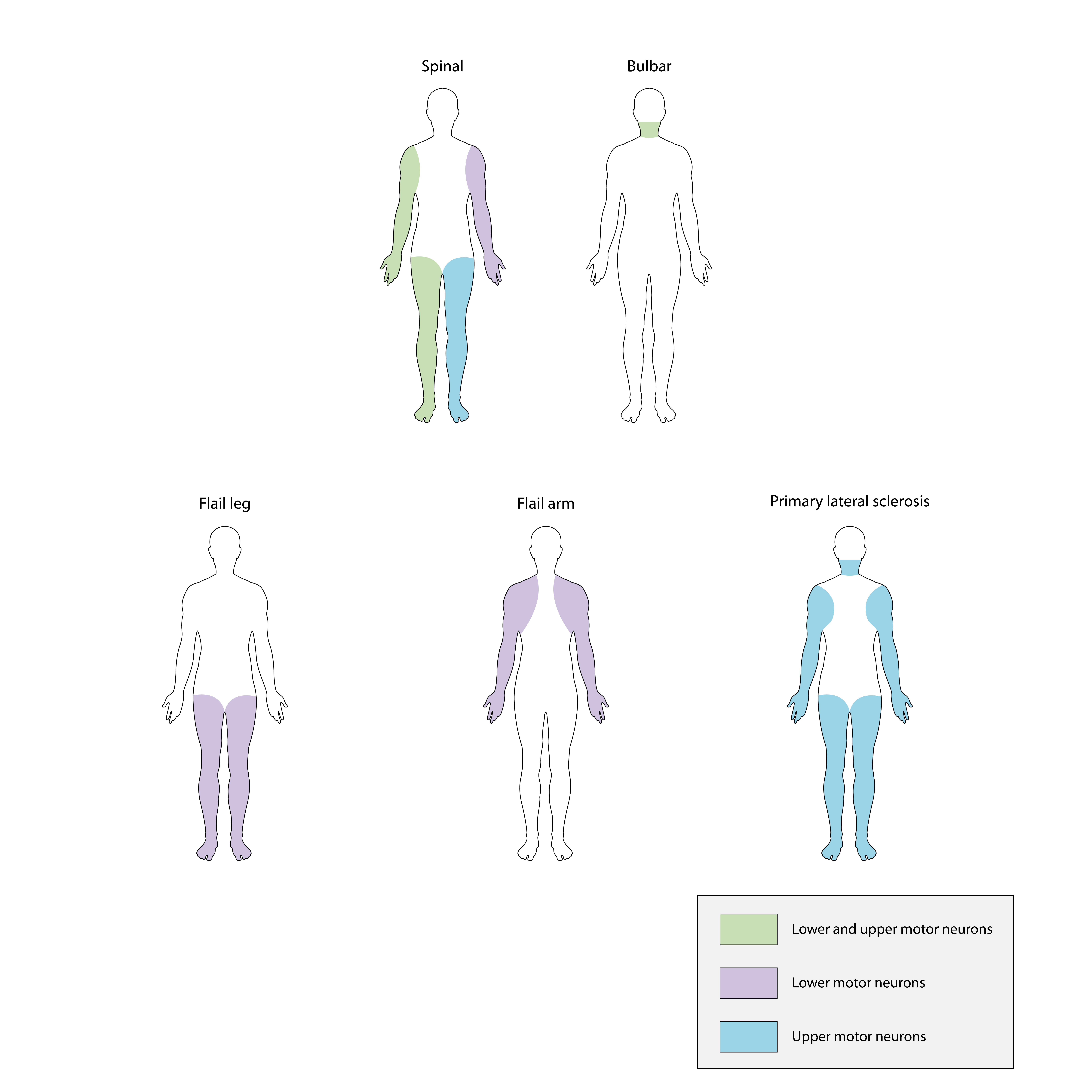
Amyotrophic lateral sclerosis (ALS), also known as motor neuron disease (MND) or‚ÄĒin the United States‚ÄĒLou Gehrig's disease (LGD), is a rare, Terminal illness, terminal neurodegenerative disease, neurodegenerative disorder that results in the progressive loss of both upper and lower motor neurons that normally control Skeletal muscle, voluntary muscle contraction. ALS is the most common form of the motor neuron diseases. ALS often presents in its early stages with gradual muscle Spasticity, stiffness, Fasciculation, twitches, Muscle weakness, weakness, and Muscle atrophy, wasting. Motor neuron loss typically continues until the abilities to eat, speak, move, and, lastly, breathe are all lost. While only 15% of people with ALS also fully develop frontotemporal dementia, an estimated 50% face at least some minor difficulties with cognitive disorder, thinking and behavior. Depending on which of the aforementioned symptoms develops first, ALS is classified as ''limb-onset'' (b ... [...More Info...] [...Related Items...] OR: [Wikipedia] [Google] [Baidu] |
Brain Stem
The brainstem (or brain stem) is the posterior stalk-like part of the brain that connects the cerebrum with the spinal cord. In the human brain the brainstem is composed of the midbrain, the pons, and the medulla oblongata. The midbrain is continuous with the thalamus of the diencephalon through the tentorial notch, and sometimes the diencephalon is included in the brainstem. The brainstem is very small, making up around only 2.6 percent of the brain's total weight. It has the critical roles of regulating heart and respiratory function, helping to control heart rate and breathing rate. It also provides the main motor and sensory nerve supply to the face and neck via the cranial nerves. Ten pairs of cranial nerves come from the brainstem. Other roles include the regulation of the central nervous system and the body's sleep cycle. It is also of prime importance in the conveyance of motor and sensory pathways from the rest of the brain to the body, and from the body back ... [...More Info...] [...Related Items...] OR: [Wikipedia] [Google] [Baidu] |
Frederick Parkes Weber
Frederick Parkes Weber (8 May 1863 – 2 June 1962) was an English dermatologist and author who practiced medicine in London. Background Weber's father, Sir Hermann David Weber (1823‚Äď1918), was a personal physician to Queen Victoria. Weber was educated at Charterhouse and Trinity College, Cambridge. He subsequently studied medicine at St. Bartholomew's Hospital, and abroad at Vienna and Paris. Career Returning to England, he worked (since 1894) at the German Hospital, Dalston (London), later he became House Physician and House Surgeon at St. Bartholomew's Hospital. He was subsequently House Physician at Brompton Hospital and Physician at Mount Vernon Hospital. Weber contributed over 1200 medical articles and wrote 23 books over a period of 50 years. In 1922, he, along with his wife, published a philosophical medical tome called ''Aspects of Death and Correlated Aspects of Life in Art, Epigram, and Poetry''. Weber was a prodigious describer of new and unique dermat ... [...More Info...] [...Related Items...] OR: [Wikipedia] [Google] [Baidu] |
Friedrich Gustav Jakob Henle
Friedrich Gustav Jakob Henle (; 9 July 1809 ‚Äď 13 May 1885) was a German physician, pathologist, and anatomist. He is credited with the discovery of the loop of Henle in the kidney. His essay, "On Miasma and Contagia," was an early argument for the germ theory of disease. He was an important figure in the development of modern medicine. Biography Henle was born in F√ľrth, Bavaria, to Simon and Rachel Diesbach Henle (H√§hnlein). He was Jewish. After studying medicine at Heidelberg and at Bonn, where he took his doctor's degree in 1832, he became prosector in anatomy to Johannes M√ľller at Berlin. During the six years he spent in that position he published a large amount of work, including three anatomical monographs on new species of animals and papers on the structure of the lymphatic system, the distribution of epithelium in the human body, the structure and development of the hair, and the formation of mucus and pus. He also developed a friendship with another ... [...More Info...] [...Related Items...] OR: [Wikipedia] [Google] [Baidu] |
Jules Baillarger
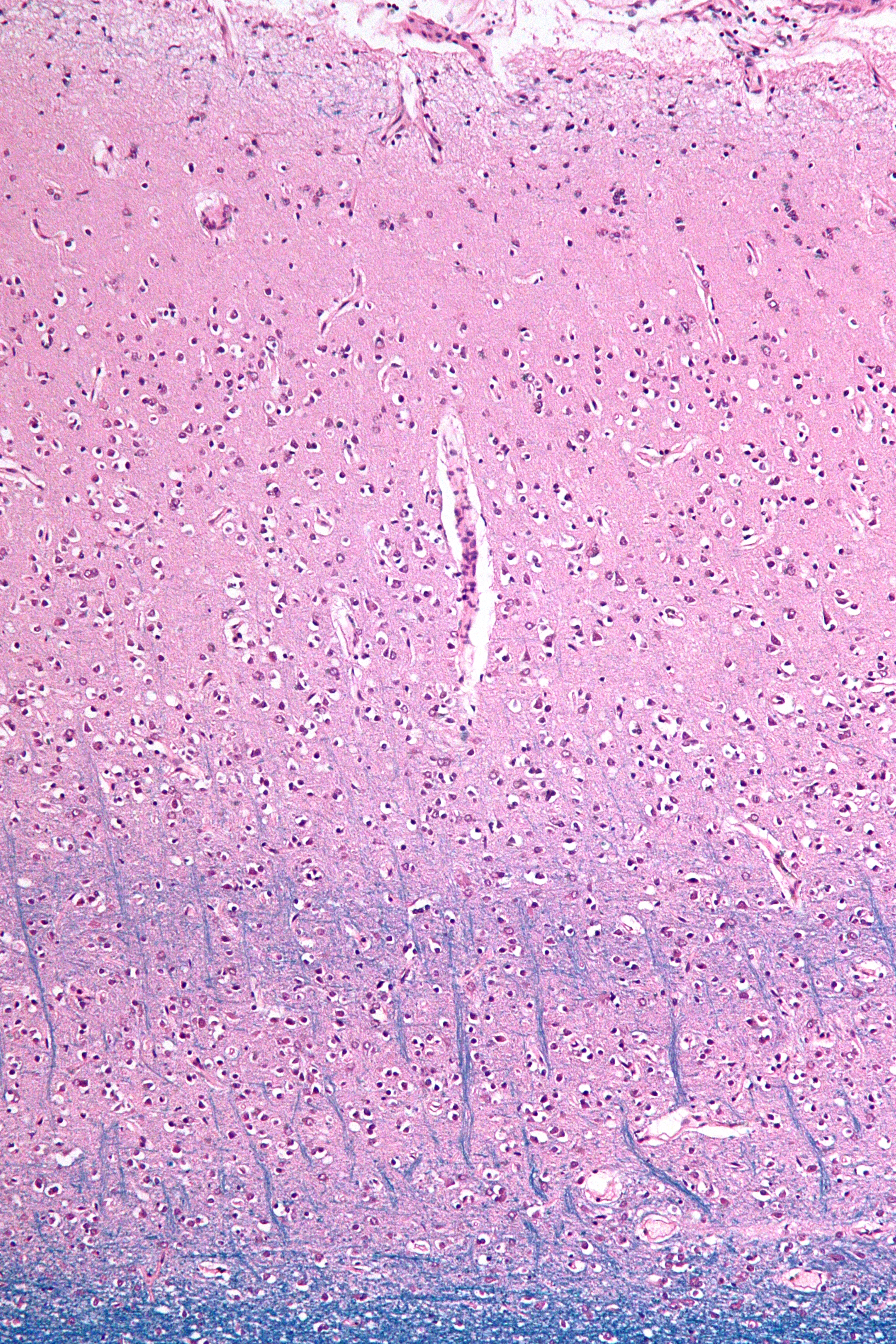
Jules Baillarger, full name Jules Gabriel Fran√ßois Baillarger (25 March 1809 ‚Äď 31 December 1890), was a French neurologist and psychiatrist. Biography Baillarger was born in Montbazon, France. He studied medicine at the University of Paris under Jean-√Čtienne Dominique Esquirol (1772‚Äď1840), and while a student worked as an intern at the Charenton mental institution. In 1840 he accepted a position at the Salp√™tri√®re, and soon after became director of a ''maison de sant√©'' in Ivry-sur-Seine. Among his assistants at Ivry was Louis-Victor Marc√© (1828‚Äď1864). With Jacques-Joseph Moreau (1804‚Äď1884) and others, he founded the influential ''Annales m√©dico-psychologiques'' (Medical-Psychological Annals). Contributions and theories In 1840 Baillarger was the first physician to discover that the cerebral cortex was divided into six layers of alternate white and grey laminae. His name is associated with the inner and outer bands of Baillarger, which are two layers of whi ... [...More Info...] [...Related Items...] OR: [Wikipedia] [Google] [Baidu] |
Charles-√Čdouard Brown-S√©quard
Charles-√Čdouard Brown-S√©quard FRS (8 April 1817 ‚Äď 2 April 1894) was a Mauritian physiologist and neurologist who, in 1850, became the first to describe what is now called Brown-S√©quard syndrome. Early life Brown-S√©quard was born at Port Louis, Mauritius, to an American father and a French mother. He attended the Royal College in Mauritius, and graduated in medicine at Paris in 1846. He then returned to Mauritius with the intention of practising there, but in 1852 he went to the United States. There he was appointed to the faculty of the Medical College of Virginia where he conducted experiments in the basement of the Egyptian Building. He was elected as a member of the American Philosophical Society in 1854. Later life He returned to Paris, and in 1859 he migrated to London, becoming physician to the National Hospital for the Paralysed and Epileptic. There he stayed for about five years, expounding his views on the pathology of the nervous system in numerous lecture ... [...More Info...] [...Related Items...] OR: [Wikipedia] [Google] [Baidu] |


