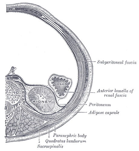
Retroperitoneal space on:
[Wikipedia]
[Google]
[Amazon]
The retroperitoneal space (retroperitoneum) is the anatomical space (sometimes a

 ;Perirenal space
It is also called the perinephric space. Bounded by the anterior and posterior leaves of the
 University of Michigan
- Lab Manual - Kidneys & Retroperitoneum ;Anterior pararenal space Bounded by the posterior layer of
potential space
In anatomy, a potential space is a space between two adjacent structures that are normally pressed together (directly apposed). Many anatomic spaces are potential spaces, which means that they are potential rather than realized (with their realiz ...
) behind (''retro'') the peritoneum
The peritoneum is the serous membrane forming the lining of the abdominal cavity or coelom in amniotes and some invertebrates, such as annelids. It covers most of the intra-abdominal (or coelomic) organs, and is composed of a layer of meso ...
. It has no specific delineating anatomical structures. Organs are retroperitoneal if they have peritoneum on their anterior side only. Structures that are not suspended by mesentery in the abdominal cavity and that lie between the parietal peritoneum and abdominal wall are classified as retroperitoneal.
This is different from organs that are not retroperitoneal, which have peritoneum on their posterior side and are suspended by mesentery in the abdominal cavity.
The retroperitoneum can be further subdivided into the following:
*Perirenal (or perinephric) space
*Anterior pararenal (or paranephric) space
*Posterior pararenal (or paranephric) space
Retroperitoneal structures
Structures that lie behind theperitoneum
The peritoneum is the serous membrane forming the lining of the abdominal cavity or coelom in amniotes and some invertebrates, such as annelids. It covers most of the intra-abdominal (or coelomic) organs, and is composed of a layer of meso ...
are termed "retroperitoneal". Organs that were once suspended within the abdominal cavity by mesentery but migrated posterior to the peritoneum during the course of embryogenesis to become retroperitoneal are considered to be secondarily retroperitoneal organs.
* Primarily retroperitoneal, meaning the structures were retroperitoneal during the entirety of development:
** urinary
*** adrenal glands
*** kidney
The kidneys are two reddish-brown bean-shaped organs found in vertebrates. They are located on the left and right in the retroperitoneal space, and in adult humans are about in length. They receive blood from the paired renal arteries; blo ...
s
*** ureter
The ureters are tubes made of smooth muscle that propel urine from the kidneys to the urinary bladder. In a human adult, the ureters are usually long and around in diameter. The ureter is lined by urothelial cells, a type of transitional epit ...
** circulatory
*** aorta
The aorta ( ) is the main and largest artery in the human body, originating from the left ventricle of the heart and extending down to the abdomen, where it splits into two smaller arteries (the common iliac arteries). The aorta distributes o ...
*** inferior vena cava
The inferior vena cava is a large vein that carries the deoxygenated blood from the lower and middle body into the right atrium of the heart. It is formed by the joining of the right and the left common iliac veins, usually at the level of th ...
** digestive
*** anal canal
* Secondarily retroperitoneal, meaning the structures initially were suspended in mesentery and later migrated behind the peritoneum during development
** the duodenum, except for the proximal first segment, which is intraperitoneal
** ascending and descending portions of the colon (but not the transverse colon, sigmoid and the cecum)
** pancreas, except for the tail, which is intraperitoneal
Subdivisions

 ;Perirenal space
It is also called the perinephric space. Bounded by the anterior and posterior leaves of the
 renal fascia
The renal fascia is a layer of connective tissue encapsulating the kidneys and the adrenal glands. It can be divided into:
*The anterior renal fascia, also called Gerota's fascia (after Dimitrie Gerota)
*The posterior renal fascia, also called Zuc ...
. It contains the following structures:
* Adrenal gland
* Kidney
The kidneys are two reddish-brown bean-shaped organs found in vertebrates. They are located on the left and right in the retroperitoneal space, and in adult humans are about in length. They receive blood from the paired renal arteries; blo ...
* Renal vessels
*Perirenal fat
The retroperitoneal space (retroperitoneum) is the anatomical space (sometimes a potential space) behind (''retro'') the peritoneum. It has no specific delineating anatomical structures. Organs are retroperitoneal if they have peritoneum on their ...
, which is also called the "adipose capsule of the kidney" and may be regarded as being part of the renal capsule
The renal capsule is a tough fibrous layer surrounding the kidney and covered in a layer of perirenal fat known as the adipose capsule of kidney. The adipose capsule is sometimes included in the structure of the renal capsule. It provides some p ...
- Lab Manual - Kidneys & Retroperitoneum ;Anterior pararenal space Bounded by the posterior layer of
peritoneum
The peritoneum is the serous membrane forming the lining of the abdominal cavity or coelom in amniotes and some invertebrates, such as annelids. It covers most of the intra-abdominal (or coelomic) organs, and is composed of a layer of meso ...
and the anterior leaf of the renal fascia
The renal fascia is a layer of connective tissue encapsulating the kidneys and the adrenal glands. It can be divided into:
*The anterior renal fascia, also called Gerota's fascia (after Dimitrie Gerota)
*The posterior renal fascia, also called Zuc ...
. It contains the following structures:
* Pancreas
The pancreas is an organ of the digestive system and endocrine system of vertebrates. In humans, it is located in the abdomen behind the stomach and functions as a gland. The pancreas is a mixed or heterocrine gland, i.e. it has both an en ...
* Ascending and descending colon
* Duodenum
;Posterior pararenal space
Bounded by the posterior leaf of the renal fascia and the muscles of the posterior abdominal wall. It contains only fat ("pararenal fat"), and is also called the "paranephric body", or "pararenal fat body".
Clinical significance
Bleeding from a blood vessel or structure in the retroperitoneal such as theaorta
The aorta ( ) is the main and largest artery in the human body, originating from the left ventricle of the heart and extending down to the abdomen, where it splits into two smaller arteries (the common iliac arteries). The aorta distributes o ...
or inferior vena cava
The inferior vena cava is a large vein that carries the deoxygenated blood from the lower and middle body into the right atrium of the heart. It is formed by the joining of the right and the left common iliac veins, usually at the level of th ...
into the retroperitoneal space can lead to a retroperitoneal hemorrhage
Retroperitoneal bleeding is an accumulation of blood in the retroperitoneal space. Signs and symptoms may include abdominal or upper leg pain, hematuria, and shock. It can be caused by major trauma or by non-traumatic mechanisms.
Signs and sympto ...
.
* Retroperitoneal fibrosis
Retroperitoneal fibrosis or Ormond's disease is a disease featuring the proliferation of fibrous tissue in the retroperitoneum, the compartment of the body containing the kidneys, aorta, renal tract, and various other structures. It may present wi ...
* Retroperitoneal lymph node dissection
Retroperitoneal lymph node dissection (RPLND) is a surgical procedure to remove abdominal lymph nodes. It is used to treat testicular cancer, as well as to help establish the exact stage and type of the cancer.
Indications
Testicular cancer met ...
It is also possible to have a neoplasm
A neoplasm () is a type of abnormal and excessive growth of tissue. The process that occurs to form or produce a neoplasm is called neoplasia. The growth of a neoplasm is uncoordinated with that of the normal surrounding tissue, and persists ...
in this area, more commonly a metastasis
Metastasis is a pathogenic agent's spread from an initial or primary site to a different or secondary site within the host's body; the term is typically used when referring to metastasis by a cancerous tumor. The newly pathological sites, then ...
; or very rarely a primary neoplasm. The most common type is a sarcoma
A sarcoma is a malignant tumor, a type of cancer that arises from transformed cells of mesenchymal ( connective tissue) origin. Connective tissue is a broad term that includes bone, cartilage, fat, vascular, or hematopoietic tissues, and sar ...
followed by lymphoma, extragonadal germ cell tumor, and Gastrointestinal stromal tumor/GIST. Examples of tumors include
** Primary retroperitoneal carcinoma
**Pseudomyxoma peritonei
Pseudomyxoma peritonei (PMP) is a clinical condition caused by cancerous cells (mucinous adenocarcinoma) that produce abundant mucin or gelatinous ascites. The tumors cause fibrosis of tissues and impede digestion or organ function, and if left unt ...
**Examples of sarcomas include:
**Soft-tissue sarcoma
A soft-tissue sarcoma (STS) is a malignant tumour, a type of cancer, that develops in soft tissue. A soft tissue sarcoma is often a painless mass that grows slowly over months or years. They may be superficial or deep-seated. Any such unexplaine ...
***liposarcoma
Liposarcomas are the most common subtype of soft tissue sarcomas, accounting for at least 20% of all sarcomas in adults. Soft tissue sarcomas are rare neoplasms with over 150 different histological subtypes or forms. Liposarcomas arise from the pr ...
*** leiomyosarcoma
*** Undifferentiated pleomorphic sarcoma, a clinically distinct sarcoma of the area
See also
*Intraperitoneal
The peritoneum is the serous membrane forming the lining of the abdominal cavity or coelom in amniotes and some invertebrates, such as annelids. It covers most of the intra-abdominal (or coelomic) organs, and is composed of a layer of mesothel ...
References
{{Authority control Abdomen