|
Angiography
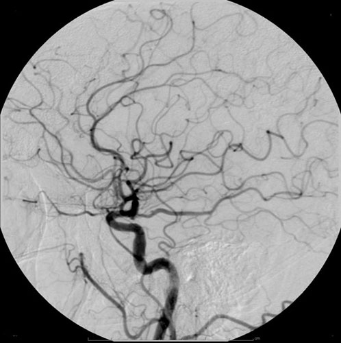
Angiography or arteriography is a medical imaging technique used to visualize the inside, or lumen, of blood vessels and organs of the body, with particular interest in the arteries, veins, and the heart chambers. Modern angiography is performed by injecting a radio-opaque contrast agent into the blood vessel and imaging using X-ray based techniques such as fluoroscopy. With time-of-flight (TOF) magnetic resonance it is no longer necessary to use a contrast. The word itself comes from the Greek words ἀνγεῖον ''angeion'' 'vessel' and γράφειν ''graphein'' 'to write, record'. The film or image of the blood vessels is called an ''angiograph'', or more commonly an ''angiogram''. Though the word can describe both an arteriogram and a venogram, in everyday usage the terms angiogram and arteriogram are often used synonymously, whereas the term venogram is used more precisely. The term angiography has been applied to radionuclide angiography and newer vascular ima ... [...More Info...] [...Related Items...] OR: [Wikipedia] [Google] [Baidu] [Amazon] |
Cerebral Angiography
Cerebral angiography is a form of angiography which provides images of blood vessels in and around the brain, thereby allowing detection of abnormalities such as arteriovenous malformations and aneurysms. It was pioneered in 1927 by the Portuguese neurologist Egas Moniz at the University of Lisbon, who also helped develop thorotrast for use in the procedure. Typically a catheter is inserted into a large artery (such as the femoral artery) and threaded through the circulatory system to the Common carotid artery, carotid artery, where a contrast agent is injected. A series of radiographs are taken as the contrast agent spreads through the brain's arterial system, then a second series as it reaches the venous system. For some applications, cerebral angiography may yield better images than less invasive methods such as computed tomography angiography and magnetic resonance angiography. In addition, cerebral angiography allows certain treatments to be performed immediately, based on ... [...More Info...] [...Related Items...] OR: [Wikipedia] [Google] [Baidu] [Amazon] |
Computed Tomography Angiography
Computed tomography angiography (also called CT angiography or CTA) is a computed tomography technique used for angiography—the visualization of arteries and veins—throughout the human body. Using contrast injected into the blood vessels, images are created to look for blockages, aneurysms (dilations of walls), Dissection (medical), dissections (tearing of walls), and stenosis (narrowing of vessel). CTA can be used to visualize the vessels of the heart, the aorta and other large blood vessels, the lungs, the kidneys, the head and neck, and the arms and legs. CTA can also be used to localise arterial or venous bleed of the gastrointestinal system. Medical uses CTA can be used to examine blood vessels in many key areas of the body including the brain, kidneys, pelvis, and the lungs. Coronary CT angiography Coronary CT angiography (CCTA) is the use of CT angiography to assess the coronary artery, arteries of the heart. The patient receives an intravenous injection of radiocont ... [...More Info...] [...Related Items...] OR: [Wikipedia] [Google] [Baidu] [Amazon] |
Radionuclide Angiography
Radionuclide angiography is an area of nuclear medicine which specialises in imaging to show the functionality of the right and left ventricles of the heart, thus allowing informed diagnostic intervention in heart failure. It involves use of a radiopharmaceutical, injected into a patient, and a gamma camera for acquisition. A MUGA scan (multigated acquisition) involves an acquisition triggered (gated) at different points of the cardiac cycle. MUGA scanning is also called equilibrium radionuclide angiocardiography, radionuclide ventriculography (RNVG), or gated blood pool imaging, as well as SYMA scanning (synchronized multigated acquisition scanning). This mode of imaging uniquely provides a cine type of image of the beating heart, and allows the interpreter to determine the efficiency of the individual heart valves and chambers. MUGA/Cine scanning represents a robust adjunct to the now more common echocardiogram. Mathematics regarding acquisition of cardiac output (''Q' ... [...More Info...] [...Related Items...] OR: [Wikipedia] [Google] [Baidu] [Amazon] |
Magnetic Resonance Angiography
Magnetic resonance angiography (MRA) is a group of techniques based on magnetic resonance imaging (MRI) to image blood vessels. Magnetic resonance angiography is used to generate images of arteries (and less commonly veins) in order to evaluate them for stenosis (abnormal narrowing), Vascular occlusion, occlusions, aneurysms (vessel wall dilatations, at risk of rupture) or other abnormalities. MRA is often used to evaluate the arteries of the neck and brain, the thoracic and abdominal aorta, the renal arteries, and the legs (the latter exam is often referred to as a "run-off"). Acquisition A variety of techniques can be used to generate the pictures of blood vessels, both artery, arteries and veins, based on flow effects or on contrast (inherent or pharmacologically generated). The most frequently applied MRA methods involve the use intravenous MRI contrast agent, contrast agents, particularly those containing gadolinium to shorten the Spin–lattice relaxation, ''T''1 of bloo ... [...More Info...] [...Related Items...] OR: [Wikipedia] [Google] [Baidu] [Amazon] |
Posterior Cerebral
The posterior cerebral artery (PCA) is one of a pair of cerebral arteries that supply oxygenated blood to the occipital lobe, as well as the medial and inferior aspects of the temporal lobe of the human brain. The two arteries originate from the distal end of the basilar artery, where it bifurcates into the left and right posterior cerebral arteries. These anastomose with the middle cerebral arteries and internal carotid arteries via the posterior communicating arteries. Structure The posterior cerebral artery is subdivided into 4 segments: P1: pre-communicating segment * Originated at the termination of the basilar artery * May give rise to the artery of Percheron if present P2: post-communicating segment * From the PCOM around the midbrain * Terminates as it enters the quadrigeminal ganglion * Gives rise to the choroidal branches (medial and lateral posterior choroidal arteries) P3: quadrigeminal segment * Courses posteromedially through the quadrigeminal cistern ... [...More Info...] [...Related Items...] OR: [Wikipedia] [Google] [Baidu] [Amazon] |
Fluoroscopy
Fluoroscopy (), informally referred to as "fluoro", is an imaging technique that uses X-rays to obtain real-time moving images of the interior of an object. In its primary application of medical imaging, a fluoroscope () allows a surgeon to see the internal anatomy, structure and physiology, function of a patient, so that the pumping action of the heart or the motion of swallowing, for example, can be watched. This is useful for both medical diagnosis, diagnosis and therapy and occurs in general radiology, interventional radiology, and image-guided surgery. In its simplest form, a fluoroscope consists of an X-ray generator, X-ray source and a fluorescence, fluorescent screen, between which a patient is placed. However, since the 1950s most fluoroscopes have included X-ray image intensifiers and cameras as well, to improve the image's visibility and make it available on a remote display screen. For many decades, fluoroscopy tended to produce live pictures that were not recorded, bu ... [...More Info...] [...Related Items...] OR: [Wikipedia] [Google] [Baidu] [Amazon] |
Radiocontrast
Radiocontrast agents are substances used to enhance the visibility of internal structures in X-ray-based imaging techniques such as computed tomography (contrast CT), projectional radiography, and fluoroscopy. Radiocontrast agents are typically iodine, or more rarely barium sulfate. The contrast agents absorb external X-rays, resulting in decreased exposure on the X-ray detector. This is different from radiopharmaceuticals used in nuclear medicine which emit radiation. Magnetic resonance imaging (MRI) functions through different principles and thus MRI contrast agents have a different mode of action. These compounds work by altering the magnetic properties of nearby hydrogen nuclei. Types and uses Radiocontrast agents used in X-ray examinations can be grouped in positive (iodinated agents, barium sulfate), and negative agents (air, carbon dioxide, methylcellulose). Iodine (circulatory system) Iodinated contrast contains iodine. It is the main type of radiocontrast used for intra ... [...More Info...] [...Related Items...] OR: [Wikipedia] [Google] [Baidu] [Amazon] |
Lucien Campeau
Lucien Campeau (June 20, 1927March 15, 2010) was a Canadian cardiologist. He was a full professor at the Université de Montréal The Université de Montréal (; UdeM; ) is a French-language public research university in Montreal, Quebec, Canada. The university's main campus is located in the Côte-des-Neiges neighborhood of Côte-des-Neiges–Notre-Dame-de-Grâce on M .... He is best known for performing the world's first transradial coronary angiogram. Campeau was one of the founding staff of the Montreal Heart Institute, joining in 1957. He is also well known for developing the Canadian Cardiovascular Society grading of angina pectoris. Education Campeau received his M.D. degree from the University of Laval in 1953 and completed a fellowship in Cardiology at Johns Hopkins Hospital from 1956 to 1957. He later became a professor at University of Montreal in 1961 and was one of the co-founders of the Montreal Heart Institute. In his lifetime, Campeau was awarded the ... [...More Info...] [...Related Items...] OR: [Wikipedia] [Google] [Baidu] [Amazon] |
Seldinger Technique
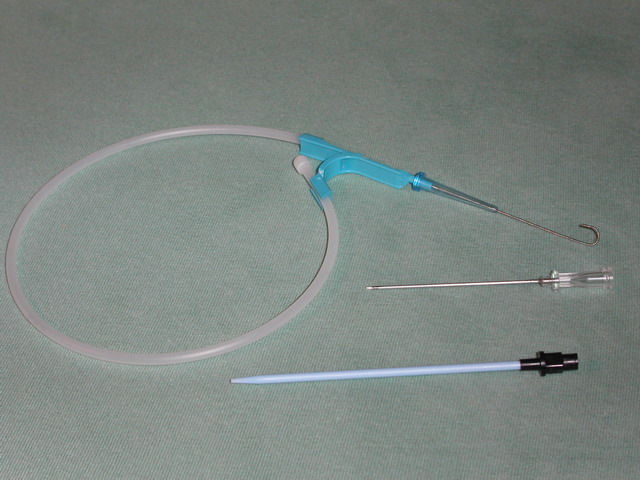
The Seldinger technique, also known as Seldinger wire technique, is a medical procedure to obtain safe access to blood vessels and other hollow organ (anatomy), organs. It is eponym, named after Sven Ivar Seldinger (1921–1998), a Sweden, Swedish radiology, radiologist who introduced the procedure in 1953. Uses The Seldinger technique is used for angiography, insertion of chest drains and central venous catheters, insertion of percutaneous endoscopic gastrostomy, PEG tubes using the push technique, insertion of the leads for an artificial pacemaker or implantable cardioverter-defibrillator, and numerous other interventional medical procedures. Complications The initial puncture is with a sharp instrument, and this may lead to hemorrhage or perforation of the organ in question. Infection is a possible complication, and hence asepsis is practiced during most Seldinger procedures. Loss of the guidewire into the cavity or blood vessel is a significant and generally preventable com ... [...More Info...] [...Related Items...] OR: [Wikipedia] [Google] [Baidu] [Amazon] |
Sousa Pereira
Sousa refers to * John Philip Sousa (1854–1932), American composer of marches Sousa also may refer to: People * Sousa (surname), including other Portuguese variants such as Souza, de Sousa, D'Souza, etc. * João Sousa, Portuguese tennis player * Paulo Sousa, Portuguese football manager * Souza (footballer, born 1975), José Ivanaldo de Souza, Brazilian football attacking midfielder * Souza (footballer, born 1977), Sergio Roberto Pereira de Souza, Brazilian football midfielder * Souza (footballer, born 1979), Willamis de Souza Silva, Brazilian former football midfielder and television pundit * Souza (footballer, born 1982), Rodrigo de Souza Cardoso, Brazilian football striker * Souza (footballer, born 1988), Elierce Barbosa de Souza, Brazilian football defensive midfielder * Souza (footballer, born 2006), João Victor de Souza Menezes, Brazilian football left-back * Sousa (Brazilian footballer), Van Basty Sousa e Silva, (born 1994), Brazilian football midfielder * Daniel ... [...More Info...] [...Related Items...] OR: [Wikipedia] [Google] [Baidu] [Amazon] |
Fausto Lopo De Carvalho
Fausto Lopo Patrício de Carvalho (15 May 1890 – 23 May 1970), more commonly known as Fausto Lopo de Carvalho, was a Portuguese pulmonologist specialising in phthisiology, and the developer of pulmonary angiography in 1931, with Egas Moniz and Almeida Lima. He was the son of eminent phthisiologist Lopo de Carvalho (founder of the first sanatorium in Portugal, in Guarda), and his wife Leopoldina dos Anjos Patrício de Carvalho. He studied at the University of Coimbra, earning a degree in medicine with the highest possible grade (20 out of a possible 20) in 1916; after completing his medical studies he worked at the Guarda Sanatorium under his father's guidance, where he prepared his thesis for a doctorate, entitled '' Artificial Pneumothorax''. He taught Medical Propaedeutics, first at the Faculty of Medicine of the University of Coimbra and later at the Faculty of Medicine of the University of Lisbon, until 1934, when he was appointed to the newly created Chair of Ch ... [...More Info...] [...Related Items...] OR: [Wikipedia] [Google] [Baidu] [Amazon] |
Reynaldo Dos Santos
Reynaldo dos Santos (3 December 1880 – 6 May 1970) was a Portuguese physician, writer, and art historian. As a physician, he was a pioneer in the fields of vascular surgery and urology; as an art historian, he published numerous works on 15th-century Portuguese art, including on the Manueline style and on the paintings of Nuno Gonçalves. Biography Reynaldo dos Santos was born in 1880 to Clemente José dos Santos (himself a physician) and Maria Amélia Pinheiro Santos, in the family home in Rua das Varinas, Vila Franca de Xira, a town in the outskirts of Lisbon. He concluded his primary and secondary studies in this town, before enrolling at the Medico-Surgical School in Lisbon, from which he graduated in 1903. Between 1902 and 1905, he was abroad in Paris and the main surgical centres of the United States, in Boston, Chicago, Rochester, Baltimore, Philadelphia, and New York. He earned his doctorate in Medicine in 1906, with his thesis titled "''Aspectos Cirúrgicos das Pan ... [...More Info...] [...Related Items...] OR: [Wikipedia] [Google] [Baidu] [Amazon] |


