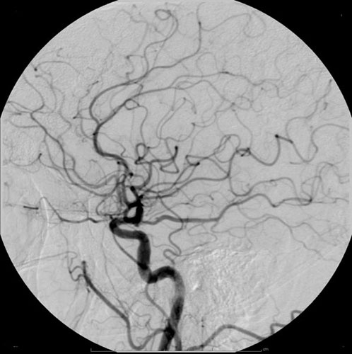
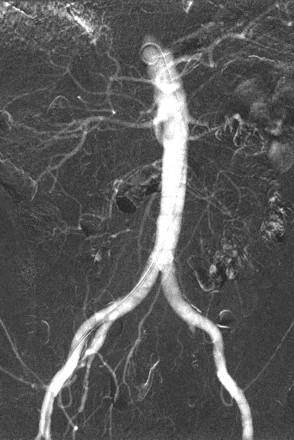
Radiocontrast on:
[Wikipedia]
[Google]
[Amazon]
Radiocontrast agents are substances used to enhance the visibility of internal structures in

 Carbon dioxide also has a role in angioplasty. It is low-risk as it is a natural product with no risk of allergic potential. However, it can be used only below the diaphragm as there is a risk of embolism in neurovascular procedures. It must be used carefully to avoid contamination with room air when injected. It is a negative contrast agent in that it displaces blood when injected intravascularly.
X-ray
An X-ray (also known in many languages as Röntgen radiation) is a form of high-energy electromagnetic radiation with a wavelength shorter than those of ultraviolet rays and longer than those of gamma rays. Roughly, X-rays have a wavelength ran ...
-based imaging techniques such as computed tomography
A computed tomography scan (CT scan), formerly called computed axial tomography scan (CAT scan), is a medical imaging technique used to obtain detailed internal images of the body. The personnel that perform CT scans are called radiographers or ...
(contrast CT
Contrast CT, or contrast-enhanced computed tomography (CECT), is CT scan, X-ray computed tomography (CT) using radiocontrast. Radiocontrasts for X-ray CT are generally Iodinated contrast, iodine-based types. This is useful to highlight structure ...
), projectional radiography
Projectional radiography, also known as conventional radiography, is a form of radiography and medical imaging that produces two-dimensional images by X-ray radiation. The image acquisition is generally performed by radiographers, and the images a ...
, and fluoroscopy
Fluoroscopy (), informally referred to as "fluoro", is an imaging technique that uses X-rays to obtain real-time moving images of the interior of an object. In its primary application of medical imaging, a fluoroscope () allows a surgeon to see t ...
. Radiocontrast agents are typically iodine, or more rarely barium sulfate
Barium sulfate (or sulphate) is the inorganic compound with the chemical formula Ba SO4. It is a white crystalline solid that is odorless and insoluble in water. It occurs in nature as the mineral barite, which is the main commercial source of ...
. The contrast agent
A contrast agent (or contrast medium) is a substance used to increase the contrast of structures or fluids within the body in medical imaging. Contrast agents absorb or alter external electromagnetism or ultrasound, which is different from radiop ...
s absorb external X-rays, resulting in decreased exposure on the X-ray detector
X-ray detectors are devices used to measure the flux, spatial distribution, spectrum, and/or other properties of X-rays.
Detectors can be divided into two major categories: imaging detectors (such as photographic plates and X-ray film (photograp ...
. This is different from radiopharmaceutical
Radiopharmaceuticals, or medicinal radiocompounds, are a group of pharmaceutical drugs containing radioactive isotopes. Radiopharmaceuticals can be used as diagnostic and therapeutic agents. Radiopharmaceuticals emit radiation themselves, which ...
s used in nuclear medicine
Nuclear medicine (nuclear radiology, nucleology), is a medical specialty involving the application of radioactivity, radioactive substances in the diagnosis and treatment of disease. Nuclear imaging is, in a sense, ''radiology done inside out'', ...
which emit radiation.
Magnetic resonance imaging
Magnetic resonance imaging (MRI) is a medical imaging technique used in radiology to generate pictures of the anatomy and the physiological processes inside the body. MRI scanners use strong magnetic fields, magnetic field gradients, and ...
(MRI) functions through different principles and thus MRI contrast agent
MRI contrast agents are contrast agents used to improve the visibility of internal body structures in magnetic resonance imaging (MRI).
The most commonly used compounds for contrast enhancement are gadolinium-based contrast agents (GBCAs). Suc ...
s have a different mode of action. These compounds work by altering the magnetic properties of nearby hydrogen nuclei.
Types and uses
Radiocontrast agents used inX-ray
An X-ray (also known in many languages as Röntgen radiation) is a form of high-energy electromagnetic radiation with a wavelength shorter than those of ultraviolet rays and longer than those of gamma rays. Roughly, X-rays have a wavelength ran ...
examinations can be grouped in positive (iodinated agents, barium sulfate), and negative agents (air, carbon dioxide, methylcellulose).
Iodine (circulatory system)

Iodinated contrast
Iodinated contrast is a form of water-soluble, intravenous radiocontrast agent containing iodine, which enhances the visibility of vascular structures and organs during radiography, radiographic procedures. Some pathologies, such as cancer, have p ...
contains iodine
Iodine is a chemical element; it has symbol I and atomic number 53. The heaviest of the stable halogens, it exists at standard conditions as a semi-lustrous, non-metallic solid that melts to form a deep violet liquid at , and boils to a vi ...
. It is the main type of radiocontrast used for intravenous administration
Intravenous therapy (abbreviated as IV therapy) is a medical technique that administers fluids, medications and nutrients directly into a person's vein. The intravenous route of administration is commonly used for rehydration or to provide nutr ...
. Iodine has a particular advantage as a contrast agent for radiography because its innermost electron ("k-shell") binding energy is 33.2 keV, similar to the average energy of x-rays used in diagnostic radiography. When the incident x-ray energy is closer to the k-edge of the atom it encounters, photoelectric absorption is more likely to occur. Its uses include:
* Contrast CT
Contrast CT, or contrast-enhanced computed tomography (CECT), is CT scan, X-ray computed tomography (CT) using radiocontrast. Radiocontrasts for X-ray CT are generally Iodinated contrast, iodine-based types. This is useful to highlight structure ...
s
* Angiography
Angiography or arteriography is a medical imaging technique used to visualize the inside, or lumen, of blood vessels and organs of the body, with particular interest in the arteries, veins, and the heart chambers. Modern angiography is perfo ...
(''arterial investigations'')
* Venography
Venography (also called phlebography or ascending phlebography) is a procedure in which an X-ray of the veins, a venogram, is taken after a special dye is injected into the bone marrow or veins. The dye has to be injected constantly via a cathete ...
(''venous investigations'')
* VCUG ('' voiding cystourethrography'')
* HSG ('' hysterosalpingogram'')
* IVU (''intravenous urography
Pyelogram (or pyelography or urography) is a form of imaging of the renal pelvis and ureter.
Types include:
* Intravenous pyelogram – In which a contrast solution is introduced through a vein into the circulatory system.
* Retrograde pyelogra ...
'')
Organic iodine molecules used for contrast include iohexol
Iohexol, sold under the trade name Iodaque among others, is a contrast agent used for X-ray imaging. This includes when visualizing arteries, veins, ventricles of the brain, the urinary system, and joints, as well as during computed tomograph ...
, iodixanol
Iodixanol, sold under the brand name Visipaque, is an iodine-containing non-ionic radiocontrast agent.
It is available as a generic medication.
Medical uses
The radio contrast agent is given intravenously for computed tomography (CT) imaging ...
and ioversol
Ioversol ( INN; trade name Optiray) is an organoiodine compound that is used as a contrast medium. It features both a high iodine content, as well as several hydrophilic groups. It is used in clinical diagnostics including arthrography, angioca ...
.
Barium sulfate (digestive system)
Barium sulfate
Barium sulfate (or sulphate) is the inorganic compound with the chemical formula Ba SO4. It is a white crystalline solid that is odorless and insoluble in water. It occurs in nature as the mineral barite, which is the main commercial source of ...
is mainly used in the imaging of the digestive system. The substance exists as a water-insoluble white powder that is made into a slurry with water and administered directly into the gastrointestinal tract
The gastrointestinal tract (GI tract, digestive tract, alimentary canal) is the tract or passageway of the Digestion, digestive system that leads from the mouth to the anus. The tract is the largest of the body's systems, after the cardiovascula ...
.
* Upper gastrointestinal series
An upper gastrointestinal series, also called a barium swallow, barium study, or barium meal, is a series of radiographs used to examine the gastrointestinal tract for abnormalities. A contrast medium, usually a radiocontrast agent such as bariu ...
* Barium enema (''large bowel investigation'') and DCBE (''double contrast barium enema'').
* Barium swallow (''oesophageal investigation'')
* Barium meal (''stomach investigation'') and double contrast barium meal
* Barium follow through (''stomach and small bowel investigation'')
* CT pneumocolon / virtual colonoscopy
Barium sulfate, an insoluble white powder, is typically used for enhancing contrast in the GI tract. Depending on how it is to be administered the compound is mixed with water, thickeners, de-clumping agents, and flavourings to make the contrast agent. As the barium sulfate does not dissolve, this type of contrast agent is an opaque white mixture. It is only used in the digestive tract; it is usually swallowed as a barium sulfate suspension
Barium sulfate suspension, often simply called barium, is a contrast agent used during X-rays. Specifically it is used to improve visualization of the gastrointestinal tract (esophagus, stomach, intestines) on radiography, plain X-ray or compu ...
or administered as an enema. After the examination, it leaves the body with the feces
Feces (also known as faeces American and British English spelling differences#ae and oe, or fæces; : faex) are the solid or semi-solid remains of food that was not digested in the small intestine, and has been broken down by bacteria in the ...
.
Air
Air can be used as a contrast material because it is less radio-opaque than the tissues it is defining. A double contrast barium enema, or DCBE, uses both air and barium together. In an air arthrogram, where air alone is used as a contrast medium, the injection of air into a joint cavity allows the cartilage covering the ends of the bones to be visualized. Before the advent of modernneuroimaging
Neuroimaging is the use of quantitative (computational) techniques to study the neuroanatomy, structure and function of the central nervous system, developed as an objective way of scientifically studying the healthy human brain in a non-invasive ...
techniques, air or other gases were used as contrast agents employed to displace the cerebrospinal fluid
Cerebrospinal fluid (CSF) is a clear, colorless Extracellular fluid#Transcellular fluid, transcellular body fluid found within the meninges, meningeal tissue that surrounds the vertebrate brain and spinal cord, and in the ventricular system, ven ...
in the brain while performing a pneumoencephalography
Pneumoencephalography (sometimes abbreviated PEG; also referred to as an "air study") was a common medical procedure in which most of the cerebrospinal fluid (CSF) was drained from around the brain by means of a lumbar puncture and replaced with ...
. Sometimes called an "air study", this once common yet highly-unpleasant procedure was used to enhance the outline of structures in the brain, looking for shape distortions caused by the presence of lesions.
Carbon dioxide
 Carbon dioxide also has a role in angioplasty. It is low-risk as it is a natural product with no risk of allergic potential. However, it can be used only below the diaphragm as there is a risk of embolism in neurovascular procedures. It must be used carefully to avoid contamination with room air when injected. It is a negative contrast agent in that it displaces blood when injected intravascularly.
Discontinued agents
Thorotrast
Thorotrast was a contrast agent based onthorium dioxide
Thorium dioxide (ThO2), also called thorium(IV) oxide, is a crystalline solid, often white or yellow in colour. Also known as thoria, it is mainly a by-product of lanthanide and uranium production. Thorianite is the name of the mineralogical for ...
, which is radioactive
Radioactive decay (also known as nuclear decay, radioactivity, radioactive disintegration, or nuclear disintegration) is the process by which an unstable atomic nucleus loses energy by radiation. A material containing unstable nuclei is conside ...
. It was first introduced in 1929. While it provided good image enhancement, its use was abandoned in the late 1950s since it turned out to be carcinogenic
A carcinogen () is any agent that promotes the development of cancer. Carcinogens can include synthetic chemicals, naturally occurring substances, physical agents such as ionizing and non-ionizing radiation, and Biological agent, biologic agent ...
. Given that the substance remained in the bodies of those to whom it was administered, it gave a continuous radiation exposure and was associated with a risk of cancers of the liver, bile ducts and bones, as well as higher rates of hematological malignancy (leukemia and lymphoma). Thorotrast may have been administered to millions of patients prior to being disused.
Nonsoluble substances
In the past, some non water-soluble contrast agents were used. One such substance was iofendylate (trade names: Pantopaque, Myodil) which was an iodinated oil-based substance that was commonly used in myelography. Due to it being oil-based, it was recommended that the physician remove it from the patient at the end of the procedure. This was a painful and difficult step and because complete removal could not always be achieved, iofendylate's persistence in the body might sometimes lead to arachnoiditis, a potentially painful and debilitating lifelong disorder of the spine. Iofendylate's use ceased when water-soluble agents (such as metrizamide) became available in the late 1970s. Also, with the advent ofMRI
Magnetic resonance imaging (MRI) is a medical imaging technique used in radiology to generate pictures of the anatomy and the physiological processes inside the body. MRI scanners use strong magnetic fields, magnetic field gradients, and rad ...
, myelography became much less-commonly performed.
Adverse effects
Modern iodinated contrast agents – especially non-ionic compounds – are generally well tolerated. The adverse effects of radiocontrast can be subdivided into type A reactions (e.g. thyrotoxicosis), and type B reactions (hypersensitivity reactions: allergy and non-allergy reactions ormerly called anaphylactoid reactions. Patients receiving contrast via IV typically experience a hot feeling around the throat, and this hot sensation gradually moves down to the pelvic area. The documentation of adverse drug reactions to contrast media should be documented precisely so that the patient receives adequate prophylaxis if contrast medium is administered again.Contrast induced nephropathy
Iodinated contrast may be toxic to the kidneys, especially when given via the arteries prior to studies such as catheter coronary angiography. Non-ionic contrast agents, which are almost exclusively used incomputed tomography
A computed tomography scan (CT scan), formerly called computed axial tomography scan (CAT scan), is a medical imaging technique used to obtain detailed internal images of the body. The personnel that perform CT scans are called radiographers or ...
studies, have not been shown to cause CIN when given intravenously at doses needed for CT studies.
Thyroid dysfunction
Iodinated radiocontrast can induce overactivity (hyperthyroidism) and underactivity (hypothyroidism) of the thyroid gland. The risk of either condition developing after a single examination is 2–3 times that of those who have not undergone a scan with iodinated contrast. Thyroid underactivity is mediated by two phenomena called the Plummer and Wolff–Chaikoff effect, where iodine suppresses the production of thyroid hormones; this is usually temporary but there is an association with longer-term thyroid underactivity. Some other people show the opposite effect, called Jod-Basedow phenomenon, where the iodine induces overproduction of thyroid hormone; this may be the result of underlying thyroid disease (such as nodules orGraves' disease
Graves' disease, also known as toxic diffuse goiter or Basedow's disease, is an autoimmune disease that affects the thyroid. It frequently results in and is the most common cause of hyperthyroidism. It also often results in an enlarged thyro ...
) or previous iodine deficiency. Children exposed to iodinated contrast during pregnancy may develop hypothyroidism after birth and monitoring of the thyroid function is recommended.
See also
* * *References
External links
* {{Use dmy dates, date=August 2019 Nephrotoxins