|
Hyperkeratosis
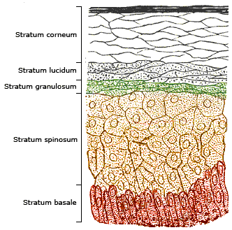
Hyperkeratosis is thickening of the stratum corneum (the outermost layer of the epidermis, or skin), often associated with the presence of an abnormal quantity of keratin,Kumar, Vinay; Fausto, Nelso; Abbas, Abul (2004) ''Robbins & Cotran Pathologic Basis of Disease'' (7th ed.). Saunders. Page 1230. . and also usually accompanied by an increase in the granular layer. As the corneum layer normally varies greatly in thickness in different sites, some experience is needed to assess minor degrees of hyperkeratosis. It can be caused by vitamin A deficiency or chronic exposure to arsenic. Hyperkeratosis can also be caused by B-Raf inhibitor drugs such as Vemurafenib and Dabrafenib.Niezgoda, Anna; Niezgoda, Piotr; Czajkowski, Rafal (2015) ''Novel Approaches to Treatment of Advanced Melanoma: A Review of Targeted Therapy and Immunotherapy'' BioMed Research International It can be treated with urea-containing creams, which dissolve the intercellular matrix of the cells of the stratum co ... [...More Info...] [...Related Items...] OR: [Wikipedia] [Google] [Baidu] |
Epidermolytic Hyperkeratosis
Epidermolytic ichthyosis (EI), also known as bullous epidermis ichthyosis (BEI), epidermolytic hyperkeratosis (EHK), bullous congenital ichthyosiform erythroderma (BCIE), bullous ichthyosiform erythrodermaFreedberg, et al. (2003). ''Fitzpatrick's Dermatology in General Medicine''. (6th ed.). McGraw-Hill. . or bullous congenital ichthyosiform erythroderma Brocq, is a rare and severe form of ichthyosis that affects around 1 in 300,000 people. It is caused by a genetic mutation, and thus cannot be completely cured without some form of gene therapy. While some research has been done into possible gene therapy treatments, the work hasn't yet been successfully developed to the stage where it can be routinely given to patients. The condition involves the clumping of keratin filaments.James, William; Berger, Timothy; Elston, Dirk (2005). ''Andrews' Diseases of the Skin: Clinical Dermatology''. (10th ed.). Saunders. . Presentation Epidermolytic hyperkeratosis is a skin disorder that is ... [...More Info...] [...Related Items...] OR: [Wikipedia] [Google] [Baidu] |
Focal Acral Hyperkeratosis
Palmoplantar keratodermas are a heterogeneous group of disorders characterized by abnormal thickening of the stratum corneum of the palms and soles. Autosomal recessive, dominant, X-linked, and acquired forms have all been described. Types Clinically, three distinct patterns of palmoplantar keratoderma may be identified: diffuse, focal, and punctate. Diffuse Diffuse palmoplantar keratoderma is a type of palmoplantar keratoderma that is characterized by an even, thick, symmetric hyperkeratosis over the whole of the palm and sole, usually evident at birth or in the first few months of life. Restated, diffuse palmoplantar keratoderma is an autosomal dominant disorder in which hyperkeratosis is confined to the palms and soles. The two major types can have a similar clinical appearance: *''Diffuse epidermolytic palmoplantar keratoderma'' (also known as "Palmoplantar keratoderma cum degeneratione granulosa Vörner," "Vörner's epidermolytic palmoplantar keratoderma", and "Vörn ... [...More Info...] [...Related Items...] OR: [Wikipedia] [Google] [Baidu] |
Multiple Minute Digitate Hyperkeratosis
Multiple minute digitate hyperkeratosis (also known as "Digitate keratoses," "Disseminated spiked hyperkeratosis," "Familial disseminated piliform hyperkeratosis," and "Minute aggregate keratosis") is a rare cutaneous condition, with about half of cases being familial, inherited in an autosomal dominant fashion, while the other half are sporadic. This disease has a unique histology, so a biopsy and further tests should be done to confirm the diagnosis and rule out other disorders and malignancy. See also * Epidermis (skin), Epidermis * Skin lesion References Epidermal nevi, neoplasms, and cysts {{Epidermal-growth-stub ... [...More Info...] [...Related Items...] OR: [Wikipedia] [Google] [Baidu] |
Keratoderma
Keratoderma is a hornlike skin condition. Classification The keratodermas are classified into the following subgroups:Freedberg, et al. (2003). ''Fitzpatrick's Dermatology in General Medicine''. (6th ed.). McGraw-Hill. . Congenital * Simple keratodermas ** Diffuse palmoplantar keratodermas *** Diffuse epidermolytic palmoplantar keratoderma *** Diffuse nonepidermolytic palmoplantar keratoderma *** mal de Meleda ** Focal palmoplantar keratoderma *** Striate palmoplantar keratoderma ** Punctate palmoplantar keratoderma *** Keratosis punctata palmaris et plantaris *** Spiny keratoderma *** Focal acral hyperkeratosis * Complex keratodermas ** Diffuse palmoplantar keratoderma *** Erythrokeratodermia variabilis *** Palmoplantar keratoderma of Sybert *** Olmsted syndrome *** Naegeli–Franceschetti–Jadassohn syndrome ** Focal palmoplantar keratoderma *** Papillon–Lefèvre syndrome *** Pachyonychia congenita type I *** Pachyonychia congenita type II *** Focal palmoplantar ... [...More Info...] [...Related Items...] OR: [Wikipedia] [Google] [Baidu] |
Urea-containing Cream
Urea, also known as carbamide-containing cream, is used as a medication and applied to the skin to treat dryness and itching such as may occur in psoriasis, dermatitis, or ichthyosis. It may also be used to soften nails. In adults side effects are generally few. It may occasionally cause skin irritation. Urea works in part by loosening dried skin. Preparations generally contain 5 to 50% urea. Urea containing creams have been used since the 1940s. It is on the World Health Organization's List of Essential Medicines. It is available over the counter. Medical uses Urea cream is indicated for debridement and promotion of normal healing of skin areas with hyperkeratosis, particularly where healing is inhibited by local skin infection, skin necrosis, fibrinous or itching debris or eschar. Specific condition with hyperkeratosis where urea cream is useful include: * Dry skin and rough skin * Dermatitis * Psoriasis * Ichthyosis * Eczema * Keratosis * Keratoderma * Corns * Calluses * ... [...More Info...] [...Related Items...] OR: [Wikipedia] [Google] [Baidu] |
Stratum Corneum
The stratum corneum (Latin for 'horny layer') is the outermost layer of the epidermis. The human stratum corneum comprises several levels of flattened corneocytes that are divided into two layers: the ''stratum disjunctum'' and ''stratum compactum''. The skin's protective acid mantle and lipid barrier sit on top of the stratum disjunctum. The stratum disjunctum is the uppermost and loosest layer of skin. The stratum compactum is the comparatively deeper, more compacted and more cohesive part of the stratum corneum. The corneocytes of the stratum disjunctum are larger, more rigid and more hydrophobic than that of the stratum compactum. The stratum corneum is the dead tissue that performs protective and adaptive physiological functions including mechanical shear, impact resistance, water flux and hydration regulation, microbial proliferation and invasion regulation, initiation of inflammation through cytokine activation and dendritic cell activity, and selective permeability to exc ... [...More Info...] [...Related Items...] OR: [Wikipedia] [Google] [Baidu] |
Keratin
Keratin () is one of a family of structural fibrous proteins also known as ''scleroproteins''. Alpha-keratin (α-keratin) is a type of keratin found in vertebrates. It is the key structural material making up scales, hair, nails, feathers, horns, claws, hooves, and the outer layer of skin among vertebrates. Keratin also protects epithelial cells from damage or stress. Keratin is extremely insoluble in water and organic solvents. Keratin monomers assemble into bundles to form intermediate filaments, which are tough and form strong unmineralized epidermal appendages found in reptiles, birds, amphibians, and mammals. Excessive keratinization participate in fortification of certain tissues such as in horns of cattle and rhinos, and armadillos' osteoderm. The only other biological matter known to approximate the toughness of keratinized tissue is chitin. Keratin comes in two types, the primitive, softer forms found in all vertebrates and harder, derived forms found only amon ... [...More Info...] [...Related Items...] OR: [Wikipedia] [Google] [Baidu] |
Ichthyosis
Ichthyosis is a family of genetic skin disorders characterized by dry, thickened, scaly skin. The more than 20 types of ichthyosis range in severity of symptoms, outward appearance, underlying genetic cause and mode of inheritance (e.g., dominant, recessive, autosomal or X-linked). Ichthyosis comes from the Greek ἰχθύς ''ichthys'', literally "fish", since dry, scaly skin is the defining feature of all forms of ichthyosis. The severity of symptoms can vary enormously, from the mildest, most common, types such as ichthyosis vulgaris, which may be mistaken for normal dry skin, up to life-threatening conditions such as harlequin-type ichthyosis. Ichthyosis vulgaris accounts for more than 95% of cases. Types Many types of ichthyoses exist, and an exact diagnosis may be difficult. Types of ichthyoses are classified by their appearance, if they are syndromic or not, and by mode of inheritance. For example, non-syndromic ichthyoses that are inherited recessively come under the um ... [...More Info...] [...Related Items...] OR: [Wikipedia] [Google] [Baidu] |
Desquamation
Desquamation occurs when the outermost layer of a tissue, such as the skin, is shed. The term is . Physiologic desquamation Keratinocytes are the predominant cells of the epidermis, the outermost layer of the skin. Living keratinocytes reside in the basal, spinous, or granular layers of the epidermis. The outermost layer of the epidermis is called the Stratum corneum and it is composed of terminally differentiated keratinocytes, the Corneocytes. In the absence of disease, desquamation occurs when corneocytes are individually shed unnoticeably from the surface of the skin. Typically the time it takes for a corneocyte to be formed and then shed is about 14 weeks but this time can vary depending on the anatomical location that the skin is covering. For example, desquamation occurs more slowly at acral (palm and sole) surfaces and more rapidly where the skin is thin, such as the eyelids. Normal desquamation can be visualized by immersing skin in warm or hot water. This induces the out ... [...More Info...] [...Related Items...] OR: [Wikipedia] [Google] [Baidu] |
Keratosis Pilaris
Keratosis pilaris (KP; also follicular keratosis, lichen pilaris, or colloquially chicken skin) is a common, autosomal- dominant, genetic condition of the skin's hair follicles characterized by the appearance of possibly itchy, small, gooseflesh-like bumps, with varying degrees of reddening or inflammation. It most often appears on the outer sides of the upper arms (the forearms can also be affected), thighs, face, back, and buttocks; KP can also occur on the hands, and tops of legs, sides, or any body part except glabrous (hairless) skin (like the palms or soles of feet). Often the lesions can appear on the face, which may be mistaken for acne or folliculitis. The several types of KP have been associated with pregnancy, type 1 diabetes mellitus, obesity, dry skin, allergic diseases (e.g., atopic dermatitis), and rarely cancer. Many rarer types of the disorder are part of inherited genetic syndromes. The cause of KP is not completely understood. As of 2018, KP is thought t ... [...More Info...] [...Related Items...] OR: [Wikipedia] [Google] [Baidu] |
Epidermis (skin)
The epidermis is the outermost of the three layers that comprise the skin, the inner layers being the dermis and hypodermis. The epidermis layer provides a barrier to infection from environmental pathogens and regulates the amount of water released from the body into the atmosphere through transepidermal water loss. The epidermis is composed of multiple layers of flattened cells that overlie a base layer (stratum basale) composed of columnar cells arranged perpendicularly. The layers of cells develop from stem cells in the basal layer. The human epidermis is a familiar example of epithelium, particularly a stratified squamous epithelium. The word epidermis is derived through Latin , itself and . Something related to or part of the epidermis is termed epidermal. Structure Cellular components The epidermis primarily consists of keratinocytes ( proliferating basal and differentiated suprabasal), which comprise 90% of its cells, but also contains melanocytes, Langerhans c ... [...More Info...] [...Related Items...] OR: [Wikipedia] [Google] [Baidu] |
BRAF (gene)
BRAF is a human gene that encodes a protein called B-Raf. The gene is also referred to as proto-oncogene B-Raf and v-Raf murine sarcoma viral oncogene homolog B, while the protein is more formally known as serine/threonine-protein kinase B-Raf. The B-Raf protein is involved in sending signals inside cells which are involved in directing cell growth. In 2002, it was shown to be mutated in some human cancers. Certain other inherited ''BRAF'' mutations cause birth defects. Drugs that treat cancers driven by ''BRAF'' mutations have been developed. Two of these drugs, vemurafenib Vemurafenib (INN, marketed as Zelboraf) is an inhibitor of the B-Raf enzyme developed by Plexxikon (now part of Daiichi-Sankyo) and Genentech for the treatment of late-stage melanoma.; The name "vemurafenib" comes from V600E mutated BRAF in ... and dabrafenib are approved by FDA for treatment of late-stage melanoma. Vemurafenib was the first approved drug to come out of fragment-based drug discovery. ... [...More Info...] [...Related Items...] OR: [Wikipedia] [Google] [Baidu] |


