Skulls Virus on:
[Wikipedia]
[Google]
[Amazon]
The skull is a bone protective cavity for the brain. The skull is composed of four types of bone i.e., cranial bones, facial bones, ear ossicles and hyoid bone. However two parts are more prominent: the cranium and the mandible. In humans, these two parts are the neurocranium and the viscerocranium (

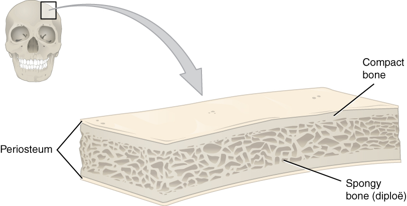

 The human skull is the bone structure that forms the
 The skull also contains sinuses, air-filled cavities known as paranasal sinuses, and numerous foramina. The sinuses are lined with respiratory epithelium. Their known functions are the lessening of the weight of the skull, the aiding of resonance to the voice and the warming and moistening of the air drawn into the nasal cavity.
The foramina are openings in the skull. The largest of these is the


 The temporal
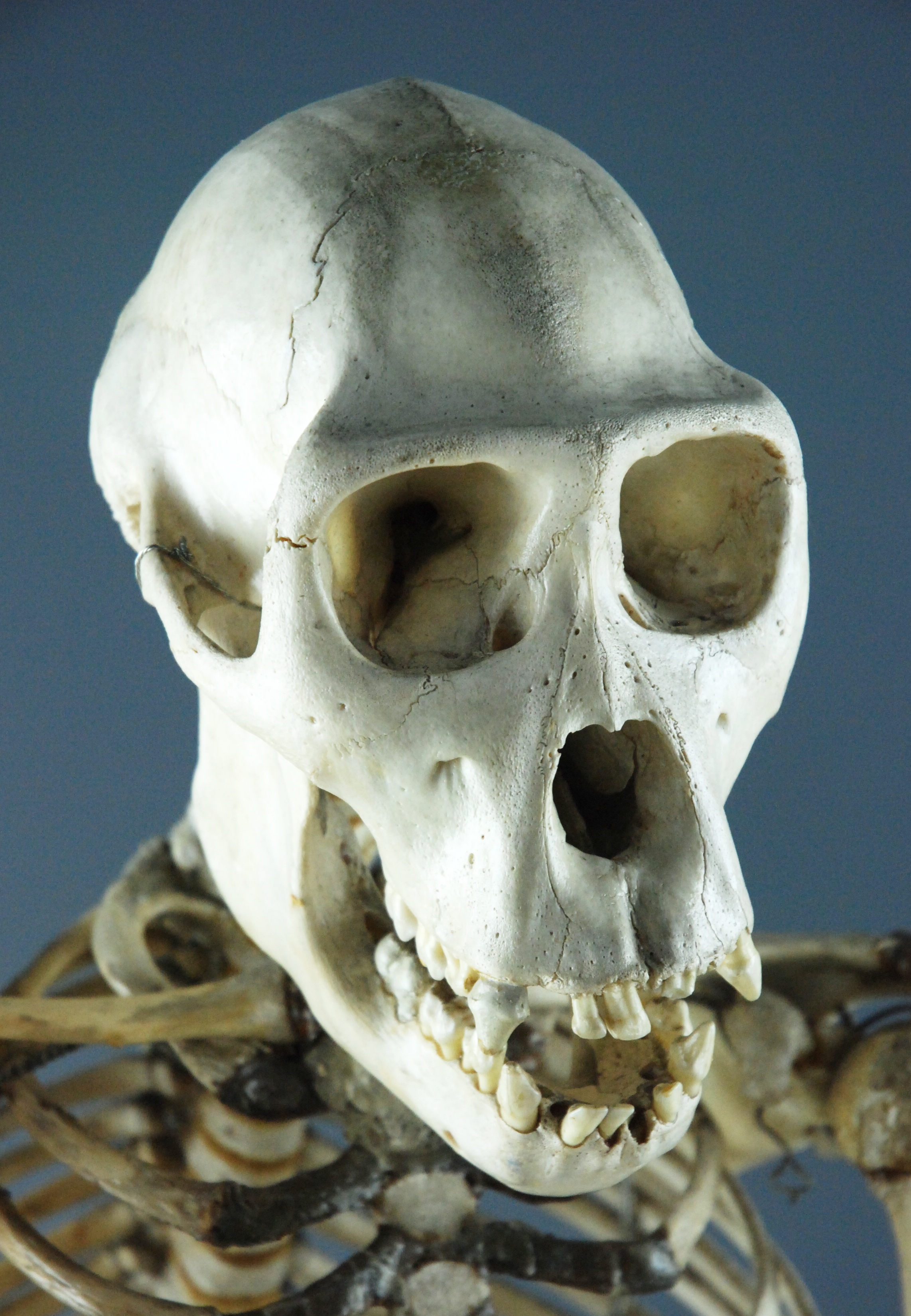
 There are four types of amniote skull, classified by the number and location of their temporal fenestrae. These are:
* Anapsida – no openings
* Synapsida – one low opening (beneath the postorbital and squamosal bones)
* Euryapsida – one high opening (above the postorbital and squamosal bones); euryapsids actually evolved from a diapsid configuration, losing their lower temporal fenestra.
* Diapsida – two openings
Evolutionarily, they are related as follows:
* Amniota
**Class Synapsida
***Order Therapsida
****Class Mammalia – mammals
**(Unranked)
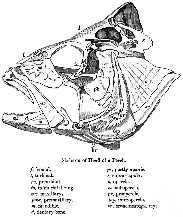
 The skull of fishes is formed from a series of only loosely connected bones. Lampreys and sharks only possess a cartilaginous endocranium, with both the upper and lower jaws being separate elements. Bony fishes have additional dermal bone, forming a more or less coherent
 The skulls of the earliest tetrapods closely resembled those of their
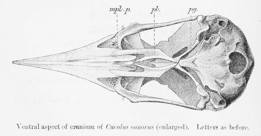 Birds have a diapsid skull, as in reptiles, with a prelacrimal fossa (present in some reptiles). The skull has a single occipital condyle. The skull consists of five major bones: the frontal (top of head), parietal (back of head), premaxillary and nasal (top beak), and the mandible (bottom beak). The skull of a normal bird usually weighs about 1% of the bird's total bodyweight. The eye occupies a considerable amount of the skull and is surrounded by a sclerotic eye-ring, a ring of tiny bones. This characteristic is also seen in reptiles.
 Living
 The skull is a complex structure; its bones are formed both by intramembranous and
WPATH Clarification on Medical Necessity of Treatment, Sex Reassignment, and Insurance Coverage in the U.S.A.
(2008).World Professional Association for Transgender Health.
Standards of Care for the Health of Transsexual, Transgender, and Gender Nonconforming People, Version 7.
'' pg. 58 (2011).
 Artificial cranial deformation is a largely historical practice of some cultures. Cords and wooden boards would be used to apply pressure to an infant's skull and alter its shape, sometimes quite significantly. This procedure would begin just after birth and would be carried on for several years.
facial skeleton
The facial skeleton comprises the ''facial bones'' that may attach to build a portion of the skull. The remainder of the skull is the braincase.
In human anatomy and development, the facial skeleton is sometimes called the ''membranous viscerocr ...
) that includes the mandible as its largest bone. The skull forms the anterior-most portion of the skeleton
A skeleton is the structural frame that supports the body of an animal. There are several types of skeletons, including the exoskeleton, which is the stable outer shell of an organism, the endoskeleton, which forms the support structure inside ...
and is a product of cephalisation
Cephalization is an evolutionary trend in which, over many generations, the mouth, sense organs, and nerve ganglia become concentrated at the front end of an animal, producing a head region. This is associated with movement and bilateral symmetr ...
—housing the brain, and several sensory
Sensory may refer to:
Biology
* Sensory ecology, how organisms obtain information about their environment
* Sensory neuron, nerve cell responsible for transmitting information about external stimuli
* Sensory perception, the process of acquiri ...
structures such as the eyes, ears, nose, and mouth. In humans these sensory structures are part of the facial skeleton.
Functions of the skull include protection of the brain, fixing the distance between the eyes to allow stereoscopic vision, and fixing the position of the ears to enable sound localisation of the direction and distance of sounds. In some animals, such as horned ungulates (mammals with hooves), the skull also has a defensive function by providing the mount (on the frontal bone) for the horns.
The English word ''skull'' is probably derived from Old Norse , while the Latin word comes from the Greek root (). The human skull fully develops two years after birth.The junctions of the skull bones are joined by structures called sutures.
The skull is made up of a number of fused flat bones, and contains many foramina, fossae, processes
A process is a series or set of activities that interact to produce a result; it may occur once-only or be recurrent or periodic.
Things called a process include:
Business and management
*Business process, activities that produce a specific se ...
, and several cavities or sinuses. In zoology there are openings in the skull called fenestrae.
Structure
Humans


head
A head is the part of an organism which usually includes the ears, brain, forehead, cheeks, chin, eyes, nose, and mouth, each of which aid in various sensory functions such as sight, hearing, smell, and taste. Some very simple animals may ...
in the human skeleton
The human skeleton is the internal framework of the human body. It is composed of around 270 bones at birth – this total decreases to around 206 bones by adulthood after some bones get fused together. The bone mass in the skeleton makes up a ...
. It supports the structures of the face and forms a cavity for the brain. Like the skulls of other vertebrates, it protects the brain from injury.
The skull consists of three parts, of different embryological origin—the neurocranium, the sutures, and the facial skeleton
The facial skeleton comprises the ''facial bones'' that may attach to build a portion of the skull. The remainder of the skull is the braincase.
In human anatomy and development, the facial skeleton is sometimes called the ''membranous viscerocr ...
(also called the ''membraneous viscerocranium''). The neurocranium (or ''braincase'') forms the protective cranial cavity
The cranial cavity, also known as intracranial space, is the space within the skull that accommodates the brain. The skull minus the mandible is called the ''cranium''. The cavity is formed by eight cranial bones known as the neurocranium that in ...
that surrounds and houses the brain and brainstem
The brainstem (or brain stem) is the posterior stalk-like part of the brain that connects the cerebrum with the spinal cord. In the human brain the brainstem is composed of the midbrain, the pons, and the medulla oblongata. The midbrain is cont ...
. The upper areas of the cranial bones form the calvaria (skullcap). The membranous viscerocranium includes the mandible.
The sutures are fairly rigid joints between bones of the neurocranium.
The facial skeleton is formed by the bones supporting the face.
Bones
Except for the mandible, all of the bones of the skull are joined by sutures— synarthrodial (immovable) joints formed by bony ossification, withSharpey's fibres
Sharpey's fibres (bone fibres, or perforating fibres) are a Matrix (biology), matrix of connective tissue consisting of bundles of strong predominantly type I Collagen, collagen fibres connecting periosteum to bone. They are part of the outer fibr ...
permitting some flexibility. Sometimes there can be extra bone pieces within the suture known as wormian bones or ''sutural bones''. Most commonly these are found in the course of the lambdoid suture.
The human skull is generally considered to consist of twenty-two bones—eight cranial bones and fourteen facial skeleton bones. In the neurocranium these are the occipital bone
The occipital bone () is a neurocranium, cranial dermal bone and the main bone of the occiput (back and lower part of the skull). It is trapezoidal in shape and curved on itself like a shallow dish. The occipital bone overlies the occipital lobe ...
, two temporal bones, two parietal bones, the sphenoid, ethmoid and frontal bones.
The bones of the facial skeleton
The facial skeleton comprises the ''facial bones'' that may attach to build a portion of the skull. The remainder of the skull is the braincase.
In human anatomy and development, the facial skeleton is sometimes called the ''membranous viscerocr ...
(14) are the vomer, two inferior nasal conchae, two nasal bone
The nasal bones are two small oblong bones, varying in size and form in different individuals; they are placed side by side at the middle and upper part of the face and by their junction, form the bridge of the upper one third of the nose.
Eac ...
s, two maxilla, the mandible, two palatine bone
In anatomy, the palatine bones () are two irregular bones of the facial skeleton in many animal species, located above the uvula in the throat. Together with the maxillae, they comprise the hard palate. (''Palate'' is derived from the Latin ''pa ...
s, two zygomatic bones, and two lacrimal bones. Some sources count a paired bone as one, or the maxilla as having two bones (as its parts); some sources include the hyoid bone
The hyoid bone (lingual bone or tongue-bone) () is a horseshoe-shaped bone situated in the anterior midline of the neck between the chin and the thyroid cartilage. At rest, it lies between the base of the mandible and the third cervical vertebr ...
or the three ossicles
The ossicles (also called auditory ossicles) are three bones in either middle ear that are among the smallest bones in the human body. They serve to transmit sounds from the air to the fluid-filled labyrinth (cochlea). The absence of the auditory ...
of the middle ear but the overall general consensus of the number of bones in the human skull is the stated twenty-two.
Some of these bones—the occipital, parietal, frontal, in the neurocranium, and the nasal, lacrimal, and vomer, in the facial skeleton are flat bones.
Cavities and foramina
 The skull also contains sinuses, air-filled cavities known as paranasal sinuses, and numerous foramina. The sinuses are lined with respiratory epithelium. Their known functions are the lessening of the weight of the skull, the aiding of resonance to the voice and the warming and moistening of the air drawn into the nasal cavity.
The foramina are openings in the skull. The largest of these is the
 foramen magnum
The foramen magnum ( la, great hole) is a large, oval-shaped opening in the occipital bone of the skull. It is one of the several oval or circular openings (foramina) in the base of the skull. The spinal cord, an extension of the medulla oblon ...
that allows the passage of the spinal cord as well as nerve
A nerve is an enclosed, cable-like bundle of nerve fibers (called axons) in the peripheral nervous system.
A nerve transmits electrical impulses. It is the basic unit of the peripheral nervous system. A nerve provides a common pathway for the e ...
s and blood vessels.
Processes
The manyprocesses
A process is a series or set of activities that interact to produce a result; it may occur once-only or be recurrent or periodic.
Things called a process include:
Business and management
*Business process, activities that produce a specific se ...
of the skull include the mastoid process and the zygomatic processes.
Other vertebrates
Fenestrae
 The temporal
 fenestrae
A fenestra (fenestration; plural fenestrae or fenestrations) is any small opening or pore, commonly used as a term in the biological sciences. It is the Latin word for "window", and is used in various fields to describe a pore in an anatomical st ...
are anatomical features of the skulls of several types of amniote
Amniotes are a clade of tetrapod vertebrates that comprises sauropsids (including all reptiles and birds, and extinct parareptiles and non-avian dinosaurs) and synapsids (including pelycosaurs and therapsids such as mammals). They are disti ...
s, characterised by bilaterally symmetrical holes (fenestrae) in the temporal bone. Depending on the lineage of a given animal, two, one, or no pairs of temporal fenestrae may be present, above or below the postorbital and squamosal bones. The upper temporal fenestrae are also known as the supratemporal fenestrae, and the lower temporal fenestrae are also known as the infratemporal fenestrae. The presence and morphology of the temporal fenestra are critical for taxonomic classification of the synapsids, of which mammals are part.
Physiological speculation associates it with a rise in metabolic rates and an increase in jaw musculature. The earlier amniotes of the Carboniferous did not have temporal fenestrae but two more advanced lines did: the synapsids (mammal-like reptiles) and the diapsids (most reptiles and later birds). As time progressed, diapsids' and synapsids' temporal fenestrae became more modified and larger to make stronger bites and more jaw muscles. Dinosaurs, which are diapsids, have large advanced openings, and their descendants, the birds, have temporal fenestrae which have been modified. Synapsids, possess one fenestral opening in the skull, situated to the rear of the orbit. In their descendants, the cynodonts, the orbit fused with the fenestral opening after the latter had started expanding within the therapsids. Thus most mammals also have this. Later, primates separated their orbit from '' temporal fossa'' by the postorbital bar with haplorhines
Haplorhini (), the haplorhines (Greek for "simple-nosed") or the "dry-nosed" primates, is a suborder of primates containing the tarsiers and the simians (Simiiformes or anthropoids), as sister of the Strepsirrhini ("moist-nosed"). The name is some ...
later evolving the postorbital septum
The ''postorbital'' is one of the bones in vertebrate skulls which forms a portion of the dermal skull roof and, sometimes, a ring about the orbit. Generally, it is located behind the postfrontal and posteriorly to the orbital fenestra. In some ve ...
.
=Classification
=
 There are four types of amniote skull, classified by the number and location of their temporal fenestrae. These are:
* Anapsida – no openings
* Synapsida – one low opening (beneath the postorbital and squamosal bones)
* Euryapsida – one high opening (above the postorbital and squamosal bones); euryapsids actually evolved from a diapsid configuration, losing their lower temporal fenestra.
* Diapsida – two openings
Evolutionarily, they are related as follows:
* Amniota
**Class Synapsida
***Order Therapsida
****Class Mammalia – mammals
**(Unranked)
 Sauropsida
Sauropsida ("lizard faces") is a clade of amniotes, broadly equivalent to the class Reptilia. Sauropsida is the sister taxon to Synapsida, the other clade of amniotes which includes mammals as its only modern representatives. Although early syna ...
– reptiles and birds
***Class Reptilia
****Subclass Parareptilia
*****Infraclass Anapsida
****Subclass Eureptilia
*****Infraclass Diapsida
******Class Aves
Birds are a group of warm-blooded vertebrates constituting the class Aves (), characterised by feathers, toothless beaked jaws, the laying of hard-shelled eggs, a high metabolic rate, a four-chambered heart, and a strong yet lightweigh ...
*****Infraclass Euryapsida
Bones
The jugal is a skull bone found in most reptiles, amphibians, and birds. In mammals, the jugal is often called the zygomatic bone or malar bone. The prefrontal bone is a bone separating the lacrimal and frontal bones in many tetrapod skulls.Fish

 The skull of fishes is formed from a series of only loosely connected bones. Lampreys and sharks only possess a cartilaginous endocranium, with both the upper and lower jaws being separate elements. Bony fishes have additional dermal bone, forming a more or less coherent
 skull roof
The skull roof, or the roofing bones of the skull, are a set of bones covering the brain, eyes and nostrils in bony fishes and all land-living vertebrates. The bones are derived from dermal bone and are part of the dermatocranium.
In comparati ...
in lungfish and holost
Holostei is a group of ray-finned bony fish. It is divided into two major clades, the Halecomorphi, represented by a single living species, the bowfin (''Amia calva''), as well as the Ginglymodi, the sole living representatives being the gars (Le ...
fish. The lower jaw defines a chin.
The simpler structure is found in jawless fish, in which the cranium is normally represented by a trough-like basket of cartilaginous elements only partially enclosing the brain, and associated with the capsules for the inner ears and the single nostril. Distinctively, these fish have no jaws.
Cartilaginous fish
Chondrichthyes (; ) is a class that contains the cartilaginous fishes that have skeletons primarily composed of cartilage. They can be contrasted with the Osteichthyes or ''bony fishes'', which have skeletons primarily composed of bone tissue ...
, such as sharks and rays, have also simple, and presumably primitive, skull structures. The cranium is a single structure forming a case around the brain, enclosing the lower surface and the sides, but always at least partially open at the top as a large fontanelle
A fontanelle (or fontanel) (colloquially, soft spot) is an anatomical feature of the infant human skull comprising soft membranous gaps ( sutures) between the cranial bones that make up the calvaria of a fetus or an infant. Fontanelles allow f ...
. The most anterior part of the cranium includes a forward plate of cartilage, the rostrum, and capsules to enclose the olfactory
The sense of smell, or olfaction, is the special sense through which smells (or odors) are perceived. The sense of smell has many functions, including detecting desirable foods, hazards, and pheromones, and plays a role in taste.
In humans, it ...
organs. Behind these are the orbits, and then an additional pair of capsules enclosing the structure of the inner ear
The inner ear (internal ear, auris interna) is the innermost part of the vertebrate ear. In vertebrates, the inner ear is mainly responsible for sound detection and balance. In mammals, it consists of the bony labyrinth, a hollow cavity in the ...
. Finally, the skull tapers towards the rear, where the foramen magnum lies immediately above a single condyle, articulating with the first vertebra. There are, in addition, at various points throughout the cranium, smaller foramina for the cranial nerves. The jaws consist of separate hoops of cartilage, almost always distinct from the cranium proper.
In ray-finned fish, there has also been considerable modification from the primitive pattern. The roof of the skull is generally well formed, and although the exact relationship of its bones to those of tetrapods is unclear, they are usually given similar names for convenience. Other elements of the skull, however, may be reduced; there is little cheek region behind the enlarged orbits, and little, if any bone in between them. The upper jaw is often formed largely from the premaxilla, with the maxilla itself located further back, and an additional bone, the symplectic, linking the jaw to the rest of the cranium.
Although the skulls of fossil lobe-finned fish resemble those of the early tetrapods, the same cannot be said of those of the living lungfishes. The skull roof
The skull roof, or the roofing bones of the skull, are a set of bones covering the brain, eyes and nostrils in bony fishes and all land-living vertebrates. The bones are derived from dermal bone and are part of the dermatocranium.
In comparati ...
is not fully formed, and consists of multiple, somewhat irregularly shaped bones with no direct relationship to those of tetrapods. The upper jaw is formed from the pterygoids and vomers alone, all of which bear teeth. Much of the skull is formed from cartilage
Cartilage is a resilient and smooth type of connective tissue. In tetrapods, it covers and protects the ends of long bones at the joints as articular cartilage, and is a structural component of many body parts including the rib cage, the neck an ...
, and its overall structure is reduced.
Tetrapods
 The skulls of the earliest tetrapods closely resembled those of their
 ancestor
An ancestor, also known as a forefather, fore-elder or a forebear, is a parent or (recursively) the parent of an antecedent (i.e., a grandparent, great-grandparent, great-great-grandparent and so forth). ''Ancestor'' is "any person from whom ...
s amongst the lobe-finned fishes. The skull roof
The skull roof, or the roofing bones of the skull, are a set of bones covering the brain, eyes and nostrils in bony fishes and all land-living vertebrates. The bones are derived from dermal bone and are part of the dermatocranium.
In comparati ...
is formed of a series of plate-like bones, including the maxilla, frontals, parietals, and lacrimals, among others. It is overlaying the endocranium
The endocranium in comparative anatomy is a part of the skull base in vertebrates and it represents the basal, inner part of the cranium. The term is also applied to the outer layer of the dura mater in human anatomy.
Structure
Structurally, t ...
, corresponding to the cartilaginous skull in sharks and rays. The various separate bones that compose the temporal bone of humans are also part of the skull roof series. A further plate composed of four pairs of bones forms the roof of the mouth; these include the vomer and palatine bone
In anatomy, the palatine bones () are two irregular bones of the facial skeleton in many animal species, located above the uvula in the throat. Together with the maxillae, they comprise the hard palate. (''Palate'' is derived from the Latin ''pa ...
s. The base of the cranium is formed from a ring of bones surrounding the foramen magnum
The foramen magnum ( la, great hole) is a large, oval-shaped opening in the occipital bone of the skull. It is one of the several oval or circular openings (foramina) in the base of the skull. The spinal cord, an extension of the medulla oblon ...
and a median bone lying further forward; these are homologous
Homology may refer to:
Sciences
Biology
*Homology (biology), any characteristic of biological organisms that is derived from a common ancestor
*Sequence homology, biological homology between DNA, RNA, or protein sequences
* Homologous chrom ...
with the occipital bone
The occipital bone () is a neurocranium, cranial dermal bone and the main bone of the occiput (back and lower part of the skull). It is trapezoidal in shape and curved on itself like a shallow dish. The occipital bone overlies the occipital lobe ...
and parts of the sphenoid in mammals. Finally, the lower jaw is composed of multiple bones, only the most anterior of which (the dentary) is homologous with the mammalian mandible.
In living tetrapods, a great many of the original bones have either disappeared or fused into one another in various arrangements.
Birds
 Birds have a diapsid skull, as in reptiles, with a prelacrimal fossa (present in some reptiles). The skull has a single occipital condyle. The skull consists of five major bones: the frontal (top of head), parietal (back of head), premaxillary and nasal (top beak), and the mandible (bottom beak). The skull of a normal bird usually weighs about 1% of the bird's total bodyweight. The eye occupies a considerable amount of the skull and is surrounded by a sclerotic eye-ring, a ring of tiny bones. This characteristic is also seen in reptiles.
Amphibians
 Living
 amphibian
Amphibians are tetrapod, four-limbed and ectothermic vertebrates of the Class (biology), class Amphibia. All living amphibians belong to the group Lissamphibia. They inhabit a wide variety of habitats, with most species living within terres ...
s typically have greatly reduced skulls, with many of the bones either absent or wholly or partly replaced by cartilage. In mammals and birds, in particular, modifications of the skull occurred to allow for the expansion of the brain. The fusion between the various bones is especially notable in birds, in which the individual structures may be difficult to identify.
Development
 The skull is a complex structure; its bones are formed both by intramembranous and
 endochondral ossification
Endochondral ossification is one of the two essential processes during fetal development of the mammalian skeletal system by which bone tissue is produced. Unlike intramembranous ossification, the other process by which bone tissue is produced, c ...
. The skull roof
The skull roof, or the roofing bones of the skull, are a set of bones covering the brain, eyes and nostrils in bony fishes and all land-living vertebrates. The bones are derived from dermal bone and are part of the dermatocranium.
In comparati ...
bones, comprising the bones of the facial skeleton
The facial skeleton comprises the ''facial bones'' that may attach to build a portion of the skull. The remainder of the skull is the braincase.
In human anatomy and development, the facial skeleton is sometimes called the ''membranous viscerocr ...
and the sides and roof of the neurocranium, are dermal bones formed by intramembranous ossification, though the temporal bones are formed by endochondral ossification. The endocranium
The endocranium in comparative anatomy is a part of the skull base in vertebrates and it represents the basal, inner part of the cranium. The term is also applied to the outer layer of the dura mater in human anatomy.
Structure
Structurally, t ...
, the bones supporting the brain (the occipital, sphenoid, and ethmoid) are largely formed by endochondral ossification. Thus frontal and parietal bones are purely membranous. The geometry of the skull base and its fossae, the anterior
Standard anatomical terms of location are used to unambiguously describe the anatomy of animals, including humans. The terms, typically derived from Latin or Greek roots, describe something in its standard anatomical position. This position prov ...
, middle
Middle or The Middle may refer to:
* Centre (geometry), the point equally distant from the outer limits.
Places
* Middle (sheading), a subdivision of the Isle of Man
* Middle Bay (disambiguation)
* Middle Brook (disambiguation)
* Middle Creek (d ...
and posterior cranial fossae changes rapidly. The anterior cranial fossa changes especially during the first trimester of pregnancy and skull defects can often develop during this time.
At birth, the human skull is made up of 44 separate bony elements. During development, many of these bony elements gradually fuse together into solid bone (for example, the frontal bone). The bones of the roof of the skull are initially separated by regions of dense connective tissue
Connective tissue is one of the four primary types of animal tissue, along with epithelial tissue, muscle tissue, and nervous tissue. It develops from the mesenchyme derived from the mesoderm the middle embryonic germ layer. Connective tiss ...
called fontanelles. There are six fontanelles: one anterior (or frontal), one posterior (or occipital), two sphenoid (or anterolateral), and two mastoid (or posterolateral). At birth, these regions are fibrous and moveable, necessary for birth and later growth. This growth can put a large amount of tension on the "obstetrical hinge", which is where the squamous and lateral parts of the occipital bone
The occipital bone () is a neurocranium, cranial dermal bone and the main bone of the occiput (back and lower part of the skull). It is trapezoidal in shape and curved on itself like a shallow dish. The occipital bone overlies the occipital lobe ...
meet. A possible complication of this tension is rupture of the great cerebral vein. As growth and ossification progress, the connective tissue of the fontanelles is invaded and replaced by bone creating sutures. The five sutures are the two squamous sutures, one coronal, one lambdoid, and one sagittal suture. The posterior fontanelle usually closes by eight weeks, but the anterior fontanel can remain open up to eighteen months. The anterior fontanelle is located at the junction of the frontal and parietal bones; it is a "soft spot" on a baby's forehead. Careful observation will show that you can count a baby's heart rate by observing the pulse pulsing softly through the anterior fontanelle.
The skull in the neonate is large in proportion to other parts of the body. The facial skeleton is one seventh of the size of the calvaria. (In the adult it is half the size). The base of the skull is short and narrow, though the inner ear
The inner ear (internal ear, auris interna) is the innermost part of the vertebrate ear. In vertebrates, the inner ear is mainly responsible for sound detection and balance. In mammals, it consists of the bony labyrinth, a hollow cavity in the ...
is almost adult size.
Clinical significance
Craniosynostosis
Craniosynostosis is a condition in which one or more of the fibrous sutures in a young infant's skull prematurely fuses by turning into bone (ossification), thereby changing the growth pattern of the skull. Because the skull cannot expand perpe ...
is a condition in which one or more of the fibrous sutures in an infant skull prematurely fuses, and changes the growth pattern of the skull. Because the skull cannot expand perpendicular to the fused suture, it grows more in the parallel direction. Sometimes the resulting growth pattern provides the necessary space for the growing brain, but results in an abnormal head shape and abnormal facial features. In cases in which the compensation does not effectively provide enough space for the growing brain, craniosynostosis results in increased intracranial pressure leading possibly to visual impairment, sleeping impairment, eating difficulties, or an impairment of mental development.
A copper beaten skull Copper beaten skull is a phenomenon wherein intense intracranial pressure disfigures the internal surface of the skull.http://radiopaedia.org/articles/copper-beaten-skull The name comes from the fact that the inner skull has the appearance of havi ...
is a phenomenon wherein intense intracranial pressure disfigures the internal surface of the skull. The name comes from the fact that the inner skull has the appearance of having been beaten with a ball-peen hammer, such as is often used by coppersmiths. The condition is most common in children.
Injuries and treatment
Injuries to the brain can be life-threatening. Normally the skull protects the brain from damage through its hard unyieldingness; the skull is one of the least deformable structures found in nature with it needing the force of about 1 ton to reduce the diameter of the skull by 1 cm. In some cases, however, of head injury, there can be raised intracranial pressure through mechanisms such as asubdural haematoma
A subdural hematoma (SDH) is a type of bleeding in which a collection of blood—usually but not always associated with a traumatic brain injury—gathers between the inner layer of the dura mater and the arachnoid mater of the meninges surround ...
. In these cases the raised intracranial pressure can cause herniation of the brain out of the foramen magnum
The foramen magnum ( la, great hole) is a large, oval-shaped opening in the occipital bone of the skull. It is one of the several oval or circular openings (foramina) in the base of the skull. The spinal cord, an extension of the medulla oblon ...
("coning") because there is no space for the brain to expand; this can result in significant brain damage
Neurotrauma, brain damage or brain injury (BI) is the destruction or degeneration of brain cells. Brain injuries occur due to a wide range of internal and external factors. In general, brain damage refers to significant, undiscriminating t ...
or death unless an urgent operation is performed to relieve the pressure. This is why patients with concussion
A concussion, also known as a mild traumatic brain injury (mTBI), is a head injury that temporarily affects brain functioning. Symptoms may include loss of consciousness (LOC); memory loss; headaches; difficulty with thinking, concentration, ...
must be watched extremely carefully. Repeated concussions can activate the structure of skull bones as the brain's protective covering.
Dating back to Neolithic times, a skull operation called trepanning was sometimes performed. This involved drilling a ''burr'' hole in the cranium. Examination of skulls from this period reveals that the patients sometimes survived for many years afterward. It seems likely that trepanning was also performed purely for ritualistic or religious reasons. Nowadays this procedure is still used but is normally called a craniectomy.
In March 2013, for the first time in the U.S., researchers replaced a large percentage of a patient's skull with a precision, 3D-printed
3D printing or additive manufacturing is the construction of a three-dimensional object from a CAD model or a digital 3D model. It can be done in a variety of processes in which material is deposited, joined or solidified under computer co ...
polymer implant
Implant can refer to:
Medicine
*Implant (medicine), or specifically:
** Brain implant
** Breast implant
**Buttock implant
**Cochlear implant
**Contraceptive implant
**Dental implant
** Fetal tissue implant
**Implantable cardioverter-defibrillator ...
. About 9 months later, the first complete cranium replacement with a 3D-printed plastic insert was performed on a Dutch woman. She had been suffering from hyperostosis, which increased the thickness of her skull and compressed her brain.
A study conducted in 2018 by the researchers of Harvard Medical School in Boston, funded by National Institutes of Health (NIH), suggested that instead of travelling via blood, there are "tiny channels" in the skull through which the immune cells combined with the bone marrow
Bone marrow is a semi-solid tissue found within the spongy (also known as cancellous) portions of bones. In birds and mammals, bone marrow is the primary site of new blood cell production (or haematopoiesis). It is composed of hematopoietic ce ...
reach the areas of inflammation after an injury to the brain tissues.
Transgender procedures
Surgical alteration of sexually dimorphic skull features may be carried out as a part of facial feminization surgery, a set of reconstructive surgical procedures that can alter male facial features to bring them closer in shape and size to typical female facial features. These procedures can be an important part of the treatment of transgender people forgender dysphoria
Gender dysphoria (GD) is the distress a person experiences due to a mismatch between their gender identitytheir personal sense of their own genderand their sex assigned at birth. The diagnostic label gender identity disorder (GID) was used until ...
.World Professional Association for Transgender HealthWPATH Clarification on Medical Necessity of Treatment, Sex Reassignment, and Insurance Coverage in the U.S.A.
(2008).World Professional Association for Transgender Health.
Standards of Care for the Health of Transsexual, Transgender, and Gender Nonconforming People, Version 7.
'' pg. 58 (2011).
Society and culture
Osteology
Like the face, the skull and teeth can also indicate a person's life history and origin. Forensic scientists andarchaeologist
Archaeology or archeology is the scientific study of human activity through the recovery and analysis of material culture. The archaeological record consists of artifacts, architecture, biofacts or ecofacts, sites, and cultural landscap ...
s use quantitative and qualitative traits to estimate what the bearer of the skull looked like. When a significant amount of bones are found, such as at Spitalfields
Spitalfields is a district in the East End of London and within the London Borough of Tower Hamlets. The area is formed around Commercial Street (on the A1202 London Inner Ring Road) and includes the locale around Brick Lane, Christ Church, ...
in the UK and Jōmon shell mounds in Japan, osteologists can use traits, such as the proportions of length, height and width, to know the relationships of the population of the study with other living or extinct populations.
The German physician Franz Joseph Gall in around 1800 formulated the theory of phrenology
Phrenology () is a pseudoscience which involves the measurement of bumps on the skull to predict mental traits.Wihe, J. V. (2002). "Science and Pseudoscience: A Primer in Critical Thinking." In ''Encyclopedia of Pseudoscience'', pp. 195–203. C ...
, which attempted to show that specific features of the skull are associated with certain personality traits or intellectual capabilities of its owner. His theory is now considered to be pseudoscientific
Pseudoscience consists of statements, beliefs, or practices that claim to be both scientific and factual but are incompatible with the scientific method. Pseudoscience is often characterized by contradictory, exaggerated or unfalsifiable claim ...
.
Sexual dimorphism
In the mid-nineteenth century,anthropologist
An anthropologist is a person engaged in the practice of anthropology. Anthropology is the study of aspects of humans within past and present societies. Social anthropology, cultural anthropology and philosophical anthropology study the norms and ...
s found it crucial to distinguish between male and female skulls. An anthropologist of the time, James McGrigor Allan
James McGrigor Allan (1827, Bristol - 1916, Epsom) was a British anthropologist and writer.
Biography
McGrigor was the son of Colin Allan, at one time chief medical officer of Halifax, Nova Scotia, and Jane Gibbon. He opposed women's right to vot ...
, argued that the female brain was similar to that of an animal. This allowed anthropologists to declare that women were in fact more emotional and less rational than men. McGrigor then concluded that women's brains were more analogous to infants, thus deeming them inferior at the time. To further these claims of female inferiority and silence the feminists of the time, other anthropologists joined in on the studies of the female skull. These cranial measurements are the basis of what is known as craniology. These cranial measurements were also used to draw a connection between women and black people.
Research has shown that while in early life there is little difference between male and female skulls, in adulthood male skulls tend to be larger and more robust than female skulls, which are lighter and smaller, with a cranial capacity about 10 percent less than that of the male. However, later studies show that women's skulls are slightly thicker and thus men may be more susceptible to head injury than women. However, other studies shows that men's skulls are slightly thicker in certain areas. As well as some studies showing that females are more susceptible to head injury (concussion) than males. Men's skulls have also been shown to maintain density with age, which may aid in preventing head injury, while women's skull density slightly decreases with age.
Male skulls can have more prominent supraorbital ridge
The brow ridge, or supraorbital ridge known as superciliary arch in medicine, is a bony ridge located above the eye sockets of all primates. In humans, the eyebrows are located on their lower margin.
Structure
The brow ridge is a nodule or crest ...
s, a more prominent glabella, and more prominent temporal lines
The parietal bones () are two bones in the Human skull, skull which, when joined at a fibrous joint, form the sides and roof of the Human skull, cranium. In humans, each bone is roughly quadrilateral in form, and has two surfaces, four borders, an ...
. Female skulls generally have rounder orbits, and narrower jaws. Male skulls on average have larger, broader palates, squarer orbits, larger mastoid processes, larger sinuses, and larger occipital condyles than those of females. Male mandibles typically have squarer chins and thicker, rougher muscle attachments than female mandibles.
Craniometry
The cephalic index is the ratio of the width of the head, multiplied by 100 and divided by its length (front to back). The index is also used to categorize animals, especially dogs and cats. The width is usually measured just below the parietal eminence, and the length from the glabella to the occipital point. Humans may be: * ''Dolichocephalic'' — long-headed * ''Mesaticephalic'' — medium-headed * ''Brachycephalic'' — short-headedTerminology
*Chondrocranium
The chondrocranium (or ''cartilaginous neurocranium'') is the primitive cartilage, cartilaginous skeleton, skeletal structure of the fetal skull that grows to envelop the rapidly growing embryonic brain.Salentijn, L. ''Biology of Mineralized Tissue ...
, a primitive cartilaginous skeletal structure
* Endocranium
The endocranium in comparative anatomy is a part of the skull base in vertebrates and it represents the basal, inner part of the cranium. The term is also applied to the outer layer of the dura mater in human anatomy.
Structure
Structurally, t ...
* Epicranium
* Pericranium, a membrane that lines the outer surface of the cranium
History
Trepanning, a practice in which a hole is created in the skull, has been described as the oldest surgical procedure for which there isarchaeological
Archaeology or archeology is the scientific study of human activity through the recovery and analysis of material culture. The archaeological record consists of artifacts, architecture, biofacts or ecofacts, sites, and cultural landscap ...
evidence, found in the forms of cave paintings and human remains. At one burial site in France dated to 6500 BCE, 40 out of 120 prehistoric
Prehistory, also known as pre-literary history, is the period of human history between the use of the first stone tools by hominins 3.3 million years ago and the beginning of recorded history with the invention of writing systems. The use of ...
skulls found had trepanation holes.
Additional images
See also
* Craniometry * Crystal skull * Head and neck anatomy * Human skull symbolism *Memento mori
''Memento mori'' (Latin for 'remember that you ave todie'