|
Great Cerebral Vein
The great cerebral vein is one of the large blood vessels in the skull draining the cerebrum of the brain. It is also known as the "vein of Galen", named for its discoverer, the Greek physician Galen. However, it is not the only vein with this eponym. Structure The great cerebral vein is considered one of the deep cerebral veins. Other deep cerebral veins are the internal cerebral veins, formed by the union of the superior thalamostriate vein and the superior choroid vein at the interventricular foramina. The internal cerebral veins can be seen on the superior surfaces of the caudate nuclei and thalami just under the corpus callosum. The veins at the anterior poles of the thalami merge posterior to the pineal gland to form the great cerebral vein. Most of the blood in the deep cerebral veins collects into the great cerebral vein. This comes from the inferior side of the posterior end of the corpus callosum and empties ie similarities, there are also differences between these ... [...More Info...] [...Related Items...] OR: [Wikipedia] [Google] [Baidu] |
Telencephalon
The cerebrum, telencephalon or endbrain is the largest part of the brain containing the cerebral cortex (of the two cerebral hemispheres), as well as several subcortical structures, including the hippocampus, basal ganglia, and olfactory bulb. In the human brain, the cerebrum is the uppermost region of the central nervous system. The cerebrum develops prenatally from the forebrain (prosencephalon). In mammals, the dorsal telencephalon, or pallium, develops into the cerebral cortex, and the ventral telencephalon, or subpallium, becomes the basal ganglia. The cerebrum is also divided into approximately symmetric left and right cerebral hemispheres. With the assistance of the cerebellum, the cerebrum controls all voluntary actions in the human body. Structure The cerebrum is the largest part of the brain. Depending upon the position of the animal it lies either in front or on top of the brainstem. In humans, the cerebrum is the largest and best-developed of the five major divi ... [...More Info...] [...Related Items...] OR: [Wikipedia] [Google] [Baidu] |
Caudate Nuclei
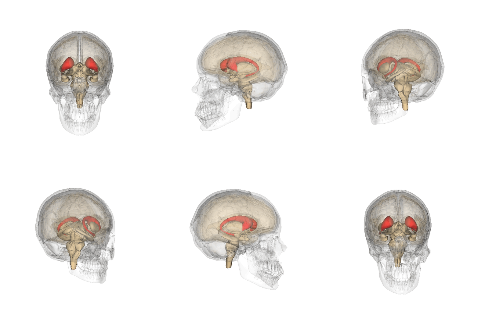
The caudate nucleus is one of the structures that make up the corpus striatum, which is a component of the basal ganglia in the human brain. While the caudate nucleus has long been associated with motor processes due to its role in Parkinson's disease, it plays important roles in various other nonmotor functions as well, including procedural learning, associative learning and inhibitory control of action, among other functions. The caudate is also one of the brain structures which compose the reward system and functions as part of the Prefrontal cortex, cortico–basal ganglia–thalamus, thalamic loop. Structure Together with the putamen, the caudate forms the dorsal striatum, which is considered a single functional structure; anatomically, it is separated by a large white matter tract, the internal capsule, so it is sometimes also referred to as two structures: the medial dorsal striatum (the caudate) and the lateral dorsal striatum (the putamen). In this vein, the two are f ... [...More Info...] [...Related Items...] OR: [Wikipedia] [Google] [Baidu] |
Congenital Disorder
A birth defect, also known as a congenital disorder, is an abnormal condition that is present at birth regardless of its cause. Birth defects may result in disabilities that may be physical, intellectual, or developmental. The disabilities can range from mild to severe. Birth defects are divided into two main types: structural disorders in which problems are seen with the shape of a body part and functional disorders in which problems exist with how a body part works. Functional disorders include metabolic and degenerative disorders. Some birth defects include both structural and functional disorders. Birth defects may result from genetic or chromosomal disorders, exposure to certain medications or chemicals, or certain infections during pregnancy. Risk factors include folate deficiency, drinking alcohol or smoking during pregnancy, poorly controlled diabetes, and a mother over the age of 35 years old. Many are believed to involve multiple factors. Birth defects may be vi ... [...More Info...] [...Related Items...] OR: [Wikipedia] [Google] [Baidu] |
Venous Sinuses
The dural venous sinuses (also called dural sinuses, cerebral sinuses, or cranial sinuses) are venous channels found between the endosteal and meningeal layers of dura mater in the brain. They receive blood from the cerebral veins, receive cerebrospinal fluid (CSF) from the subarachnoid space via arachnoid granulations, and mainly empty into the internal jugular vein. Venous sinuses Structure The walls of the dural venous sinuses are composed of dura mater lined with endothelium, a specialized layer of flattened cells found in blood vessels. They differ from other blood vessels in that they lack a full set of vessel layers (e.g. tunica media) characteristic of arteries and veins. It also lacks valves (in veins; with exception of materno-fetal blood circulation i.e. placental artery and pulmonary arteries both of which carry deoxygenated blood). Clinical relevance The sinuses can be injured by trauma in which damage to the dura mater, may result in blood clot formation (thr ... [...More Info...] [...Related Items...] OR: [Wikipedia] [Google] [Baidu] |
Subarachnoid Space
In anatomy, the meninges (, ''singular:'' meninx ( or ), ) are the three membranes that envelop the brain and spinal cord. In mammals, the meninges are the dura mater, the arachnoid mater, and the pia mater. Cerebrospinal fluid is located in the subarachnoid space between the arachnoid mater and the pia mater. The primary function of the meninges is to protect the central nervous system. Structure Dura mater The dura mater ( la, tough mother) (also rarely called ''meninx fibrosa'' or ''pachymeninx'') is a thick, durable membrane, closest to the skull and vertebrae. The dura mater, the outermost part, is a loosely arranged, fibroelastic layer of cells, characterized by multiple interdigitating cell processes, no extracellular collagen, and significant extracellular spaces. The middle region is a mostly fibrous portion. It consists of two layers: the endosteal layer, which lies closest to the skull, and the inner meningeal layer, which lies closer to the brain. It contains large ... [...More Info...] [...Related Items...] OR: [Wikipedia] [Google] [Baidu] |
Jugular Foramen
A jugular foramen is one of the two (left and right) large foramina (openings) in the base of the skull, located behind the carotid canal. It is formed by the temporal bone and the occipital bone. It allows many structures to pass, including the inferior petrosal sinus, three cranial nerves, the sigmoid sinus, and meningeal arteries. Structure The jugular foramen is formed in front by the petrous portion of the temporal bone, and behind by the occipital bone. It is generally slightly larger on the right side than on the left side. Contents The jugular foramen may be subdivided into three compartments, each with their own contents. * The ''anterior'' compartment transmits the inferior petrosal sinus. * The ''intermediate'' compartment transmits the glossopharyngeal nerve, the vagus nerve, and the accessory nerve. * The ''posterior'' compartment transmits the sigmoid sinus (becoming the internal jugular vein), and some meningeal branches from the occipital artery and ascending ... [...More Info...] [...Related Items...] OR: [Wikipedia] [Google] [Baidu] |
Jugular Vein
The jugular veins are veins that take deoxygenated blood from the head back to the heart via the superior vena cava. The internal jugular vein descends next to the internal carotid artery and continues posteriorly to the sternocleidomastoid muscle. Structure and Function There are two sets of jugular veins: external and internal. The left and right external jugular veins drain into the subclavian veins. The internal jugular veins join with the subclavian veins more medially to form the brachiocephalic veins. Finally, the left and right brachiocephalic veins join to form the superior vena cava, which delivers deoxygenated blood to the right atrium of the heart. The Jugular veins help carry blood from the heart to and from the brain. An average human brain weighs about 3 pounds, and gets about 15%-20% of the blood that the heart pumps out. It is important for the brain to get enough blood for many reasons. The jugu ... [...More Info...] [...Related Items...] OR: [Wikipedia] [Google] [Baidu] |
Sigmoid Sinus
The sigmoid sinuses (sigma- or s-shaped hollow curve), also known as the , are venous sinuses within the skull that receive blood from posterior dural venous sinus veins. Structure The sigmoid sinus is a dural venous sinus situated within the dura mater. The sigmoid sinus receives blood from the transverse sinuses, which track the posterior wall of the cranial cavity, travels inferiorly along the parietal bone, temporal bone and occipital bone, and converges with the inferior petrosal sinuses to form the internal jugular vein. Each sigmoid sinus begins beneath the temporal bone and follows a tortuous course to the jugular foramen, at which point the sinus becomes continuous with the internal jugular vein. Function The sigmoid sinus receives blood from the transverse sinuses, which receive blood from the posterior aspect of the skull. Along its course, the sigmoid sinus also receives blood from the cerebral veins, cerebellar veins, diploic veins, and emissary veins. See als ... [...More Info...] [...Related Items...] OR: [Wikipedia] [Google] [Baidu] |
Falx Cerebri
The falx cerebri (also known as the cerebral falx) is a large, crescent-shaped fold of dura mater that descends vertically into the longitudinal fissure between the cerebral hemispheres of the human brain,Saladin K. "Anatomy & Physiology: The Unity of Form and Function. New York: McGraw Hill, 2014. Print. pp 512, 770-773 separating the two hemispheres and supporting dural sinuses that provide venous and CSF drainage to the brain. The falx cerebri is often subject to age-related calcification, and a site of falcine meningiomas. The falx cerebri is named for its sickle-like form. Anatomy The falx cerebri is a strong, crescent-shaped sheet lying in the sagittal plane. It is a dural formation (one of four dural partitions of the brain along with the falx cerebelli, tentorium cerebelli, and diaphragma sellae); it is formed through invagination of the dura mater into the longitudinal fissure between the cerebral hemispheres. Anteriorly, the falx cerebri is narrower, thinner, and m ... [...More Info...] [...Related Items...] OR: [Wikipedia] [Google] [Baidu] |
Superior Sagittal Sinus
The superior sagittal sinus (also known as the superior longitudinal sinus), within the human head, is an unpaired area along the attached margin of the falx cerebri. It allows blood to drain from the lateral aspects of anterior cerebral hemispheres to the confluence of sinuses. Cerebrospinal fluid drains through arachnoid granulations into the superior sagittal sinus and is returned to venous circulation. Structure Commencing at the foramen cecum, through which it receives emissary veins from the nasal cavity, it runs from anterior to posterior, grooving the inner surface of the frontal, the adjacent margins of the two parietal lobes, and the superior division of the cruciate eminence of the occipital lobe. Near the internal occipital protuberance, it drains into the confluence of sinuses and deviates to either side (usually the right). At this point it is continued as the corresponding transverse sinus. The superior sagittal sinus is usually divided into three parts: anterior ... [...More Info...] [...Related Items...] OR: [Wikipedia] [Google] [Baidu] |
Pineal Gland
The pineal gland, conarium, or epiphysis cerebri, is a small endocrine gland in the brain of most vertebrates. The pineal gland produces melatonin, a serotonin-derived hormone which modulates sleep, sleep patterns in both circadian rhythm, circadian and Season, seasonal cycles. The shape of the gland resembles a pine cone, which gives it its name. The pineal gland is located in the epithalamus, near the center of the brain, between the two cerebral hemisphere, hemispheres, tucked in a groove where the two halves of the thalamus join. The pineal gland is one of the neuroendocrinology, neuroendocrine Circumventricular organs, secretory circumventricular organs in which capillaries are mostly Vascular permeability, permeable to solutes in the blood. Nearly all vertebrate species possess a pineal gland. The most important exception is a primitive vertebrate, the hagfish. Even in the hagfish, however, there may be a "pineal equivalent" structure in the dorsal diencephalon. The lanc ... [...More Info...] [...Related Items...] OR: [Wikipedia] [Google] [Baidu] |
Corpus Callosum
The corpus callosum (Latin for "tough body"), also callosal commissure, is a wide, thick nerve tract, consisting of a flat bundle of commissural fibers, beneath the cerebral cortex in the brain. The corpus callosum is only found in placental mammals. It spans part of the longitudinal fissure, connecting the left and right cerebral hemispheres, enabling communication between them. It is the largest white matter structure in the human brain, about in length and consisting of 200–300 million axonal projections. A number of separate nerve tracts, classed as subregions of the corpus callosum, connect different parts of the hemispheres. The main ones are known as the genu, the rostrum, the trunk or body, and the splenium. Structure The corpus callosum forms the floor of the longitudinal fissure that separates the two cerebral hemispheres. Part of the corpus callosum forms the roof of the lateral ventricles. The corpus callosum has four main parts – individual nerve tracts ... [...More Info...] [...Related Items...] OR: [Wikipedia] [Google] [Baidu] |


