|
Chondrocranium
The chondrocranium (or ''cartilaginous neurocranium'') is the primitive cartilaginous skeletal structure of the fetal skull that grows to envelop the rapidly growing embryonic brain.Salentijn, L. ''Biology of Mineralized Tissues: Prenatal Skull Development'', Columbia University College of Dental Medicine post-graduate dental lecture series, 2007 The chondrocranium in different species can vary greatly, but in general it is made up of five components, the sphenoids, the mesethmoid, the occipitals, the optic capsules and the nasal capsules. In humans, the chondrocranium begins forming at 28 days from mesenchymal condensations and is fully formed between week 7 and 9 of fetal development. While the majority of the chondrocranium is succeeded by the bony skull, some components do persist into adulthood. In cartilaginous fishes (e.g. sharks and rays) and agnathans (e.g. lampreys and hagfish), the chondrocranium persists throughout life.Kent, G.C & Miller, L. (1997): Comparative Anatomy ... [...More Info...] [...Related Items...] OR: [Wikipedia] [Google] [Baidu] |
Neurocranium
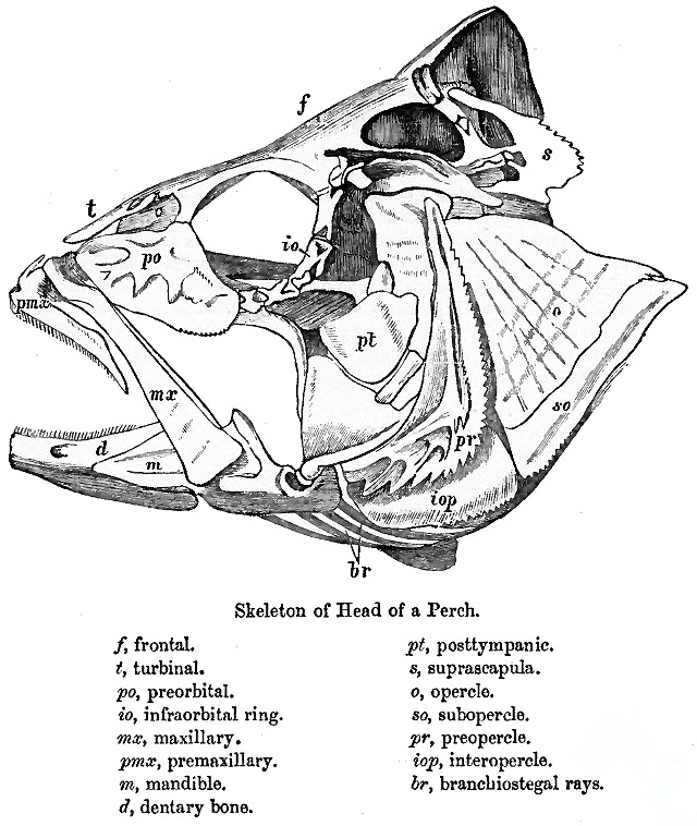
In human anatomy, the neurocranium, also known as the braincase, brainpan, or brain-pan is the upper and back part of the skull, which forms a protective case around the brain. In the human skull, the neurocranium includes the calvaria or skullcap. The remainder of the skull is the facial skeleton. In comparative anatomy, neurocranium is sometimes used synonymously with endocranium or chondrocranium. Structure The neurocranium is divided into two portions: * the membranous part, consisting of flat bones, which surround the brain; and * the cartilaginous part, or chondrocranium, which forms bones of the base of the skull. In humans, the neurocranium is usually considered to include the following eight bones: * 1 ethmoid bone * 1 frontal bone * 1 occipital bone * 2 parietal bones * 1 sphenoid bone * 2 temporal bones The ossicles (three on each side) are usually not included as bones of the neurocranium. There may variably also be extra sutural bones present. Below the ... [...More Info...] [...Related Items...] OR: [Wikipedia] [Google] [Baidu] |
Neural Crest
Neural crest cells are a temporary group of cells unique to vertebrates that arise from the embryonic ectoderm germ layer, and in turn give rise to a diverse cell lineage—including melanocytes, craniofacial cartilage and bone, smooth muscle, peripheral and enteric neurons and glia. After gastrulation, neural crest cells are specified at the border of the neural plate and the non-neural ectoderm. During neurulation, the borders of the neural plate, also known as the neural folds, converge at the dorsal midline to form the neural tube. Subsequently, neural crest cells from the roof plate of the neural tube undergo an epithelial to mesenchymal transition, delaminating from the neuroepithelium and migrating through the periphery where they differentiate into varied cell types. The emergence of neural crest was important in vertebrate evolution because many of its structural derivatives are defining features of the vertebrate clade. Underlying the development of neura ... [...More Info...] [...Related Items...] OR: [Wikipedia] [Google] [Baidu] |
Endocranium
The endocranium in comparative anatomy is a part of the skull base in vertebrates and it represents the basal, inner part of the cranium. The term is also applied to the outer layer of the dura mater in human anatomy. Structure Structurally, the endocranium consists of a boxlike shape, open at the top. The posterior margin exhibit the '' foramen magnum'', an opening for the spinal cord. The floor of the endocranium has several paired openings for the cranial nerves, and the anterior margin holds a spongy construction, allowing for the external nasal nerves to pass through. Romer, A.S. & T.S. Parsons. 1977. ''The Vertebrate Body.'' 5th ed. Saunders, Philadelphia. (6th ed. 1985) All bones of the structure derive from the cranial neural crest during fetal development. Endocranial elements in humans In humans and other mammals, the endocranium forms during fetal development as a cartilaginous neurocranium, that ossifies from several centers. Several of these bones merge, and in ... [...More Info...] [...Related Items...] OR: [Wikipedia] [Google] [Baidu] |
Hagfish
Hagfish, of the class Myxini (also known as Hyperotreti) and order Myxiniformes , are eel-shaped, slime-producing marine fish (occasionally called slime eels). They are the only known living animals that have a skull but no vertebral column, although hagfish do have rudimentary vertebrae. Along with lampreys, hagfish are jawless; the two form the sister group to jawed vertebrates, and living hagfish remain similar to hagfish from around 300 million years ago. The classification of hagfish had been controversial. The issue was whether the hagfish was a degenerate type of vertebrate-fish that through evolution had lost its vertebrae (the original scheme) and was most closely related to lampreys, or whether hagfish represent a stage that precedes the evolution of the vertebral column (the alternative scheme) as is the case with lancelets. Recent DNA evidence has supported the original scheme. The original scheme groups hagfish and lampreys together as cyclostomes (or histori ... [...More Info...] [...Related Items...] OR: [Wikipedia] [Google] [Baidu] |
Agnatha
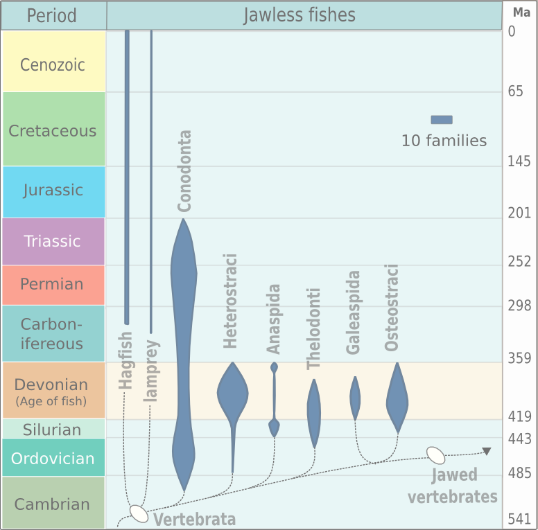
Agnatha (, Ancient Greek 'without jaws') is an infraphylum of jawless fish in the phylum Chordata, subphylum Vertebrata, consisting of both present ( cyclostomes) and extinct (conodonts and ostracoderms) species. Among recent animals, cyclostomes are sister to all vertebrates with jaws, known as gnathostomes. Recent molecular data, both from rRNA and from mtDNA as well as embryological data, strongly supports the hypothesis that living agnathans, the cyclostomes, are monophyletic. The oldest fossil agnathans appeared in the Cambrian, and two groups still survive today: the lampreys and the hagfish, comprising about 120 species in total. Hagfish are considered members of the subphylum Vertebrata, because they secondarily lost vertebrae; before this event was inferred from molecular and developmental data, the group Craniata was created by Linnaeus (and is still sometimes used as a strictly morphological descriptor) to reference hagfish plus vertebrates. While ... [...More Info...] [...Related Items...] OR: [Wikipedia] [Google] [Baidu] |
Dermal Bone
A dermal bone or investing bone or membrane bone is a bony structure derived from intramembranous ossification forming components of the vertebrate skeleton including much of the skull, jaws, gill covers, shoulder girdle and fin spines rays ( lepidotrichia), and the shell (of tortoises and turtles). In contrast to endochondral bone, dermal bone does not form from cartilage that then calcifies, and it is often ornamented. Dermal bone is formed within the dermis and grows by accretion only – the outer portion of the bone is deposited by osteoblasts. The function of some dermal bone is conserved throughout vertebrates, although there is variation in shape and in the number of bones in the skull roof and postcranial structures. In bony fish, dermal bone is found in the fin rays and scales. A special example of dermal bone is the clavicle The clavicle, or collarbone, is a slender, S-shaped long bone approximately 6 inches (15 cm) long that serves as a strut between ... [...More Info...] [...Related Items...] OR: [Wikipedia] [Google] [Baidu] |
Anatomical Terms Of Location
Standard anatomical terms of location are used to unambiguously describe the anatomy of animals, including humans. The terms, typically derived from Latin or Greek roots, describe something in its standard anatomical position. This position provides a definition of what is at the front ("anterior"), behind ("posterior") and so on. As part of defining and describing terms, the body is described through the use of anatomical planes and anatomical axes. The meaning of terms that are used can change depending on whether an organism is bipedal or quadrupedal. Additionally, for some animals such as invertebrates, some terms may not have any meaning at all; for example, an animal that is radially symmetrical will have no anterior surface, but can still have a description that a part is close to the middle ("proximal") or further from the middle ("distal"). International organisations have determined vocabularies that are often used as standard vocabularies for subdisciplines o ... [...More Info...] [...Related Items...] OR: [Wikipedia] [Google] [Baidu] |
Mesoderm
The mesoderm is the middle layer of the three germ layers that develops during gastrulation in the very early development of the embryo of most animals. The outer layer is the ectoderm, and the inner layer is the endoderm.Langman's Medical Embryology, 11th edition. 2010. The mesoderm forms mesenchyme, mesothelium, non-epithelial blood cells and coelomocytes. Mesothelium lines coeloms. Mesoderm forms the muscles in a process known as myogenesis, septa (cross-wise partitions) and mesenteries (length-wise partitions); and forms part of the gonads (the rest being the gametes). Myogenesis is specifically a function of mesenchyme. The mesoderm differentiates from the rest of the embryo through intercellular signaling, after which the mesoderm is polarized by an organizing center. The position of the organizing center is in turn determined by the regions in which beta-catenin is protected from degradation by GSK-3. Beta-catenin acts as a co-factor that alters the activity of ... [...More Info...] [...Related Items...] OR: [Wikipedia] [Google] [Baidu] |
Notochord
In anatomy, the notochord is a flexible rod which is similar in structure to the stiffer cartilage. If a species has a notochord at any stage of its life cycle (along with 4 other features), it is, by definition, a chordate. The notochord consists of inner, vacuolated cells covered by fibrous and elastic sheaths, lies along the anteroposterior axis (''front to back''), is usually closer to the dorsal than the ventral surface of the embryo, and is composed of cells derived from the mesoderm. The most commonly cited functions of the notochord are: as a midline tissue that provides directional signals to surrounding tissue during development, as a skeletal (structural) element, and as a vertebral precursor. In lancelets the notochord persists throughout life as the main structural support of the body. In tunicates the notochord is present only in the larval stage, being completely absent in the adult animal. In these invertebrate chordates, the notochord is not vacuolated ... [...More Info...] [...Related Items...] OR: [Wikipedia] [Google] [Baidu] |
Embryology
Embryology (from Greek ἔμβρυον, ''embryon'', "the unborn, embryo"; and -λογία, '' -logia'') is the branch of animal biology that studies the prenatal development of gametes (sex cells), fertilization, and development of embryos and fetuses. Additionally, embryology encompasses the study of congenital disorders that occur before birth, known as teratology. Early embryology was proposed by Marcello Malpighi, and known as preformationism, the theory that organisms develop from pre-existing miniature versions of themselves. Aristotle proposed the theory that is now accepted, epigenesis. Epigenesis is the idea that organisms develop from seed or egg in a sequence of steps. Modern embryology, developed from the work of Karl Ernst von Baer, though accurate observations had been made in Italy by anatomists such as Aldrovandi and Leonardo da Vinci in the Renaissance. Comparative embryology Preformationism and epigenesis As recently as the 18th century, the prev ... [...More Info...] [...Related Items...] OR: [Wikipedia] [Google] [Baidu] |
Lamprey
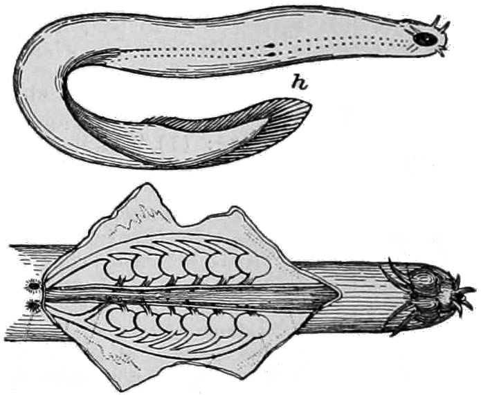
Lampreys (sometimes inaccurately called lamprey eels) are an ancient extant lineage of jawless fish of the order Petromyzontiformes , placed in the superclass Cyclostomata. The adult lamprey may be characterized by a toothed, funnel-like sucking mouth. The common name "lamprey" is probably derived from Latin , which may mean "stone licker" ( "to lick" + "stone"), though the etymology is uncertain. ''Lamprey'' is sometimes seen for the plural form. There are about 38 known extant species of lampreys and five known extinct species. Parasitic carnivorous species are the most well-known, and feed by boring into the flesh of other fish to suck their blood; but only 18 species of lampreys engage in this micropredatory lifestyle. Of the 18 carnivorous species, nine migrate from saltwater to freshwater to breed (some of them also have freshwater populations), and nine live exclusively in freshwater. All non-carnivorous forms are freshwater species. Adults of the non-carnivorous ... [...More Info...] [...Related Items...] OR: [Wikipedia] [Google] [Baidu] |
Batoidea
Batoidea is a superorder of cartilaginous fishes, commonly known as rays. They and their close relatives, the sharks, comprise the subclass Elasmobranchii. Rays are the largest group of cartilaginous fishes, with well over 600 species in 26 families. Rays are distinguished by their flattened bodies, enlarged pectoral fins that are fused to the head, and gill slits that are placed on their ventral surfaces. Anatomy Batoids are flat-bodied, and, like sharks, are cartilaginous fish, meaning they have a boneless skeleton made of a tough, elastic cartilage. Most batoids have five ventral slot-like body openings called gill slits that lead from the gills, but the Hexatrygonidae have six. Batoid gill slits lie under the pectoral fins on the underside, whereas a shark's are on the sides of the head. Most batoids have a flat, disk-like body, with the exception of the guitarfishes and sawfishes, while most sharks have a spindle-shaped body. Many species of batoid have developed th ... [...More Info...] [...Related Items...] OR: [Wikipedia] [Google] [Baidu] |
.jpg)


2.jpg)