ILO Classification on:
[Wikipedia]
[Google]
[Amazon]
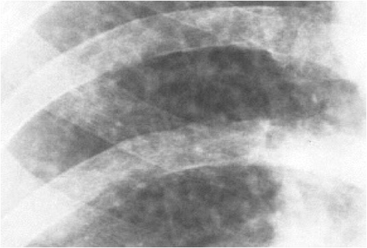
The ILO International Classification of Radiographs of Pneumoconioses is a system of classifying chest radiographs (
NIOSH Roentgenographic Interpretation form
The ILO Classification system pertains to pulmonary parenchymal abnormalities (''small and large opacities''), pleural changes (''pleural plaques, calcification, and diffuse pleural thickening'') and other features associated, or sometimes confused, with occupational lung disease. The "Complete Set" of standard x-rays consists of 22 radiographs: two illustrating normal profusion, fifteen of differing profusion category and shape/size of small opacity (see below), three illustrating large opacity, one of "u"-sized small opacity, and one of various pleural abnormalities. The "Quad Set" consists of 14 radiographs, nine of the most commonly used standards from the Complete Set, plus five additional composite reproductions of quadrant sections from the other radiographs in the Complete Set. The film sets were new to coincide with the ILO (2000) Guidelines; the digital set is new and coincides with the 2011 Guidelines.
 :''Small Opacities'': The reader will categorize small opacities according to shape and size. The small, rounded opacities are ''p'' (up to about 1.5 mm), ''q'' (about 1.5 mm to about 3 mm), or ''r'' (exceeding about 3mm and up to about 10 mm). Small, irregular opacities are classified by width as ''s'', ''t'', or ''u'' (same respective sizes as for small, rounded opacities).
:''Lung Zones'': Each lung is mentally subdivided by the reader into 3 evenly spaced zones: ''upper'', ''middle'', and ''lower''. The zones in which the small parenchymal opacities appear are recorded.
:''Profusion'': Using the Standard X-rays, the profusion (concentration) of small opacities is classified on a 4-point major category scale (0, 1, 2, or 3), with each major category divided into three, giving 12 ordered subcategories of increasing profusion: 0/-, 0/0, 0/1, 1/0, 1/1, 1/2, 2/1, 2/2, 2/3, 3/2, 3/3, and 3/+. Category 0 refers to the absence of small opacity and category 3 represents the most profuse. The major category (first number) represents the profusion felt to best fit the subject x-ray, and the minor category (second number) represents either the profusion seriously considered as an alternative, or if none, the same profusion as the major category. For example, if the reader thinks the x-ray being read has profusion most like the standard x-ray for category 1, but serious considered category 2 as an alternative description of the profusion, then the reading is 1/2.
 :''Large opacities'': A large opacity is defined as any opacity greater than 1 cm in diameter. They are classified as Category A (for one or more large opacities whose combined longest dimension does not exceed about 50 mm), category B (for one or more large opacities whose combined longest dimension exceeds 50 mm but does not exceed the equivalent area of the right upper lung zone), or category C (for one or more large opacities whose combined longest dimension exceed the equivalent area of the right upper lung zone).
*''Pleural Abnormalities:''
:
X-rays
X-rays (or rarely, ''X-radiation'') are a form of high-energy electromagnetic radiation. In many languages, it is referred to as Röntgen radiation, after the German scientist Wilhelm Conrad Röntgen, who discovered it in 1895 and named it ' ...
) for persons with a (or, rarely, more than one) form of pneumoconiosis
Pneumoconiosis is the general term for a class of interstitial lung disease where inhalation of dust ( for example, ash dust, lead particles, pollen grains etc) has caused interstitial fibrosis. The three most common types are asbestosis, sili ...
. The intent is to provide a standardized, uniform method of interpreting and describing abnormalities in chest x-rays that are thought to be caused by prolonged dust inhalation. In use, it provides a system for both epidemiological
Epidemiology is the study and analysis of the distribution (who, when, and where), patterns and determinants of health and disease conditions in a defined population.
It is a cornerstone of public health, and shapes policy decisions and evide ...
comparisons of many individuals exposed to dust and evaluation of an individual's potential disease relative to established standards.
History
Since 1946, theInternational Labour Organization
The International Labour Organization (ILO) is a United Nations agency whose mandate is to advance social and economic justice by setting international labour standards. Founded in October 1919 under the League of Nations, it is the first and o ...
has been a specialized agency of the United Nations
The United Nations (UN) is an intergovernmental organization whose stated purposes are to maintain international peace and security, develop friendly relations among nations, achieve international cooperation, and be a centre for harmonizi ...
, with objectives including establishing and overseeing international labor standards
International labour law is the body of rules spanning public and private international law which concern the rights and duties of employees, employers, trade unions and governments in regulating Work (human activity) and the workplace. The Interna ...
and labor rights. The International Labour Office ("ILO") is the Organization’s research body and publishing house. Since 1950, the ILO has periodically published guidelines on how to classify chest X-rays for pneumoconiosis. The purpose of the Classification was to describe and codify radiographic abnormalities of the pneumoconioses in a simple, systematic, and reproducible manner, aiding international comparisons of data, epidemiology, screening and surveillance, clinical purposes, and medical research. The most recent edition of the Guidelines, completed in 2011, replaced the 2000 revised edition.
In 1974, after studies of surveillance programs for coal miners revealed unacceptable degrees of interreader variability, the National Institute for Occupational Safety and Health (NIOSH
The National Institute for Occupational Safety and Health (NIOSH, ) is the United States federal agency responsible for conducting research and making recommendations for the prevention of work-related injury and illness. NIOSH is part of the ...
),began the "B" reader program (so named because of the ''Black'' lung or Coal Workers' X-ray Surveillance Program), with the intent to train and certify physicians in the ILO Classification system. The "B" reader certification examination went into full operation in 1978. A physician must pass the certification examination to be a "B" reader.
Basic Description
The ILO Classification system includes the printed Guidelines and sets of standard radiographs, available in both film and, as of 2011, digital forms. The reader compares the subject chest X-ray (only the appearances seen on postero-anterior, or PA, chest x-ray) with those of the standard set. The standard radiographs provide differing types ("shape and size") and severity ("profusion") of abnormalities seen in persons with pneumoconiosis, including Coal Workers’ Pneumoconiosis,silicosis
Silicosis is a form of occupational lung disease caused by inhalation of crystalline silica dust. It is marked by inflammation and scarring in the form of nodular lesions in the upper lobes of the lungs. It is a type of pneumoconiosis. Silicosi ...
, and asbestosis
Asbestosis is long-term inflammation and scarring of the lungs due to asbestos fibers. Symptoms may include shortness of breath, cough, wheezing, and chest tightness. Complications may include lung cancer, mesothelioma, and pulmonary heart diseas ...
. The reader then classifies the subject x-ray, often recording the findings on thNIOSH Roentgenographic Interpretation form
The ILO Classification system pertains to pulmonary parenchymal abnormalities (''small and large opacities''), pleural changes (''pleural plaques, calcification, and diffuse pleural thickening'') and other features associated, or sometimes confused, with occupational lung disease. The "Complete Set" of standard x-rays consists of 22 radiographs: two illustrating normal profusion, fifteen of differing profusion category and shape/size of small opacity (see below), three illustrating large opacity, one of "u"-sized small opacity, and one of various pleural abnormalities. The "Quad Set" consists of 14 radiographs, nine of the most commonly used standards from the Complete Set, plus five additional composite reproductions of quadrant sections from the other radiographs in the Complete Set. The film sets were new to coincide with the ILO (2000) Guidelines; the digital set is new and coincides with the 2011 Guidelines.
Methodology
*''Technical Quality (formerly Film Quality):'' :In the current ILO Classification system, the reader is first asked to grade radiographic quality. There are four technical grades: (1) Good; (2) Acceptable, with no technical defect likely to impair classification; (3) Acceptable, with some technical defect but still adequate; and (4) Unacceptable. Quality defects include over- or under-exposure, underinflation, artifacts, improper positioning, and others. *''Parenchymal Abnormalities:'' :''Small Opacities'': The reader will categorize small opacities according to shape and size. The small, rounded opacities are ''p'' (up to about 1.5 mm), ''q'' (about 1.5 mm to about 3 mm), or ''r'' (exceeding about 3mm and up to about 10 mm). Small, irregular opacities are classified by width as ''s'', ''t'', or ''u'' (same respective sizes as for small, rounded opacities).
:''Lung Zones'': Each lung is mentally subdivided by the reader into 3 evenly spaced zones: ''upper'', ''middle'', and ''lower''. The zones in which the small parenchymal opacities appear are recorded.
:''Profusion'': Using the Standard X-rays, the profusion (concentration) of small opacities is classified on a 4-point major category scale (0, 1, 2, or 3), with each major category divided into three, giving 12 ordered subcategories of increasing profusion: 0/-, 0/0, 0/1, 1/0, 1/1, 1/2, 2/1, 2/2, 2/3, 3/2, 3/3, and 3/+. Category 0 refers to the absence of small opacity and category 3 represents the most profuse. The major category (first number) represents the profusion felt to best fit the subject x-ray, and the minor category (second number) represents either the profusion seriously considered as an alternative, or if none, the same profusion as the major category. For example, if the reader thinks the x-ray being read has profusion most like the standard x-ray for category 1, but serious considered category 2 as an alternative description of the profusion, then the reading is 1/2.
 :''Large opacities'': A large opacity is defined as any opacity greater than 1 cm in diameter. They are classified as Category A (for one or more large opacities whose combined longest dimension does not exceed about 50 mm), category B (for one or more large opacities whose combined longest dimension exceeds 50 mm but does not exceed the equivalent area of the right upper lung zone), or category C (for one or more large opacities whose combined longest dimension exceed the equivalent area of the right upper lung zone).
*''Pleural Abnormalities:''
:
:''Large opacities'': A large opacity is defined as any opacity greater than 1 cm in diameter. They are classified as Category A (for one or more large opacities whose combined longest dimension does not exceed about 50 mm), category B (for one or more large opacities whose combined longest dimension exceeds 50 mm but does not exceed the equivalent area of the right upper lung zone), or category C (for one or more large opacities whose combined longest dimension exceed the equivalent area of the right upper lung zone).
*''Pleural Abnormalities:''
:Pleural
The pleural cavity, pleural space, or interpleural space is the potential space between the pleurae of the pleural sac that surrounds each lung. A small amount of serous pleural fluid is maintained in the pleural cavity to enable lubrication ...
abnormalities are reported with respect to ''type'' (pleural plaques or diffuse pleural thickening), ''location'' (chest wall, diaphragm, or other), ''presence of calcification'', ''width'' (only of ''in profile'' pleural thickening seen along the chest wall edge), and ''extent'' (combined distance for involved chest wall).
*''Any Other Abnormality:''
:There are 29 "obligatory" symbols representing important features related to dust diseases of the lungs and other etiologies. These symbols are: aa atherosclerotic aorta; at significant apical pleural thickening; ax coalescence of small opacities; bu bulla(e); ca cancer
Cancer is a group of diseases involving abnormal cell growth with the potential to invade or spread to other parts of the body. These contrast with benign tumors, which do not spread. Possible signs and symptoms include a lump, abnormal bl ...
; cg calcified granuloma
A granuloma is an aggregation of macrophages that forms in response to chronic inflammation. This occurs when the immune system attempts to isolate foreign substances that it is otherwise unable to eliminate. Such substances include infectious ...
; cn calcification of small pneumoconiotic opacities; co abnormal cardiac shape or size; cp cor pulmonale
Pulmonary heart disease, also known as cor pulmonale, is the enlargement and failure of the right ventricle of the heart as a response to increased vascular resistance (such as from pulmonic stenosis) or high blood pressure in the lungs.
Chronic ...
; cv cavity; di marked distortion of an intrathoracic structure; ef pleural effusion
A pleural effusion is accumulation of excessive fluid in the pleural space, the potential space that surrounds each lung.
Under normal conditions, pleural fluid is secreted by the parietal pleural capillaries at a rate of 0.6 millilitre per ...
; em emphysema
Emphysema, or pulmonary emphysema, is a lower respiratory tract disease, characterised by air-filled spaces ( pneumatoses) in the lungs, that can vary in size and may be very large. The spaces are caused by the breakdown of the walls of the a ...
; es eggshell calcification of hilar lymph node; fr rib fracture(s); hi enlargement of non-calcified hilar nodes; ho honeycombing; id ill-defined diaphragm border; ih ill-defined heart border; kl septal (Kerley) lines; me mesothelioma (pleural). pa plate atelectasis
Atelectasis is the collapse or closure of a lung resulting in reduced or absent gas exchange. It is usually unilateral, affecting part or all of one lung. It is a condition where the alveoli are deflated down to little or no volume, as distinct ...
; pb parenchymal bands; pi pleural thickening of an interlobar fissure; px pneumothorax
A pneumothorax is an abnormal collection of air in the pleural space between the lung and the chest wall. Symptoms typically include sudden onset of sharp, one-sided chest pain and shortness of breath. In a minority of cases, a one-way valve ...
; ra rounded atelectasis; rp rheumatoid pneumoconiosis; tb tuberculosis
Tuberculosis (TB) is an infectious disease usually caused by ''Mycobacterium tuberculosis'' (MTB) bacteria. Tuberculosis generally affects the lungs, but it can also affect other parts of the body. Most infections show no symptoms, in w ...
; and od other disease or significant abnormality. Finally, the reader comments on any other abnormal features of the chest radiograph or other relevant information.
References
{{DEFAULTSORT:Ilo Classification Projectional radiography