fixator muscle on:
[Wikipedia]
[Google]
[Amazon]
 Synergist muscles also called ''fixators'', act around a joint to help the action of an
 There are a number of terms used in the naming of muscles including those relating to size, shape, action, location, their orientation, and their number of heads.
;By size: ''brevis'' means short; ''longus'' means long; ''major'' means large; ''maximus'' means largest; ''minor'' means small, and ''minimus'' smallest. These terms are often used after the particular muscle such as

 Muscles may also be described by the direction that the muscle fibres run, in their muscle architecture.
* Fusiform muscles have fibres that run parallel to the length of the muscle, and are spindle-shaped. For example, the pronator teres muscle of the

Anatomical terminology
Anatomical terminology is a specialized system of terms used by anatomists, zoologists, and health professionals, such as doctors, surgeons, and pharmacists, to describe the structures and functions of the body.
This terminology incorpor ...
is used to uniquely describe aspects of skeletal muscle
Skeletal muscle (commonly referred to as muscle) is one of the three types of vertebrate muscle tissue, the others being cardiac muscle and smooth muscle. They are part of the somatic nervous system, voluntary muscular system and typically are a ...
, cardiac muscle
Cardiac muscle (also called heart muscle or myocardium) is one of three types of vertebrate muscle tissues, the others being skeletal muscle and smooth muscle. It is an involuntary, striated muscle that constitutes the main tissue of the wall o ...
, and smooth muscle
Smooth muscle is one of the three major types of vertebrate muscle tissue, the others being skeletal and cardiac muscle. It can also be found in invertebrates and is controlled by the autonomic nervous system. It is non- striated, so-called bec ...
such as their actions, structure, size, and location.
Types
There are three types ofmuscle tissue
Muscle is a soft tissue, one of the four basic types of animal tissue. There are three types of muscle tissue in vertebrates: skeletal muscle, cardiac muscle, and smooth muscle. Muscle tissue gives skeletal muscles the ability to contract. ...
in the body: skeletal, smooth, and cardiac.
Skeletal muscle
Skeletal muscle
Skeletal muscle (commonly referred to as muscle) is one of the three types of vertebrate muscle tissue, the others being cardiac muscle and smooth muscle. They are part of the somatic nervous system, voluntary muscular system and typically are a ...
, or "voluntary muscle", is a striated muscle tissue
Striated muscle tissue is a muscle tissue that features repeating functional units called sarcomeres. Under the microscope, sarcomeres are visible along muscle fibers, giving a striated appearance to the tissue. The two types of striated muscle a ...
that primarily joins to bone
A bone is a rigid organ that constitutes part of the skeleton in most vertebrate animals. Bones protect the various other organs of the body, produce red and white blood cells, store minerals, provide structure and support for the body, ...
with tendon
A tendon or sinew is a tough band of fibrous connective tissue, dense fibrous connective tissue that connects skeletal muscle, muscle to bone. It sends the mechanical forces of muscle contraction to the skeletal system, while withstanding tensi ...
s. Skeletal muscle enables movement of bones, and maintains posture. The widest part of a muscle that pulls on the tendons is known as the belly.
Muscle slip
A muscle slip is a slip of muscle that can either be an anatomical variant, or a branching of a muscle as inrib
In vertebrate anatomy, ribs () are the long curved bones which form the rib cage, part of the axial skeleton. In most tetrapods, ribs surround the thoracic cavity, enabling the lungs to expand and thus facilitate breathing by expanding the ...
connections of the serratus anterior muscle
The serratus anterior is a muscle of the chest. It originates at the side of the chest from the upper 8 or 9 ribs; it inserts along the entire length of the anterior aspect of the medial border of the scapula. It is innervated by the long tho ...
.
Smooth muscle
Smooth muscle
Smooth muscle is one of the three major types of vertebrate muscle tissue, the others being skeletal and cardiac muscle. It can also be found in invertebrates and is controlled by the autonomic nervous system. It is non- striated, so-called bec ...
is involuntary and found in parts of the body where it conveys action without conscious intent. The majority of this type of muscle tissue is found in the digestive and urinary system
The human urinary system, also known as the urinary tract or renal system, consists of the kidneys, ureters, urinary bladder, bladder, and the urethra. The purpose of the urinary system is to eliminate waste from the body, regulate blood volume ...
s where it acts by propelling forward food, chyme
Chyme or chymus (; ) is the semi-fluid mass of partly digested food that is expelled by the stomach, through the pyloric valve, into the duodenum (the beginning of the small intestine).
Chyme results from the mechanical and chemical breakdown ...
, and feces
Feces (also known as faeces American and British English spelling differences#ae and oe, or fæces; : faex) are the solid or semi-solid remains of food that was not digested in the small intestine, and has been broken down by bacteria in the ...
in the former and urine
Urine is a liquid by-product of metabolism in humans and many other animals. In placental mammals, urine flows from the Kidney (vertebrates), kidneys through the ureters to the urinary bladder and exits the urethra through the penile meatus (mal ...
in the latter. Other places smooth muscle can be found are within the uterus
The uterus (from Latin ''uterus'', : uteri or uteruses) or womb () is the hollow organ, organ in the reproductive system of most female mammals, including humans, that accommodates the embryonic development, embryonic and prenatal development, f ...
, where it helps facilitate birth
Birth is the act or process of bearing or bringing forth offspring, also referred to in technical contexts as parturition. In mammals, the process is initiated by hormones which cause the muscular walls of the uterus to contract, expelling the f ...
, and the eye
An eye is a sensory organ that allows an organism to perceive visual information. It detects light and converts it into electro-chemical impulses in neurons (neurones). It is part of an organism's visual system.
In higher organisms, the ey ...
, where the pupillary sphincter controls pupil
The pupil is a hole located in the center of the iris of the eye that allows light to strike the retina.Cassin, B. and Solomon, S. (1990) ''Dictionary of Eye Terminology''. Gainesville, Florida: Triad Publishing Company. It appears black becau ...
size.
Cardiac muscle
Cardiac muscle
Cardiac muscle (also called heart muscle or myocardium) is one of three types of vertebrate muscle tissues, the others being skeletal muscle and smooth muscle. It is an involuntary, striated muscle that constitutes the main tissue of the wall o ...
is specific to the heart
The heart is a muscular Organ (biology), organ found in humans and other animals. This organ pumps blood through the blood vessels. The heart and blood vessels together make the circulatory system. The pumped blood carries oxygen and nutrie ...
. It is also involuntary in its movement, and is additionally self-excitatory, contracting without outside stimuli.
Actions of skeletal muscle
As well asanatomical terms of motion
Motion, the process of movement, is described using specific anatomical terms. Motion includes movement of organs, joints, limbs, and specific sections of the body. The terminology used describes this motion according to its direction relativ ...
, which describe the motion made by a muscle, unique terminology is used to describe the action of a set of muscles.
Agonists and antagonists
Agonist muscles and antagonist muscles are muscles that cause or inhibit a movement. Agonist muscles are also called prime movers since they produce most of the force, and control of an action. Agonists cause a movement to occur through their own activation. For example, thetriceps brachii
The triceps, or triceps brachii (Latin for "three-headed muscle of the arm"), is a large muscle on the back of the upper limb of many vertebrates. It consists of three parts: the medial, lateral, and long head. All three heads cross the elbow jo ...
contracts, producing a shortening (concentric) contraction, during the up phase of a push-up ( elbow extension). During the down phase of a push-up, the same triceps brachii actively controls elbow flexion while producing a lengthening (eccentric) contraction. It is still the agonist, because while resisting gravity during relaxing, the triceps brachii continues to be the prime mover, or controller, of the joint action.
Another example is the dumb-bell curl at the elbow. The elbow flexor group is the agonist, shortening during the lifting phase ( elbow flexion). During the lowering phase the elbow flexor muscles lengthen, remaining the agonists because they are controlling the load and the movement (elbow extension). For both the lifting and lowering phase, the "elbow extensor" muscles are the antagonists (see below). They lengthen during the dumbbell lifting phase and shorten during the dumbbell lowering phase. Here it is important to understand that it is common practice to give a name to a muscle group (e.g. elbow flexors) based on the joint action they produce during a shortening contraction. However, this naming convention does not mean they are only agonists during shortening. This term typically describes the function of skeletal muscle
Skeletal muscle (commonly referred to as muscle) is one of the three types of vertebrate muscle tissue, the others being cardiac muscle and smooth muscle. They are part of the somatic nervous system, voluntary muscular system and typically are a ...
s.
Antagonist muscles are simply the muscles that produce an opposing joint torque to the agonist muscles. This torque can aid in controlling a motion. The opposing torque can slow movement down - especially in the case of a ballistic movement
Ballistic movement can be defined as muscle contractions that exhibit maximum velocities and accelerations over a very short period of time. They exhibit high firing rates, high force production, and very brief contraction times.
Physiology
Muscl ...
. For example, during a very rapid (ballistic) discrete movement of the elbow, such as throwing a dart, the triceps muscles will be activated very briefly and strongly (in a "burst") to rapidly accelerate the extension movement at the elbow, followed almost immediately by a "burst" of activation to the elbow flexor muscles that decelerates the elbow movement to arrive at a quick stop. To use an automotive analogy, this would be similar to pressing the accelerator pedal rapidly and then immediately pressing the brake. Antagonism is not an intrinsic property of a particular muscle or muscle group; it is a role that a muscle plays depending on which muscle is currently the agonist. During slower joint actions that involve gravity, just as with the agonist muscle, the antagonist muscle can shorten and lengthen. Using the example of the triceps brachii during a push-up, the elbow flexor muscles are the antagonists at the elbow during both the up phase and down phase of the movement. During the dumbbell curl, the elbow extensors are the antagonists for both the lifting and lowering phases.
Antagonistic pairs
Antagonist and agonist muscles often occur in pairs, called antagonistic pairs. As one muscle contracts, the other relaxes. An example of an antagonistic pair is thebiceps
The biceps or biceps brachii (, "two-headed muscle of the arm") is a large muscle that lies on the front of the upper arm between the shoulder and the elbow. Both heads of the muscle arise on the scapula and join to form a single muscle bel ...
and triceps
The triceps, or triceps brachii (Latin for "three-headed muscle of the arm"), is a large muscle on the ventral, back of the upper limb of many vertebrates. It consists of three parts: the medial, lateral, and long head. All three heads cross the ...
; to contract, the triceps relaxes while the biceps contracts to lift the arm. "Reverse motions" need antagonistic pairs located in opposite sides of a joint or bone, including abductor-adductor pairs and flexor-extensor pairs. These consist of an extensor muscle
In anatomy, extension is a movement of a joint that increases the angle between two bones or body surfaces at a joint. Extension usually results in straightening of the bones or body surfaces involved. For example, extension is produced by extend ...
, which "opens" the joint (by increasing the angle between the two bones) and a flexor muscle, which does the opposite by decreasing the angle between two bones.
However, muscles do not always work this way; sometimes agonists and antagonists contract at the same time to produce force, as per Lombard's paradox. Also, sometimes during a joint action controlled by an agonist muscle, the antagonist will be slightly activated, naturally. This occurs normally and is not considered to be a problem unless it is excessive or uncontrolled and disturbs the control of the joint action. This is called agonist/antagonist co-activation and serves to mechanically stiffen the joint.
Not all muscles are paired in this way. An example of an exception is the deltoid Deltoid (delta-shaped) can refer to:
* The deltoid muscle, a muscle in the shoulder
* Kite (geometry), also known as a deltoid, a type of quadrilateral
* A deltoid curve, a three-cusped hypocycloid
* A leaf shape
* The deltoid tuberosity, a part o ...
.
Synergists
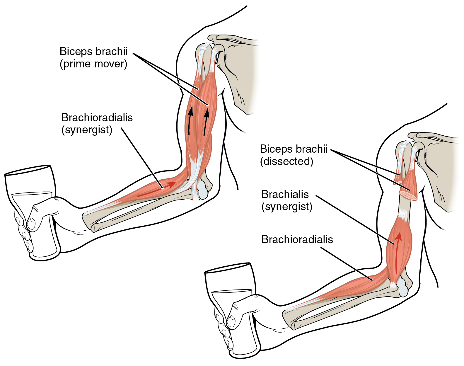
 Synergist muscles also called ''fixators'', act around a joint to help the action of an
 agonist muscle
Anatomical terminology is used to uniquely describe aspects of skeletal muscle, cardiac muscle, and smooth muscle such as their actions, structure, size, and location.
Types
There are three types of muscle tissue in the body: skeletal, smooth, a ...
. Synergist muscles can also act to counter or neutralize the force of an agonist and are also known as neutralizers when they do this. As neutralizers they help to cancel out or neutralize extra motion produced from the agonists to ensure that the force generated works within the desired plane of motion.
Muscle fibers can only contract up to 40% of their fully stretched length. Thus the short fibers of pennate muscle
A pennate or pinnate muscle (also called a penniform muscle) is a type of skeletal muscle with fascicles that attach obliquely (in a slanting position) to its tendon. This type of muscle generally allows higher force production but a smaller ra ...
s are more suitable where power rather than range of contraction is required. This limitation in the range of contraction affects all muscles, and those that act over several joints may be unable to shorten sufficiently to produce the full range of movement at all of them simultaneously (active insufficiency, e.g., the fingers cannot be fully flexed when the wrist is also flexed). Likewise, the opposing muscles may be unable to stretch sufficiently to allow such movement to take place (passive insufficiency). For both these reasons, it is often essential to use other synergists, in this type of action to fix certain of the joints so that others can be moved effectively, e.g., fixation of the wrist during full flexion of the fingers in clenching the fist. Synergists are muscles that facilitate the fixation action.
There is an important difference between a ''helping synergist'' muscle and a ''true synergist'' muscle. A true synergist muscle is one that only neutralizes an undesired joint action, whereas a helping synergist is one that neutralizes an undesired action but also assists with the desired action.
Neutralizer action
A muscle that fixes or holds a bone so that the agonist can carry out the intended movement is said to have a neutralizing action. A good famous example of this are thehamstrings
A hamstring () is any one of the three posterior thigh muscles in human anatomy between the hip and the knee: from anatomical_terms_of_location#Medial_and_lateral, medial to anatomical_terms_of_location#Medial_and_lateral, lateral, the semimembra ...
; the semitendinosus
The semitendinosus () is a long superficial muscle in the back of the thigh. It is so named because it has a very long tendon of insertion. It lies posteromedially in the thigh, superficial to the semimembranosus.
Structure
The semitendinosus, ...
and semimembranosus muscle
The semimembranosus muscle () is the most medial of the three hamstring muscles in the thigh. It is so named because it has a flat tendon of origin. It lies posteromedially in the thigh, deep to the semitendinosus muscle. It Anatomical terms of mo ...
s perform knee flexion and knee internal rotation
Motion, the process of movement, is described using specific anatomical terms. Motion includes movement of organs, joints, limbs, and specific sections of the body. The terminology used describes this motion according to its direction relativ ...
whereas the biceps femoris
The biceps femoris () is a muscle of the thigh located to the posterior, or back. As its name implies, it consists of two heads; the long head is considered part of the hamstring muscle group, while the short head is sometimes excluded from this ...
carries out knee flexion and knee external rotation. For the knee to flex while not rotating in either direction, all three muscles contract to stabilize the knee while it moves in the desired way.
Composite muscle
'' Composite'' or ''hybrid'' muscles have more than one set of fibers that perform the same function, and are usually supplied by different nerves for different set of fibers. For example, the tongue itself is a composite muscle made up of various components like longitudinal, transverse, horizontal muscles with different parts innervated from a different nerve supply.Muscle naming
 There are a number of terms used in the naming of muscles including those relating to size, shape, action, location, their orientation, and their number of heads.
;By size: ''brevis'' means short; ''longus'' means long; ''major'' means large; ''maximus'' means largest; ''minor'' means small, and ''minimus'' smallest. These terms are often used after the particular muscle such as
 gluteus maximus
The gluteus maximus is the main extensor muscle of the hip in humans. It is the largest and outermost of the three gluteal muscles and makes up a large part of the shape and appearance of each side of the hips. It is the single largest muscle in ...
, and gluteus minimus.
;By shape: ''deltoid'' means triangular; ''quadratus'' means having four sides; ''rhomboideus'' means having a rhomboid
Traditionally, in two-dimensional geometry, a rhomboid is a parallelogram in which adjacent sides are of unequal lengths and angles are non-right angled.
The terms "rhomboid" and "parallelogram" are often erroneously conflated with each oth ...
shape; ''teres'' means round or cylindrical, ''trapezius'' means having a trapezoid
In geometry, a trapezoid () in North American English, or trapezium () in British English, is a quadrilateral that has at least one pair of parallel sides.
The parallel sides are called the ''bases'' of the trapezoid. The other two sides are ...
shape, ''rectus'' means straight. Examples are the pronator teres, the pronator quadratus and the rectus abdominis
The rectus abdominis muscle, () also known as the "abdominal muscle" or simply better known as the "abs", is a pair of segmented skeletal muscle on the ventral aspect of a person, person's abdomen. The paired muscle is separated at the midline b ...
.
;By action: '' abductor'' moving away from the midline; '' adductor'' moving towards the midline; '' depressor'' moving downwards; ''elevator
An elevator (American English) or lift (Commonwealth English) is a machine that vertically transports people or freight between levels. They are typically powered by electric motors that drive traction cables and counterweight systems suc ...
'' moving upwards; ''flexor
In anatomy, flexor is a muscle that contracts to perform flexion (from the Latin verb ''flectere'', to bend), a movement that decreases the angle between the bones converging at a joint. For example, one's elbow joint flexes when one brin ...
'' moving that decreases an angle; '' extensor'' moving that increase an angle or straightens; '' pronator'' moving to face down; ''supinator
In human anatomy, the supinator is a broad muscle in the posterior compartment of the forearm, curved around the upper third of the radius (bone), radius. Its function is to supination, supinate the forearm.
Structure
The supinator consists of tw ...
'' moving to face upwards; '' Internal rotator'' rotating towards the body; '' external rotator'' rotating away from the body.
Form

Insertion and origin
The insertion and origin of a muscle are the two places where it is anchored, one at each end. The connective tissue of the attachment is called an enthesis.Origin
The origin of a muscle is thebone
A bone is a rigid organ that constitutes part of the skeleton in most vertebrate animals. Bones protect the various other organs of the body, produce red and white blood cells, store minerals, provide structure and support for the body, ...
, typically proximal, which has greater mass and is more stable during a contraction than a muscle's insertion. For example, with the latissimus dorsi muscle
The latissimus dorsi () is a large, flat muscle on the back that stretches to the sides, behind the arm, and is partly covered by the trapezius on the back near the midline.
The word latissimus dorsi (plural: ''latissimi dorsi'') comes from ...
, the origin site is the torso, and the insertion is the arm. When this muscle contracts, normally the arm moves due to having less mass than the torso. This is the case when grabbing objects lighter than the body, as in the typical use of a lat pull down machine. This can be reversed however, such as in a chin up where the torso moves up to meet the arm.
The head of a muscle, also called ''caput musculi'' is the part at the end of a muscle at its origin, where it attaches to a fixed bone. Some muscles such as the biceps
The biceps or biceps brachii (, "two-headed muscle of the arm") is a large muscle that lies on the front of the upper arm between the shoulder and the elbow. Both heads of the muscle arise on the scapula and join to form a single muscle bel ...
have more than one head.
Insertion
The insertion of a muscle is the structure that it attaches to and tends to be moved by the contraction of the muscle. This may be abone
A bone is a rigid organ that constitutes part of the skeleton in most vertebrate animals. Bones protect the various other organs of the body, produce red and white blood cells, store minerals, provide structure and support for the body, ...
, a tendon
A tendon or sinew is a tough band of fibrous connective tissue, dense fibrous connective tissue that connects skeletal muscle, muscle to bone. It sends the mechanical forces of muscle contraction to the skeletal system, while withstanding tensi ...
or the subcutaneous dermal connective tissue
Connective tissue is one of the four primary types of animal tissue, a group of cells that are similar in structure, along with epithelial tissue, muscle tissue, and nervous tissue. It develops mostly from the mesenchyme, derived from the mesod ...
. Insertions are usually connections of muscle via tendon
A tendon or sinew is a tough band of fibrous connective tissue, dense fibrous connective tissue that connects skeletal muscle, muscle to bone. It sends the mechanical forces of muscle contraction to the skeletal system, while withstanding tensi ...
to bone. The insertion is a bone that tends to be distal, have less mass, and greater motion than the origin during a contraction.
Intrinsic and extrinsic muscles
Intrinsic muscles have their origin in the part of the body that they act on, and are contained within that part. Extrinsic muscles have their origin outside of the part of the body that they act on. Examples are the intrinsic and extrinsic muscles of the tongue, and those of the hand.Muscle fibres
 Muscles may also be described by the direction that the muscle fibres run, in their muscle architecture.
* Fusiform muscles have fibres that run parallel to the length of the muscle, and are spindle-shaped. For example, the pronator teres muscle of the
 forearm
The forearm is the region of the upper limb between the elbow and the wrist. The term forearm is used in anatomy to distinguish it from the arm, a word which is used to describe the entire appendage of the upper limb, but which in anatomy, techn ...
.
* Unipennate muscles have fibres that run the entire length of only one side of a muscle, like a quill pen
A quill is a writing tool made from a moulted flight feather (preferably a primary wing-feather) of a large bird. Quills were used for writing with ink before the invention of the dip pen/metal- nibbed pen, the fountain pen, and, eventually, ...
. For example, the fibularis muscles.
* Bipennate muscles consist of two rows of oblique muscle fibres, facing in opposite diagonal directions, converging on a central tendon
A tendon or sinew is a tough band of fibrous connective tissue, dense fibrous connective tissue that connects skeletal muscle, muscle to bone. It sends the mechanical forces of muscle contraction to the skeletal system, while withstanding tensi ...
. Bipennate muscle is stronger than both unipennate muscle and fusiform muscle, due to a larger physiological cross-sectional area. Bipennate muscle shortens less than unipennate muscle but develops greater tension when it does, translated into greater power but less range of motion. Pennate muscle
A pennate or pinnate muscle (also called a penniform muscle) is a type of skeletal muscle with fascicles that attach obliquely (in a slanting position) to its tendon. This type of muscle generally allows higher force production but a smaller ra ...
s generally also tire easily. Examples of bipennate muscles are the rectus femoris muscle
The rectus femoris muscle is one of the four quadriceps muscles of the human body. The others are the vastus medialis, the vastus intermedius (deep to the rectus femoris), and the vastus lateralis. All four parts of the quadriceps muscle attach ...
of the thigh
In anatomy, the thigh is the area between the hip (pelvis) and the knee. Anatomically, it is part of the lower limb.
The single bone in the thigh is called the femur. This bone is very thick and strong (due to the high proportion of bone tissu ...
, and the stapedius muscle
The stapedius is the smallest skeletal muscle
Skeletal muscle (commonly referred to as muscle) is one of the three types of vertebrate muscle tissue, the others being cardiac muscle and smooth muscle. They are part of the somatic nervous syste ...
of the middle ear
The middle ear is the portion of the ear medial to the eardrum, and distal to the oval window of the cochlea (of the inner ear).
The mammalian middle ear contains three ossicles (malleus, incus, and stapes), which transfer the vibrations ...
.
State
Hypertrophy and atrophy

Hypertrophy
Hypertrophy is the increase in the volume of an organ or tissue due to the enlargement of its component cells. It is distinguished from hyperplasia, in which the cells remain approximately the same size but increase in number. Although hypertro ...
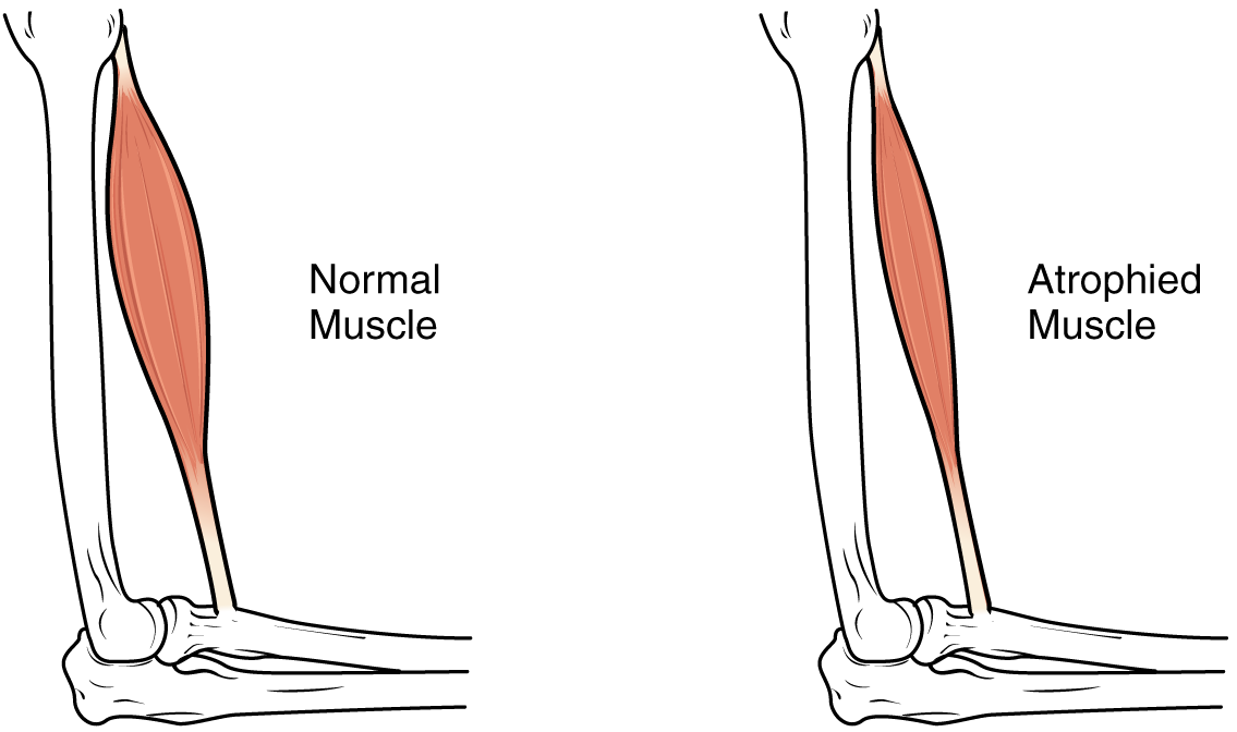
is increase in muscle size from an increase in size of individual muscle cells. This usually occurs as a result of exercise.
See also
* Reciprocal inhibition * Anatomical terms of bone * Anatomical terms of neuroanatomyReferences
;Books * * {{Portal bar, Anatomy Human anatomy Muscular system Muscle terminology Anatomical terminology