|
Composite Muscle
Composite or hybrid muscles are those muscles which have more than one set of fibers but perform the same function and are usually supplied by different nerves for different set of fibers. Examples * Brachialis: Musculocutaneous nerve is motor and radial nerve is proprioceptive. * Adductor magnus : Its adductor part by posterior division of obturator nerve and hamstring part by tibial part of sciatic nerve. * Biceps femoris: Its long head is supplied by the tibial part of sciatic nerve, whereas the short head is supplied by the common peroneal nerve. This reflects the composite derivation from the flexor and extensor musculature. * Pectineus: Its anterior set of fibers are supplied by the femoral nerve, whereas posterior set of fibers are supplied by the obturator nerve. * Flexor digitorum profundus: Its radial half of is supplied by the median nerve and the ulnar half is supplied by the ulnar nerve. * Flexor policis brevis: Supplied by recurrent branch of the median nerve and the ... [...More Info...] [...Related Items...] OR: [Wikipedia] [Google] [Baidu] |
Brachialis
The brachialis (brachialis anticus), also known as the Teichmann muscle, is a muscle in the upper arm that flexes the elbow. It lies deeper than the biceps brachii, and makes up part of the floor of the region known as the cubital fossa (elbow pit). The brachialis is the prime mover of elbow flexion generating about 50% more power than the biceps.Saladin, Kenneth S, Stephen J. Sullivan, and Christina A. Gan. Anatomy & Physiology: The Unity of Form and Function. 2015. Print. Structure The brachialis originates from the anterior surface of the distal half of the humerus, near the insertion of the deltoid muscle, which it embraces by two angular processes. Its origin extends below to within 2.5 cm of the margin of the articular surface of the humerus at the elbow joint. Its fibers converge to a thick tendon, which is inserted into the tuberosity of the ulna and the rough depression on the anterior surface of the coronoid process of the ulna. Blood supply The brachialis is supp ... [...More Info...] [...Related Items...] OR: [Wikipedia] [Google] [Baidu] |
Adductor Magnus Muscle
Adductor may refer to: * One of the anatomical terms of motion * Adductor muscle (other) * Adductor canal The adductor canal, also known as the subsartorial canal or Hunter’s canal, is an aponeurotic tunnel in the middle third of the thigh. It extends from the apex of the femoral triangle to the adductor hiatus. Structure The adductor canal extends ... {{disambiguation ... [...More Info...] [...Related Items...] OR: [Wikipedia] [Google] [Baidu] |
Biceps Femoris Muscle
The biceps femoris () is a muscle of the thigh located to the posterior, or back. As its name implies, it has two parts, one of which (the long head) forms part of the hamstrings muscle group. Structure It has two heads of origin: *the ''long head'' arises from the lower and inner impression on the posterior part of the tuberosity of the ischium. This is a common tendon origin with the semitendinosus muscle, and from the lower part of the sacrotuberous ligament. *the ''short head'', arises from the lateral lip of the linea aspera, between the adductor magnus and vastus lateralis extending up almost as high as the insertion of the gluteus maximus, from the lateral prolongation of the linea aspera to within 5 cm. of the lateral condyle; and from the lateral intermuscular septum. The two muscle heads joint together distally and unite in an intricate fashion. The fibers of the long head form a fusiform belly, which passes obliquely downward and lateralward across the sciatic ... [...More Info...] [...Related Items...] OR: [Wikipedia] [Google] [Baidu] |
Pectineus Muscle
The pectineus muscle (, from the Latin word ''pecten'', meaning comb) is a flat, quadrangular muscle, situated at the anterior (front) part of the upper and medial (inner) aspect of the thigh. The pectineus muscle is the most anterior adductor of the hip. The muscle does adduct and internally rotate the thigh but its primary function is hip flexion. It can be classified in the medial compartment of thigh (when the function is emphasized) or the anterior compartment of thigh (when the nerve is emphasized). Structure The pectineus muscle arises from the pectineal line of the pubis and to a slight extent from the surface of bone in front of it, between the iliopectineal eminence and pubic tubercle, and from the fascia covering the anterior surface of the muscle; the fibers pass downward, backward, and lateral, to be inserted into the pectineal line of the femur which leads from the lesser trochanter to the linea aspera. Relations The pectineus is in relation by its anterior s ... [...More Info...] [...Related Items...] OR: [Wikipedia] [Google] [Baidu] |
Flexor Digitorum Profundus Muscle
The flexor digitorum profundus is a muscle in the forearm of humans that flexes the fingers (also known as digits). It is considered an extrinsic hand muscle because it acts on the hand while its muscle belly is located in the forearm. Together the flexor pollicis longus, pronator quadratus, and flexor digitorum profundus form the deep layer of ventral forearm muscles.Platzer 2004, p 162 The muscle is named . Structure Flexor digitorum profundus originates in the upper 3/4 of the anterior and medial surfaces of the ulna, interosseous membrane and deep fascia of the forearm. The muscle fans out into four tendons (one to each of the second to fifth fingers) to the palmar base of the distal phalanx. Along with the flexor digitorum superficialis, it has long tendons that run down the arm and through the carpal tunnel and attach to the palmar side of the phalanges of the fingers. Flexor digitorum profundus lies deep to the superficialis, but it attaches more distally. Therefore, ... [...More Info...] [...Related Items...] OR: [Wikipedia] [Google] [Baidu] |
Flexor Policis Brevis
A flexor is a muscle that flexes a joint. In anatomy, flexion (from the Latin verb ''flectere'', to bend) is a joint movement that decreases the angle between the bones that converge at the joint. For example, one’s elbow joint flexes when one brings their hand closer to the shoulder. Flexion is typically instigated by muscle contraction of a flexor. Flexors Upper limb *of the humerus bone (the bone in the upper arm) at the shoulder ** Pectoralis major **Anterior deltoid **Coracobrachialis ** Biceps brachii * of the forearm at the elbow ** Brachialis **Brachioradialis ** Biceps brachii *of carpus (the carpal bones) at the wrist **flexor carpi radialis **flexor carpi ulnaris **palmaris longus *of the hand **flexor pollicis longus muscle **flexor pollicis brevis muscle **flexor digitorum profundus muscle **flexor digitorum superficialis muscle Lower limb Hip The hip flexors are (in descending order of importance to the action of flexing the hip joint):Platzer (2004), p 246 ... [...More Info...] [...Related Items...] OR: [Wikipedia] [Google] [Baidu] |
Iliopsoas
The iliopsoas muscle (; from lat, ile, lit=groin and grc, ψόᾱ, psóā, muscles of the loins) refers to the joined psoas major and the iliacus muscles. The two muscles are separate in the abdomen, but usually merge in the thigh. They are usually given the common name ''iliopsoas''. The iliopsoas muscle joins to the femur at the lesser trochanter. It acts as the strongest flexor of the hip. The iliopsoas muscle is supplied by the lumbar spinal nerves L1– L3 (psoas) and parts of the femoral nerve (iliacus). Structure The iliopsoas muscle is a composite muscle formed from the psoas major muscle, and the iliacus muscle. The psoas major originates along the outer surfaces of the vertebral bodies of T12 and L1– L3 and their associated intervertebral discs. The iliacus originates in the iliac fossa of the pelvis. The psoas major unites with the iliacus at the level of the inguinal ligament. It crosses the hip joint to insert on the lesser trochanter of the femur. T ... [...More Info...] [...Related Items...] OR: [Wikipedia] [Google] [Baidu] |
Digastric Muscle
The digastric muscle (also digastricus) (named ''digastric'' as it has two 'bellies') is a small muscle located under the jaw. The term "digastric muscle" refers to this specific muscle. However, other muscles that have two separate muscle bellies include the suspensory muscle of duodenum, omohyoid, occipitofrontalis. It lies below the body of the mandible, and extends, in a curved form, from the mastoid notch to the mandibular symphysis. It belongs to the suprahyoid muscles group. A broad aponeurotic layer is given off from the tendon of the digastric muscle on either side, to be attached to the body and greater cornu of the hyoid bone; this is termed the suprahyoid aponeurosis. Structure The digastricus (digastric muscle) consists of two muscular bellies united by an intermediate rounded tendon. The two bellies of the digastric muscle have different embryological origins, and are supplied by different cranial nerves. Each person has a right and left digastric muscle. ... [...More Info...] [...Related Items...] OR: [Wikipedia] [Google] [Baidu] |
Pectoralis Major
The pectoralis major () is a thick, fan-shaped or triangular convergent muscle, situated at the chest of the human body. It makes up the bulk of the chest muscles and lies under the breast. Beneath the pectoralis major is the pectoralis minor, a thin, triangular muscle. The pectoralis major's primary functions are flexion, adduction, and internal rotation of the humerus. The pectoral major may colloquially be referred to as "pecs", "pectoral muscle", or "chest muscle", because it is the largest and most superficial muscle in the chest area. Structure It arises from the anterior surface of the sternal half of the clavicle from breadth of the half of the anterior surface of the sternum, as low down as the attachment of the cartilage of the sixth or seventh rib; from the cartilages of all the true ribs, with the exception, frequently, of the first or seventh, and from the aponeurosis of the abdominal external oblique muscle. From this extensive origin the fibers converge toward the ... [...More Info...] [...Related Items...] OR: [Wikipedia] [Google] [Baidu] |
Muscular System
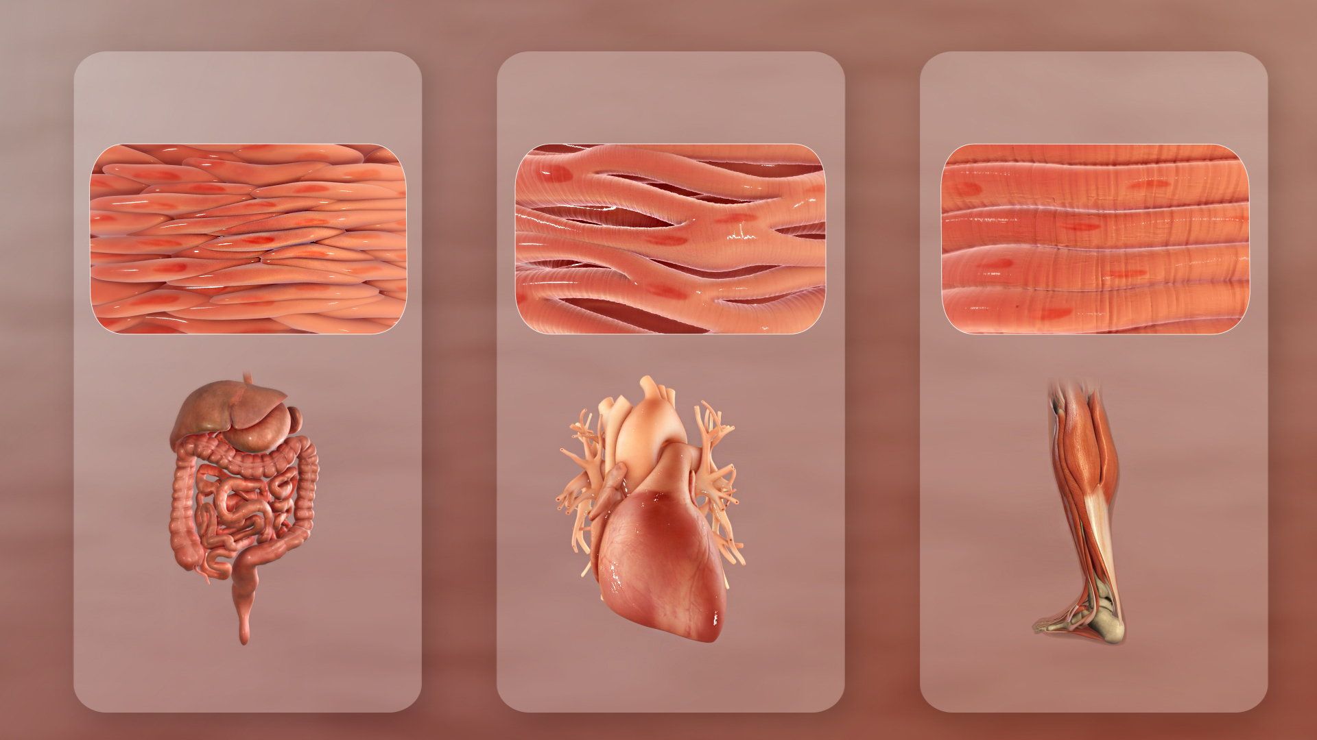
The muscular system is an organ (anatomy), organ system consisting of skeletal muscle, skeletal, smooth muscle, smooth, and cardiac muscle, cardiac muscle. It permits movement of the body, maintains posture, and circulates blood throughout the body. The muscular systems in vertebrates are controlled through the nervous system although some muscles (such as the cardiac muscle) can be completely autonomous. Together with the skeletal system in the human, it forms the musculoskeletal system, which is responsible for the movement of the human body, body. Types There are three distinct types of muscle: skeletal muscle, cardiac muscle, cardiac or heart muscle, and smooth muscle, smooth (non-striated) muscle. Muscles provide strength, balance, posture, movement, and heat for the body to keep warm. There are over 650 muscles in the human body. A kind of elastic tissue makes up each muscle, which consists of thousands, or tens of thousands, of small muscle fibers. Each fiber comprises ... [...More Info...] [...Related Items...] OR: [Wikipedia] [Google] [Baidu] |

