|
Thigh
In anatomy, the thigh is the area between the hip (pelvis) and the knee. Anatomically, it is part of the lower limb. The single bone in the thigh is called the femur. This bone is very thick and strong (due to the high proportion of bone tissue), and forms a ball and socket joint at the hip, and a modified hinge joint at the knee. Structure Bones The femur is the only bone in the thigh and serves as an attachment site for all thigh muscles. The head of the femur articulates with the acetabulum in the pelvic bone forming the hip joint, while the distal part of the femur articulates with the tibia and patella forming the knee. By most measures, the femur is the strongest and longest bone in the body. The femur is categorised as a long bone and comprises a diaphysis, the shaft (or body) and two epiphyses, the lower extremity and the upper extremity of femur, that articulate with adjacent bones in the hip and knee. Muscular compartments In cross-section, the thigh is d ... [...More Info...] [...Related Items...] OR: [Wikipedia] [Google] [Baidu] |
Posterior Fascial Compartment Of Thigh
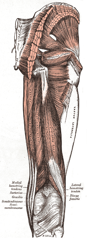
The posterior compartment of the thigh is one of the fascial compartments of thigh, fascial compartments that contains the knee anatomical terms of motion#flexion, flexors and hip anatomical terms of motion#extension, extensors known as the hamstring muscles, as well as vascular and nervous elements, particularly the sciatic nerve. Structure The posterior compartment is a fascial compartment bounded by fascia. It is separated from the anterior compartment of thigh, anterior compartment by two folds of fascia lata, deep fascia, known as the Medial intermuscular septum of thigh, medial intermuscular septum and the Lateral intermuscular septum of thigh, lateral intermuscular septum. The muscles of the posterior compartment of the thigh are the: * biceps femoris muscle, which consists of a short head and a long head. * semitendinosus muscle * semimembranosus muscle These muscles (or their tendons) apart from the short head of the biceps femoris, are commonly known as the hamstrings. ... [...More Info...] [...Related Items...] OR: [Wikipedia] [Google] [Baidu] |
Anterior Fascial Compartment Of Thigh
The anterior compartment of thigh contains muscles which extend the knee and flex the hip. Structure The anterior compartment is one of the fascial compartments of the thigh that contains groups of muscles together with their nerves and blood supply. The anterior compartment contains the sartorius muscle (the longest muscle in the body) and the quadriceps femoris group, which consists of the rectus femoris muscle and the three vasti muscles – the vastus lateralis, vastus intermedius, and the vastus medialis. The iliopsoas is sometimes considered a member of the anterior compartment muscles, as is the articularis genus muscle. The anterior compartment is separated from the posterior compartment by the lateral intermuscular septum and from the medial compartment by the medial intermuscular septum. Image:Gray430.png, Anterior aspect of right leg. Image:Illu lower extremity muscles.jpg, Muscles of leg Nerve supply The nerve of the anterior compartment of thigh is the fe ... [...More Info...] [...Related Items...] OR: [Wikipedia] [Google] [Baidu] |
Femur
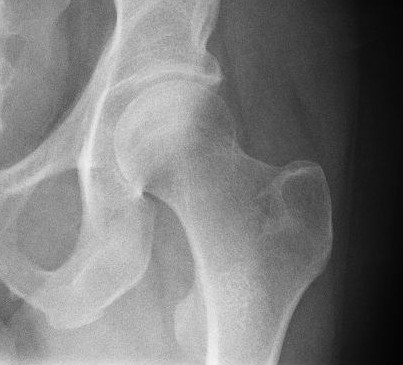
The femur (; : femurs or femora ), or thigh bone is the only long bone, bone in the thigh — the region of the lower limb between the hip and the knee. In many quadrupeds, four-legged animals the femur is the upper bone of the hindleg. The Femoral head, top of the femur fits into a socket in the pelvis called the hip joint, and the bottom of the femur connects to the shinbone (tibia) and kneecap (patella) to form the knee. In humans the femur is the largest and thickest bone in the body. Structure The femur is the only bone in the upper Human leg, leg. The two femurs converge Anatomical terms of location, medially toward the knees, where they articulate with the Anatomical terms of location, proximal ends of the tibiae. The angle at which the femora converge is an important factor in determining the femoral-tibial angle. In females, thicker pelvic bones cause the femora to converge more than in males. In the condition genu valgum, ''genu valgum'' (knock knee), the femurs conve ... [...More Info...] [...Related Items...] OR: [Wikipedia] [Google] [Baidu] |
Pelvis
The pelvis (: pelves or pelvises) is the lower part of an Anatomy, anatomical Trunk (anatomy), trunk, between the human abdomen, abdomen and the thighs (sometimes also called pelvic region), together with its embedded skeleton (sometimes also called bony pelvis or pelvic skeleton). The pelvic region of the trunk includes the bony pelvis, the pelvic cavity (the space enclosed by the bony pelvis), the pelvic floor, below the pelvic cavity, and the perineum, below the pelvic floor. The pelvic skeleton is formed in the area of the back, by the sacrum and the coccyx and anteriorly and to the left and right sides, by a pair of hip bones. The two hip bones connect the spine with the lower limbs. They are attached to the sacrum posteriorly, connected to each other anteriorly, and joined with the two femurs at the hip joints. The gap enclosed by the bony pelvis, called the pelvic cavity, is the section of the body underneath the abdomen and mainly consists of the reproductive organs and ... [...More Info...] [...Related Items...] OR: [Wikipedia] [Google] [Baidu] |
Sartorius Muscle
The sartorius muscle () is the longest muscle in the human body. It is a long, thin, superficial muscle that runs down the length of the thigh in the anterior compartment. Structure The sartorius muscle originates from the anterior superior iliac spine, and part of the notch between the anterior superior iliac spine and anterior inferior iliac spine. It runs obliquely across the upper and anterior part of the thigh in an inferomedial direction. It passes behind the medial condyle of the femur to end in a tendon. This tendon curves anteriorly to join the tendons of the gracilis and semitendinosus muscles in the pes anserinus, where it inserts into the superomedial surface of the tibia. Its upper portion forms the lateral border of the femoral triangle, and the point where it crosses adductor longus marks the apex of the triangle. Deep to sartorius and its fascia is the adductor canal, through which the saphenous nerve, femoral artery and vein, and nerve to vastus medialis pa ... [...More Info...] [...Related Items...] OR: [Wikipedia] [Google] [Baidu] |
Medial Fascial Compartment Of Thigh
The medial compartment of thigh is one of the fascial compartments of the thigh and contains the hip adductor muscles and the gracilis muscle. The obturator nerve is the primary nerve supplying this compartment. The obturator artery is the blood supply to the medial thigh. The muscles in the compartment are: * gracilis * adductor longus * adductor brevis * adductor magnus The obturator externus muscle is sometimes considered part of this group, and sometimes excluded. (Spatially, it is in this location, but functionally, it is more similar to the other lateral rotator group muscles). The pectineus The pectineus muscle (, from the Latin word ''pecten'', meaning comb) is a flat, quadrangular muscle, situated at the anterior (front) part of the upper and medial (inner) aspect of the thigh. The pectineus muscle is the most anterior adductor o ... is sometimes included in this group, and sometimes excluded. It has the same function as the others in this group, but different ... [...More Info...] [...Related Items...] OR: [Wikipedia] [Google] [Baidu] |
Hip Joint
In vertebrate anatomy, the hip, or coxaLatin ''coxa'' was used by Celsus in the sense "hip", but by Pliny the Elder in the sense "hip bone" (Diab, p 77) (: ''coxae'') in medical terminology, refers to either an anatomical region or a joint on the outer (lateral) side of the pelvis. The hip region is located lateral and anterior to the gluteal region, inferior to the iliac crest, and lateral to the obturator foramen, with muscle tendons and soft tissues overlying the greater trochanter of the femur The femur (; : femurs or femora ), or thigh bone is the only long bone, bone in the thigh — the region of the lower limb between the hip and the knee. In many quadrupeds, four-legged animals the femur is the upper bone of the hindleg. The Femo .... In adults, the three pelvic bones (ilium (bone), ilium, ischium and pubis (bone), pubis) have fused into one hip bone, which forms the superomedial/deep wall of the hip region. The hip joint, scientifically referred to as t ... [...More Info...] [...Related Items...] OR: [Wikipedia] [Google] [Baidu] |
Fascia
A fascia (; : fasciae or fascias; adjective fascial; ) is a generic term for macroscopic membranous bodily structures. Fasciae are classified as superficial, visceral or deep, and further designated according to their anatomical location. The knowledge of fascial structures is essential in surgery, as they create borders for infectious processes (for example Psoas abscess) and haematoma. An increase in pressure may result in a compartment syndrome, where a prompt fasciotomy may be necessary. For this reason, profound descriptions of fascial structures are available in anatomical literature from the 19th century. Function Fasciae were traditionally thought of as passive structures that transmit mechanical tension generated by muscular activities or external forces throughout the body. An important function of muscle fasciae is to reduce friction of muscular force. In doing so, fasciae provide a supportive and movable wrapping for nerves and blood vessels as they pass thro ... [...More Info...] [...Related Items...] OR: [Wikipedia] [Google] [Baidu] |
Knee
In humans and other primates, the knee joins the thigh with the leg and consists of two joints: one between the femur and tibia (tibiofemoral joint), and one between the femur and patella (patellofemoral joint). It is the largest joint in the human body. The knee is a modified hinge joint, which permits flexion and extension (kinesiology), extension as well as slight internal and external rotation. The knee is vulnerable to injury and to the development of osteoarthritis. It is often termed a ''compound joint'' having tibiofemoral and patellofemoral components. (The fibular collateral ligament is often considered with tibiofemoral components.) Structure The knee is a modified hinge joint, a type of synovial joint, which is composed of three functional compartments: the patellofemoral articulation, consisting of the patella, or "kneecap", and the patellar groove on the front of the femur through which it slides; and the medial and lateral tibiofemoral articulations linking the ... [...More Info...] [...Related Items...] OR: [Wikipedia] [Google] [Baidu] |
Hamstring
A hamstring () is any one of the three posterior thigh muscles in human anatomy between the hip and the knee: from medial to lateral, the semimembranosus, semitendinosus and biceps femoris. Etymology The word " ham" is derived from the Old English “ham” or “hom” meaning the hollow or bend of the knee, from a Germanic base where it meant "crooked". It gained the meaning of the leg of an animal around the 15th century. ''String'' refers to tendons, and thus the hamstrings' string-like tendons felt on either side of the back of the knee. Criteria The common criteria of any hamstring muscles are: # Muscles should originate from ischial tuberosity. # Muscles should be inserted over the knee joint, in the tibia or in the fibula. # Muscles will be innervated by the tibial branch of the sciatic nerve. # Muscle will participate in flexion of the knee joint and extension of the hip joint. Those muscles which fulfill all of the four criteria are called true hamstrings. ... [...More Info...] [...Related Items...] OR: [Wikipedia] [Google] [Baidu] |
Pelvic Bone
The hip bone (os coxae, innominate bone, pelvic bone or coxal bone) is a large flat bone, constricted in the center and expanded above and below. In some vertebrates (including humans before puberty) it is composed of three parts: the ilium, ischium, and the pubis. The two hip bones join at the pubic symphysis and together with the sacrum and coccyx (the pelvic part of the spine) comprise the skeletal component of the pelvis – the pelvic girdle which surrounds the pelvic cavity. They are connected to the sacrum, which is part of the axial skeleton, at the sacroiliac joint. Each hip bone is connected to the corresponding femur (thigh bone) (forming the primary connection between the bones of the lower limb and the axial skeleton) through the large ball and socket joint of the hip. Structure The hip bone is formed by three parts: the ilium, ischium, and pubis. At birth, these three components are separated by hyaline cartilage. They join each other in a Y-shaped portion of ... [...More Info...] [...Related Items...] OR: [Wikipedia] [Google] [Baidu] |
Upper Extremity Of Femur
The upper extremity, proximal extremity or superior epiphysis of the femur is the part of the femur closest to the pelvic bone and the trunk. It contains the following structures: * Femoral head including the fovea * Femur neck * Greater trochanter * Lesser trochanter * Intertrochanteric line * Intertrochanteric crest * Trochanteric fossa * Linea quadrata * Quadrate tubercle The head of femur, which articulates with the acetabulum of the pelvic bone, composes two-thirds of a sphere. It has a small groove or fovea, connected through the round ligament to the sides of the acetabular notch. The head of the femur is connected to the shaft through the neck or ''collum''. The neck is 4–5 cm. long and the diameter is smallest front to back and compressed at its middle. The collum forms an angle with the shaft in about 130 degrees. This angle is highly variant. In the infant it is about 150 degrees and in old age reduced to 120 degrees in average. An abnormal increase in t ... [...More Info...] [...Related Items...] OR: [Wikipedia] [Google] [Baidu] |


