|
Suspensory Muscle Of Duodenum
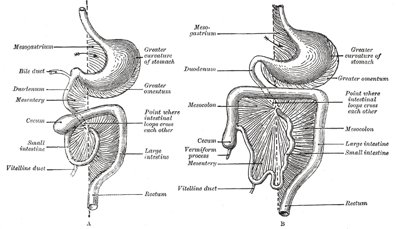
The suspensory muscle of duodenum is a thin muscle connecting the junction between the duodenum, jejunum, and duodenojejunal flexure to connective tissue surrounding the superior mesenteric artery and coeliac artery. It is also known as the ligament of Treitz. The suspensory muscle most often connects to both the third and fourth parts of the duodenum, as well as the duodenojejunal flexure, although the attachment is quite variable. The suspensory muscle marks the formal division between the first and second parts of the small intestine, the duodenum and the jejunum. This division is used to mark the difference between the upper and lower gastrointestinal tracts, which is relevant in clinical medicine as it may determine the source of bleeding in the gastrointestinal tract. The suspensory muscle is derived from mesoderm and plays a role in the embryological rotation of the gut, by offering a point of fixation for the rotating gut. It is also thought to help digestion by widenin ... [...More Info...] [...Related Items...] OR: [Wikipedia] [Google] [Baidu] |
Duodenum
The duodenum is the first section of the small intestine in most higher vertebrates, including mammals, reptiles, and birds. In fish, the divisions of the small intestine are not as clear, and the terms anterior intestine or proximal intestine may be used instead of duodenum. In mammals the duodenum may be the principal site for iron absorption. The duodenum precedes the jejunum and ileum and is the shortest part of the small intestine. In humans, the duodenum is a hollow jointed tube about 25–38 cm (10–15 inches) long connecting the stomach to the middle part of the small intestine. It begins with the duodenal bulb and ends at the suspensory muscle of duodenum. Duodenum can be divided into four parts: the first (superior), the second (descending), the third (horizontal) and the fourth (ascending) parts. Structure The duodenum is a C-shaped structure lying adjacent to the stomach. It is divided anatomically into four sections. The first part of the duodenu ... [...More Info...] [...Related Items...] OR: [Wikipedia] [Google] [Baidu] |
Mesoderm
The mesoderm is the middle layer of the three germ layers that develops during gastrulation in the very early development of the embryo of most animals. The outer layer is the ectoderm, and the inner layer is the endoderm.Langman's Medical Embryology, 11th edition. 2010. The mesoderm forms mesenchyme, mesothelium, non-epithelial blood cells and coelomocytes. Mesothelium lines coeloms. Mesoderm forms the muscles in a process known as myogenesis, septa (cross-wise partitions) and mesenteries (length-wise partitions); and forms part of the gonads (the rest being the gametes). Myogenesis is specifically a function of mesenchyme. The mesoderm differentiates from the rest of the embryo through intercellular signaling, after which the mesoderm is polarized by an organizing center. The position of the organizing center is in turn determined by the regions in which beta-catenin is protected from degradation by GSK-3. Beta-catenin acts as a co-factor that alters the activity of ... [...More Info...] [...Related Items...] OR: [Wikipedia] [Google] [Baidu] |
Anatomy
Anatomy () is the branch of biology concerned with the study of the structure of organisms and their parts. Anatomy is a branch of natural science that deals with the structural organization of living things. It is an old science, having its beginnings in prehistoric times. Anatomy is inherently tied to developmental biology, embryology, comparative anatomy, evolutionary biology, and phylogeny, as these are the processes by which anatomy is generated, both over immediate and long-term timescales. Anatomy and physiology, which study the structure and function of organisms and their parts respectively, make a natural pair of related disciplines, and are often studied together. Human anatomy is one of the essential basic sciences that are applied in medicine. The discipline of anatomy is divided into macroscopic and microscopic. Macroscopic anatomy, or gross anatomy, is the examination of an animal's body parts using unaided eyesight. Gross anatomy also includes the br ... [...More Info...] [...Related Items...] OR: [Wikipedia] [Google] [Baidu] |
Human Gastrointestinal Tract
The gastrointestinal tract (GI tract, digestive tract, alimentary canal) is the tract or passageway of the digestive system that leads from the mouth to the anus. The GI tract contains all the major organs of the digestive system, in humans and other animals, including the esophagus, stomach, and intestines. Food taken in through the mouth is digested to extract nutrients and absorb energy, and the waste expelled at the anus as feces. ''Gastrointestinal'' is an adjective meaning of or pertaining to the stomach and intestines. Most animals have a "through-gut" or complete digestive tract. Exceptions are more primitive ones: sponges have small pores (ostia) throughout their body for digestion and a larger dorsal pore ( osculum) for excretion, comb jellies have both a ventral mouth and dorsal anal pores, while cnidarians and acoels have a single pore for both digestion and excretion. The human gastrointestinal tract consists of the esophagus, stomach, and intestines, and is di ... [...More Info...] [...Related Items...] OR: [Wikipedia] [Google] [Baidu] |
Embryology
Embryology (from Greek ἔμβρυον, ''embryon'', "the unborn, embryo"; and -λογία, '' -logia'') is the branch of animal biology that studies the prenatal development of gametes (sex cells), fertilization, and development of embryos and fetuses. Additionally, embryology encompasses the study of congenital disorders that occur before birth, known as teratology. Early embryology was proposed by Marcello Malpighi, and known as preformationism, the theory that organisms develop from pre-existing miniature versions of themselves. Aristotle proposed the theory that is now accepted, epigenesis. Epigenesis is the idea that organisms develop from seed or egg in a sequence of steps. Modern embryology, developed from the work of Karl Ernst von Baer, though accurate observations had been made in Italy by anatomists such as Aldrovandi and Leonardo da Vinci in the Renaissance. Comparative embryology Preformationism and epigenesis As recently as the 18th century, the prev ... [...More Info...] [...Related Items...] OR: [Wikipedia] [Google] [Baidu] |
Right Crus Of Diaphragm
The crus of diaphragm (pl. crura), refers to one of two tendinous structures that extends below the diaphragm to the vertebral column. There is a right crus and a left crus, which together form a tether for muscular contraction. They take their name from their leg-shaped appearance – ''crus'' meaning ''leg'' in Latin. Structure The crura originate from the front of the bodies and intervertebral fibrocartilage of the lumbar vertebrae. They are tendinous and blend with the anterior longitudinal ligament of the vertebral column. * The ''right crus'', larger and longer than the left, arises from the front of the bodies and intervertebral fibrocartilages of the upper three lumbar vertebrae. * The ''left crus'' arises from the corresponding parts of the upper two lumbar vertebrae only. The medial tendinous margins of the crura pass anteriorly and medialward, and meet in the middle line to form an arch across the front of the aorta known as the median arcuate ligament; this arch is ... [...More Info...] [...Related Items...] OR: [Wikipedia] [Google] [Baidu] |
Anatomic Variation
An anatomical variation, anatomical variant, or anatomical variability is a presentation of body structure with morphological features different from those that are typically described in the majority of individuals. Anatomical variations are categorized into three types including morphometric (size or shape), consistency (present or absent), and spatial (proximal/distal or right/left). Variations are seen as normal in the sense that they are found consistently among different individuals, are mostly without symptoms, and are termed anatomical variations rather than abnormalities. Anatomical variations are mainly caused by genetics and may vary considerably between different populations. The rate of variation considerably differs between single organs, particularly in muscles. Knowledge of anatomical variations is important in order to distinguish them from pathological conditions. A very early paper published in 1898, presented anatomic variations to have a wide range and signi ... [...More Info...] [...Related Items...] OR: [Wikipedia] [Google] [Baidu] |
Mesentery
The mesentery is an organ that attaches the intestines to the posterior abdominal wall in humans and is formed by the double fold of peritoneum. It helps in storing fat and allowing blood vessels, lymphatics, and nerves to supply the intestines, among other functions. The mesocolon was thought to be a fragmented structure, with all named parts—the ascending, transverse, descending, and sigmoid mesocolons, the mesoappendix, and the mesorectum—separately terminating their insertion into the posterior abdominal wall. However, in 2012, new microscopic and electron microscopic examinations showed the mesocolon to be a single structure derived from the duodenojejunal flexure and extending to the distal mesorectal layer. Thus, the mesentery is an internal organ. Structure The mesentery of the small intestine arises from the root of the mesentery (or mesenteric root) and is the part connected with the structures in front of the vertebral column. The root is narrow, about 15 ... [...More Info...] [...Related Items...] OR: [Wikipedia] [Google] [Baidu] |
Pancreas
The pancreas is an organ of the digestive system and endocrine system of vertebrates. In humans, it is located in the abdomen behind the stomach and functions as a gland. The pancreas is a mixed or heterocrine gland, i.e. it has both an endocrine and a digestive exocrine function. 99% of the pancreas is exocrine and 1% is endocrine. As an endocrine gland, it functions mostly to regulate blood sugar levels, secreting the hormones insulin, glucagon, somatostatin, and pancreatic polypeptide. As a part of the digestive system, it functions as an exocrine gland secreting pancreatic juice into the duodenum through the pancreatic duct. This juice contains bicarbonate, which neutralizes acid entering the duodenum from the stomach; and digestive enzymes, which break down carbohydrates, proteins, and fats in food entering the duodenum from the stomach. Inflammation of the pancreas is known as pancreatitis, with common causes including chronic alcohol use and gallstone ... [...More Info...] [...Related Items...] OR: [Wikipedia] [Google] [Baidu] |
Celiac Trunk
The celiac () artery (also spelled ''coeliac''), also known as the celiac trunk or truncus coeliacus, is the first major branch of the abdominal aorta. It is about 1.25 cm in length. Branching from the aorta at thoracic vertebra 12 (T12) in humans, it is one of three anterior/ midline branches of the abdominal aorta (the others are the superior and inferior mesenteric arteries). Structure The celiac artery is the first major branch of the descending abdominal aorta, branching at a 90° angle. This occurs just below the crus of the diaphragm. This is around the first lumbar vertebra. There are three main divisions of the celiac artery, and each in turn has its own named branches: The celiac artery may also give rise to the inferior phrenic arteries. Function The celiac artery supplies oxygenated blood to the liver, stomach, abdominal esophagus, spleen, and the superior half of both the duodenum and the pancreas. These structures correspond to the embryonic foregut. (Si ... [...More Info...] [...Related Items...] OR: [Wikipedia] [Google] [Baidu] |
Esophagus
The esophagus (American English) or oesophagus (British English; both ), non-technically known also as the food pipe or gullet, is an organ in vertebrates through which food passes, aided by peristaltic contractions, from the pharynx to the stomach. The esophagus is a fibromuscular tube, about long in adults, that travels behind the trachea and heart, passes through the diaphragm, and empties into the uppermost region of the stomach. During swallowing, the epiglottis tilts backwards to prevent food from going down the larynx and lungs. The word ''oesophagus'' is from Ancient Greek οἰσοφάγος (oisophágos), from οἴσω (oísō), future form of φέρω (phérō, “I carry”) + ἔφαγον (éphagon, “I ate”). The wall of the esophagus from the lumen outwards consists of mucosa, submucosa (connective tissue), layers of muscle fibers between layers of fibrous tissue, and an outer layer of connective tissue. The mucosa is a stratified squamous epith ... [...More Info...] [...Related Items...] OR: [Wikipedia] [Google] [Baidu] |
Thoracic Diaphragm
The thoracic diaphragm, or simply the diaphragm ( grc, διάφραγμα, diáphragma, partition), is a sheet of internal skeletal muscle in humans and other mammals that extends across the bottom of the thoracic cavity. The diaphragm is the most important muscle of respiration, and separates the thoracic cavity, containing the heart and lungs, from the abdominal cavity: as the diaphragm contracts, the volume of the thoracic cavity increases, creating a negative pressure there, which draws air into the lungs. Its high oxygen consumption is noted by the many mitochondria and capillaries present; more than in any other skeletal muscle. The term ''diaphragm'' in anatomy, created by Gerard of Cremona, can refer to other flat structures such as the urogenital diaphragm or pelvic diaphragm, but "the diaphragm" generally refers to the thoracic diaphragm. In humans, the diaphragm is slightly asymmetric—its right half is higher up (superior) to the left half, since the large ... [...More Info...] [...Related Items...] OR: [Wikipedia] [Google] [Baidu] |


