|
Thoracic Diaphragm
The thoracic diaphragm, or simply the diaphragm ( grc, διάφραγμα, diáphragma, partition), is a sheet of internal skeletal muscle in humans and other mammals that extends across the bottom of the thoracic cavity. The diaphragm is the most important muscle of respiration, and separates the thoracic cavity, containing the heart and lungs, from the abdominal cavity: as the diaphragm contracts, the volume of the thoracic cavity increases, creating a negative pressure there, which draws air into the lungs. Its high oxygen consumption is noted by the many mitochondria and capillaries present; more than in any other skeletal muscle. The term ''diaphragm'' in anatomy, created by Gerard of Cremona, can refer to other flat structures such as the urogenital diaphragm or pelvic diaphragm, but "the diaphragm" generally refers to the thoracic diaphragm. In humans, the diaphragm is slightly asymmetric—its right half is higher up (superior) to the left half, since the large ... [...More Info...] [...Related Items...] OR: [Wikipedia] [Google] [Baidu] |
Septum Transversum
The septum transversum is a thick mass of cranial mesenchyme, formed in the embryo, that gives rise to parts of the thoracic diaphragm and the ventral mesentery of the foregut in the developed human being and other mammals. Origins The septum transversum originally arises as the most cranial part of the mesenchyme on day 22. During craniocaudal folding, it assumes a position cranial to the developing heart at the level of the cervical vertebrae. During subsequent weeks the dorsal end of the embryo grows much faster than its ventral counterpart resulting in an ''apparent descent'' of the ventrally located septum transversum. At week 8, it can be found at the level of the thoracic vertebrae. Nerve supply After successful craniocaudal folding the septum transversum picks up innervation from the adjacent ventral rami of spinal nerves C3, C4 and C5, thus forming the precursor of the phrenic nerve. During the descent of the septum, the phrenic nerve is carried along and assumes its d ... [...More Info...] [...Related Items...] OR: [Wikipedia] [Google] [Baidu] |
Abdominal Cavity
The abdominal cavity is a large body cavity in humans and many other animals that contains many organs. It is a part of the abdominopelvic cavity. It is located below the thoracic cavity, and above the pelvic cavity. Its dome-shaped roof is the thoracic diaphragm, a thin sheet of muscle under the lungs, and its floor is the pelvic inlet, opening into the pelvis. Structure Organs Organs of the abdominal cavity include the stomach, liver, gallbladder, spleen, pancreas, small intestine, kidneys, large intestine, and adrenal glands. Peritoneum The abdominal cavity is lined with a protective membrane termed the peritoneum. The inside wall is covered by the parietal peritoneum. The kidneys are located behind the peritoneum, in the retroperitoneum, outside the abdominal cavity. The viscera are also covered by visceral peritoneum. Between the visceral and parietal peritoneum is the peritoneal cavity, which is a potential space. It contains a serous fluid called periton ... [...More Info...] [...Related Items...] OR: [Wikipedia] [Google] [Baidu] |
Inferior Thoracic Aperture
The superior thoracic aperture, also known as the thoracic outlet, or thoracic inlet refers to the opening at the top of the thoracic cavity. It is also clinically referred to as the thoracic outlet, in the case of thoracic outlet syndrome. A lower thoracic opening is the ''inferior thoracic aperture''. Structure The superior thoracic aperture is essentially a hole surrounded by a bony ring, through which several vital structures pass. It is bounded by: the first thoracic vertebra (T1) ''posteriorly''; the first pair of ribs ''laterally'', forming lateral C-shaped curves posterior to anterior; and the costal cartilage of the first rib and the superior border of the manubrium ''anteriorly''. Dimensions The adult thoracic outlet is around 6.5 cm antero-posteriorly and 11 cm transversely. Because of the obliquity of the first pair of ribs, the aperture slopes antero-inferiorly. Relations The clavicle articulates with the manubrium to form the anterior border of the thoracic outle ... [...More Info...] [...Related Items...] OR: [Wikipedia] [Google] [Baidu] |
Central Tendon Of Diaphragm
The central tendon of the diaphragm is a thin but strong aponeurosis situated slightly anterior to the vault formed by the muscle, resulting in longer posterior muscle fibers. It is inferior to the fibrous pericardium, which fuses with the central tendon of the diaphragm via the pericardiacophrenic ligament. The caval opening (at the level of the T8 vertebra) passes through the central tendon. This transmits the inferior vena cava and right phrenic nerve. Structure The central tendon is shaped somewhat like a trefoil leaf, consisting of three divisions or leaflets separated from one another by slight indentations. The right leaflet is the largest, the middle (directed toward the xiphoid process) the next in size, and the left the smallest. The central tendon is composed of several planes of fibers, which intersect one another at various angles and unite into straight or curved bundles—an arrangement which gives it additional strength. Action during respiration During i ... [...More Info...] [...Related Items...] OR: [Wikipedia] [Google] [Baidu] |
Fibrous Tissue
Fiber or fibre (from la, fibra, links=no) is a natural or artificial substance that is significantly longer than it is wide. Fibers are often used in the manufacture of other materials. The strongest engineering materials often incorporate fibers, for example carbon fiber and ultra-high-molecular-weight polyethylene. Synthetic fibers can often be produced very cheaply and in large amounts compared to natural fibers, but for clothing natural fibers can give some benefits, such as comfort, over their synthetic counterparts. Natural fibers Natural fibers develop or occur in the fiber shape, and include those produced by plants, animals, and geological processes. They can be classified according to their origin: * Vegetable fibers are generally based on arrangements of cellulose, often with lignin: examples include cotton, hemp, jute, flax, abaca, piña, ramie, sisal, bagasse, and banana. Plant fibers are employed in the manufacture of paper and textile (cloth), ... [...More Info...] [...Related Items...] OR: [Wikipedia] [Google] [Baidu] |
Muscle Tissue
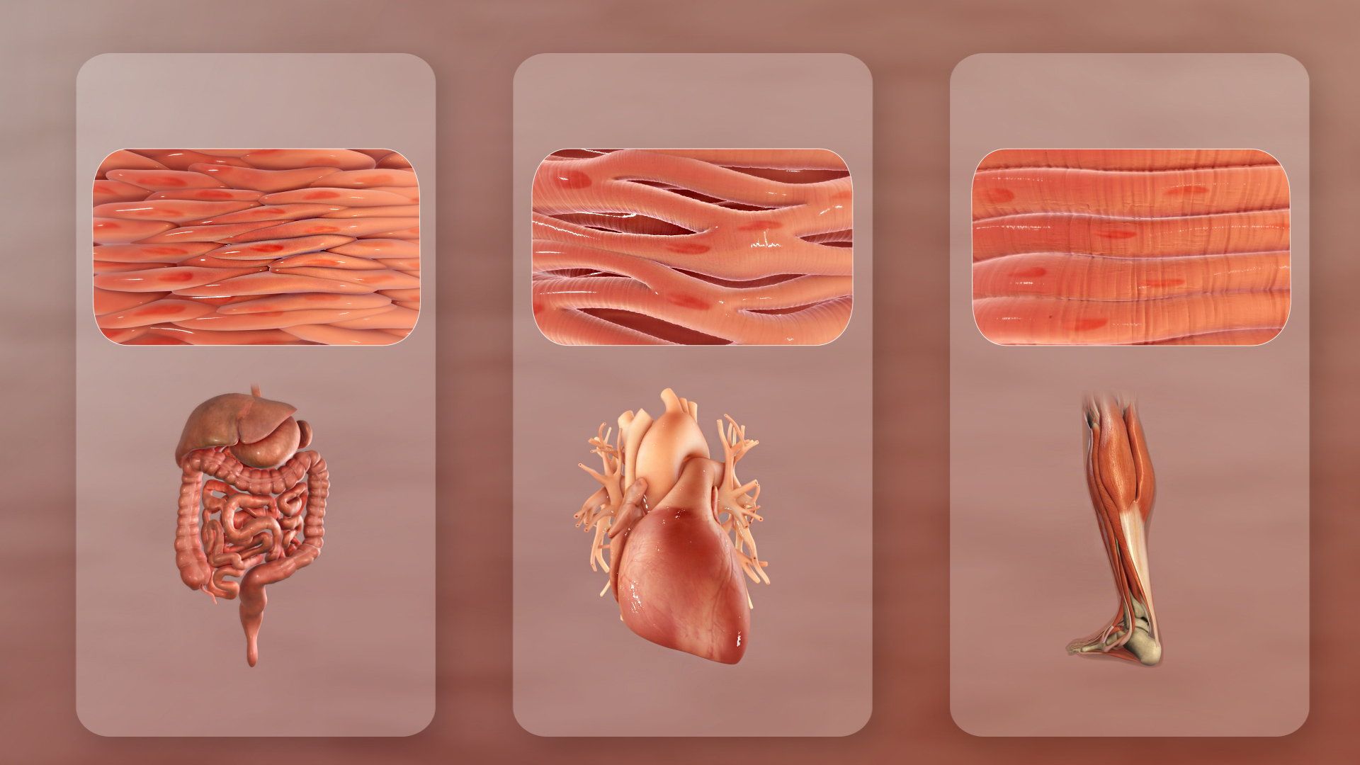
Muscle tissue (or muscular tissue) is soft tissue that makes up the different types of muscles in most animals, and give the ability of muscles to contract. Muscle tissue is formed during embryonic development, in a process known as myogenesis. Muscle tissue contains special contractile proteins called actin and myosin which contract and relax to cause movement. Among many other muscle proteins present are two regulatory proteins, troponin and tropomyosin. Muscle tissues vary with function and location in the body. In mammals the three types are: skeletal or striated muscle tissue; smooth muscle (non-striated) muscle; and cardiac muscle. Skeletal muscle tissue consists of elongated muscle cells called muscle fibers, and is responsible for movements of the body. Other tissues in skeletal muscle include tendons and perimysium. Smooth and cardiac muscle contract involuntarily, without conscious intervention. These muscle types may be activated both through the interaction ... [...More Info...] [...Related Items...] OR: [Wikipedia] [Google] [Baidu] |
Diaphragm Def 1707
Diaphragm may refer to: Anatomy * Thoracic diaphragm, a thin sheet of muscle between the thorax and the abdomen * Pelvic diaphragm or pelvic floor, a pelvic structure * Urogenital diaphragm or triangular ligament, a pelvic structure Other * Diaphragm (optics), a stop in the light path of a lens, having an aperture that regulates the amount of light that passes * Diaphragm (acoustics), a thin, semi-rigid membrane that vibrates to produce or transmit sound waves * Diaphragm (birth control), a small rubber dome placed in the vagina to wall off the cervix, thus preventing sperm from entering * Diaphragm (mechanical device), a sheet of a semi-flexible material anchored at its periphery * Diaphragm (structural system), a structural engineering system used to resist lateral loads See also * Diaphragm arch * Diaphragm pump * Diaphragm seal, a membrane that seals an enclosure * Diaphragm shutter, a type of leaf shutter consisting of a number of thin blades in a camera * Diaphragm va ... [...More Info...] [...Related Items...] OR: [Wikipedia] [Google] [Baidu] |
Reptile
Reptiles, as most commonly defined are the animals in the class Reptilia ( ), a paraphyletic grouping comprising all sauropsids except birds. Living reptiles comprise turtles, crocodilians, squamates (lizards and snakes) and rhynchocephalians ( tuatara). As of March 2022, the Reptile Database includes about 11,700 species. In the traditional Linnaean classification system, birds are considered a separate class to reptiles. However, crocodilians are more closely related to birds than they are to other living reptiles, and so modern cladistic classification systems include birds within Reptilia, redefining the term as a clade. Other cladistic definitions abandon the term reptile altogether in favor of the clade Sauropsida, which refers to all amniotes more closely related to modern reptiles than to mammals. The study of the traditional reptile orders, historically combined with that of modern amphibians, is called herpetology. The earliest known proto-reptiles originated ... [...More Info...] [...Related Items...] OR: [Wikipedia] [Google] [Baidu] |
Amphibian
Amphibians are four-limbed and ectothermic vertebrates of the class Amphibia. All living amphibians belong to the group Lissamphibia. They inhabit a wide variety of habitats, with most species living within terrestrial, fossorial, arboreal or freshwater aquatic ecosystems. Thus amphibians typically start out as larvae living in water, but some species have developed behavioural adaptations to bypass this. The young generally undergo metamorphosis from larva with gills to an adult air-breathing form with lungs. Amphibians use their skin as a secondary respiratory surface and some small terrestrial salamanders and frogs lack lungs and rely entirely on their skin. They are superficially similar to reptiles like lizards but, along with mammals and birds, reptiles are amniotes and do not require water bodies in which to breed. With their complex reproductive needs and permeable skins, amphibians are often ecological indicators; in recent decades there has been a dramat ... [...More Info...] [...Related Items...] OR: [Wikipedia] [Google] [Baidu] |
Vertebrate
Vertebrates () comprise all animal taxon, taxa within the subphylum Vertebrata () (chordates with vertebral column, backbones), including all mammals, birds, reptiles, amphibians, and fish. Vertebrates represent the overwhelming majority of the phylum Chordata, with currently about 69,963 species described. Vertebrates comprise such groups as the following: * Agnatha, jawless fish, which include hagfish and lampreys * Gnathostomata, jawed vertebrates, which include: ** Chondrichthyes, cartilaginous fish (sharks, Batoidea, rays, and Chimaeriformes, ratfish) ** Euteleostomi, bony vertebrates, which include: *** Actinopterygii, ray-fins (the majority of living Osteichthyes, bony fish) *** lobe-fins, which include: **** coelacanths and lungfish **** tetrapods (limbed vertebrates) Extant taxon, Extant vertebrates range in size from the frog species ''Paedophryne amauensis'', at as little as , to the blue whale, at up to . Vertebrates make up less than five percent of all described a ... [...More Info...] [...Related Items...] OR: [Wikipedia] [Google] [Baidu] |
Pelvic Floor
The pelvic floor or pelvic diaphragm is composed of muscle fibers of the levator ani, the coccygeus muscle, and associated connective tissue which span the area underneath the pelvis. The pelvic diaphragm is a muscular partition formed by the levatores ani and coccygei, with which may be included the parietal pelvic fascia on their upper and lower aspects. The pelvic floor separates the pelvic cavity above from the perineal region (including perineum) below. Both males and females have a pelvic floor. To accommodate the birth canal, a female's pelvic cavity is larger than a male's. Structure The right and left levator ani lie almost horizontally in the floor of the pelvis, separated by a narrow gap that transmits the urethra, vagina, and anal canal. The levator ani is usually considered in three parts: pubococcygeus, puborectalis, and iliococcygeus. The pubococcygeus, the main part of the levator, runs backward from the body of the pubis toward the coccyx and may be damaged dur ... [...More Info...] [...Related Items...] OR: [Wikipedia] [Google] [Baidu] |
Urogenital Diaphragm
Older texts have asserted the existence of a urogenital diaphragm, also called the triangular ligament, which was described as a layer of the pelvis that separates the deep perineal sac from the upper pelvis, lying between the inferior fascia of the urogenital diaphragm (perineal membrane) and superior fascia of the urogenital diaphragm. While this term is used to refer to a layer of the pelvis that separates the deep perineal sac from the upper pelvis, such a discrete border of the sac probably does not exist. While it has no official entry in Terminologia Anatomica, the term is still used occasionally to describe the muscular components of the deep perineal pouch. The urethra and the vagina, though part of the pouch, are usually said to be passing through the urogenital diaphragm, rather than part of the diaphragm itself. Some researchers still assert that such a diaphragm exists, and the term is still used in the literature. The urethral diaphragm is an anatomic landmar ... [...More Info...] [...Related Items...] OR: [Wikipedia] [Google] [Baidu] |


.png)

.png)