|
Celiac Trunk
The celiac () artery (also spelled ''coeliac''), also known as the celiac trunk or truncus coeliacus, is the first major branch of the abdominal aorta. It is about 1.25 cm in length. Branching from the aorta at thoracic vertebra 12 (T12) in humans, it is one of three anterior/ midline branches of the abdominal aorta (the others are the superior and inferior mesenteric arteries). Structure The celiac artery is the first major branch of the descending abdominal aorta, branching at a 90° angle. This occurs just below the crus of the diaphragm. This is around the first lumbar vertebra. There are three main divisions of the celiac artery, and each in turn has its own named branches: The celiac artery may also give rise to the inferior phrenic arteries. Function The celiac artery supplies oxygenated blood to the liver, stomach, abdominal esophagus, spleen, and the superior half of both the duodenum and the pancreas. These structures correspond to the embryonic foregut. ... [...More Info...] [...Related Items...] OR: [Wikipedia] [Google] [Baidu] |
Trunk (anatomy)
The torso or trunk is an anatomical term for the central part, or the core, of the body of many animals (including humans), from which the head, neck, limbs, tail and other appendages extend. The tetrapod torso — including that of a human — is usually divided into the ''thoracic'' segment (also known as the upper torso, where the forelimbs extend), the ''abdominal'' segment (also known as the "mid-section" or "midriff"), and the ''pelvic'' and '' perineal'' segments (sometimes known together with the abdomen as the lower torso, where the hindlimbs extend). Anatomy Major organs In humans, most critical organs, with the notable exception of the brain, are housed within the torso. In the upper chest, the heart and lungs are protected by the rib cage, and the abdomen contains most of the organs responsible for digestion: the stomach, which breaks down partially digested food via gastric acid; the liver, which respectively produces bile necessary for digestion; the large and smal ... [...More Info...] [...Related Items...] OR: [Wikipedia] [Google] [Baidu] |
Left Gastro-omental Artery
The left gastroepiploic artery (or left gastro-omental artery), the largest branch of the splenic artery, runs from left to right about a finger's breadth or more from the greater curvature of the stomach, between the layers of the greater omentum, and anastomoses with the right gastroepiploic (a branch of the right gastro-duodenal artery originating from the hepatic branch of the coeliac trunk). In its course it distributes: * "Gastric branches": several ascending branches to both surfaces of the stomach; * "Omental branches": descend to supply the greater omentum and anastomose with branches of the middle colic. Additional images File:Gray533.png, Branches of the celiac artery The celiac () artery (also spelled ''coeliac''), also known as the celiac trunk or truncus coeliacus, is the first major branch of the abdominal aorta. It is about 1.25 cm in length. Branching from the aorta at thoracic vertebra 12 (T12) in .... References External links * - "Stomach, Sp ... [...More Info...] [...Related Items...] OR: [Wikipedia] [Google] [Baidu] |
Perfusion
Perfusion is the passage of fluid through the circulatory system or lymphatic system to an organ or a tissue, usually referring to the delivery of blood to a capillary bed in tissue. Perfusion is measured as the rate at which blood is delivered to tissue, or volume of blood per unit time (blood flow) per unit tissue mass. The SI unit is m3/(s·kg), although for human organs perfusion is typically reported in ml/min/g. The word is derived from the French verb "perfuser" meaning to "pour over or through". All animal tissues require an adequate blood supply for health and life. Poor perfusion (malperfusion), that is, ischemia, causes health problems, as seen in cardiovascular disease, including coronary artery disease, cerebrovascular disease, peripheral artery disease, and many other conditions. Tests verifying that adequate perfusion exists are a part of a patient's assessment process that are performed by medical or emergency personnel. The most common methods include evalua ... [...More Info...] [...Related Items...] OR: [Wikipedia] [Google] [Baidu] |
Hindgut
The hindgut (or epigaster) is the posterior ( caudal) part of the alimentary canal. In mammals, it includes the distal one third of the transverse colon and the splenic flexure, the descending colon, sigmoid colon and up to the ano-rectal junction. In zoology, the term ''hindgut'' refers also to the cecum and ascending colon. Structure Blood supply Arterial supply is by the inferior mesenteric artery, and venous drainage is to the portal venous system. Lymphatic drainage is to the chyle cistern. Nerve supply The hindgut is innervated via the inferior mesenteric plexus. Sympathetic innervation is from the Lumbar splanchnic nerves (L1-L2), parasympathetic innervation is from S2-S4. Development Additional images File:Gray985.png, Abdominal part of digestive tube and its attachment to the primitive or common mesentery. Human embryo of six weeks. File:Gray1115.png, Tail end of human embryo twenty-five to twenty-nine days old. File:Illacme plenipes female with 170 segments and ... [...More Info...] [...Related Items...] OR: [Wikipedia] [Google] [Baidu] |
Midgut
The midgut is the portion of the embryo from which most of the intestines develop. After it bends around the superior mesenteric artery, it is called the "midgut loop". It comprises the portion of the alimentary canal from the end of the foregut at the opening of the bile duct to the hindgut, about two-thirds of the way through the transverse colon. In the embryo During development, the human midgut undergoes a rapid phase of growth in which the loop of midgut herniates outside of the abdominal cavity of the fetus and protrudes into the umbilical cord. This herniation is physiological (occurs normally). Later in development, the fetus's body catches up in size relative to the midgut and creates adequate room in the abdominal cavity for the entirety of the midgut to reside. The midgut loops slip back out of the umbilical cord and the physiological hernia ceases to exist. This change coincides with the termination of the yolk sac and the counterclockwise rotation of the two limbs ... [...More Info...] [...Related Items...] OR: [Wikipedia] [Google] [Baidu] |
Foregut
The foregut is the anterior part of the alimentary canal, from the mouth to the duodenum at the entrance of the bile duct. Beyond the stomach, the foregut is attached to the abdominal walls by mesentery. The foregut arises from the endoderm, developing from the folding primitive gut, and is developmentally distinct from the midgut and hindgut. Although the term “foregut” is typically used in reference to the anterior section of the primitive gut, components of the adult gut can also be described with this designation. Pain in the epigastric region, just below the intersection of the ribs, typically refers to structures in the adult foregut. Adult foregut Components * Esophagus * Respiratory tract (lower respiratory tract) * Stomach * Duodenum (up to ampulla of vater) * Liver * Gallbladder * Pancreas * Spleen – The spleen arises from the mesodermal dorsal mesentery (the foregut arises from the endoderm not mesoderm). But the spleen shares the same blood supply as many of the ... [...More Info...] [...Related Items...] OR: [Wikipedia] [Google] [Baidu] |
Pancreas
The pancreas is an organ of the digestive system and endocrine system of vertebrates. In humans, it is located in the abdomen behind the stomach and functions as a gland. The pancreas is a mixed or heterocrine gland, i.e. it has both an endocrine and a digestive exocrine function. 99% of the pancreas is exocrine and 1% is endocrine. As an endocrine gland, it functions mostly to regulate blood sugar levels, secreting the hormones insulin, glucagon, somatostatin, and pancreatic polypeptide. As a part of the digestive system, it functions as an exocrine gland secreting pancreatic juice into the duodenum through the pancreatic duct. This juice contains bicarbonate, which neutralizes acid entering the duodenum from the stomach; and digestive enzymes, which break down carbohydrates, proteins, and fats in food entering the duodenum from the stomach. Inflammation of the pancreas is known as pancreatitis, with common causes including chronic alcohol use and gallstones. Becaus ... [...More Info...] [...Related Items...] OR: [Wikipedia] [Google] [Baidu] |
Duodenum
The duodenum is the first section of the small intestine in most higher vertebrates, including mammals, reptiles, and birds. In fish, the divisions of the small intestine are not as clear, and the terms anterior intestine or proximal intestine may be used instead of duodenum. In mammals the duodenum may be the principal site for iron absorption. The duodenum precedes the jejunum and ileum and is the shortest part of the small intestine. In humans, the duodenum is a hollow jointed tube about 25–38 cm (10–15 inches) long connecting the stomach to the middle part of the small intestine. It begins with the duodenal bulb and ends at the suspensory muscle of duodenum. Duodenum can be divided into four parts: the first (superior), the second (descending), the third (horizontal) and the fourth (ascending) parts. Structure The duodenum is a C-shaped structure lying adjacent to the stomach. It is divided anatomically into four sections. The first part of the duodenum lies ... [...More Info...] [...Related Items...] OR: [Wikipedia] [Google] [Baidu] |
Spleen
The spleen is an organ found in almost all vertebrates. Similar in structure to a large lymph node, it acts primarily as a blood filter. The word spleen comes .σπλήν Henry George Liddell, Robert Scott, ''A Greek-English Lexicon'', on Perseus Digital Library The spleen plays very important roles in regard to s (erythrocytes) and the . It removes old red blood cells and holds a reserve of blood, which can be valuable in case of |
Esophagus
The esophagus (American English) or oesophagus (British English; both ), non-technically known also as the food pipe or gullet, is an organ in vertebrates through which food passes, aided by peristaltic contractions, from the pharynx to the stomach. The esophagus is a fibromuscular tube, about long in adults, that travels behind the trachea and heart, passes through the diaphragm, and empties into the uppermost region of the stomach. During swallowing, the epiglottis tilts backwards to prevent food from going down the larynx and lungs. The word ''oesophagus'' is from Ancient Greek οἰσοφάγος (oisophágos), from οἴσω (oísō), future form of φέρω (phérō, “I carry”) + ἔφαγον (éphagon, “I ate”). The wall of the esophagus from the lumen outwards consists of mucosa, submucosa (connective tissue), layers of muscle fibers between layers of fibrous tissue, and an outer layer of connective tissue. The mucosa is a stratified squamous epithel ... [...More Info...] [...Related Items...] OR: [Wikipedia] [Google] [Baidu] |
Stomach
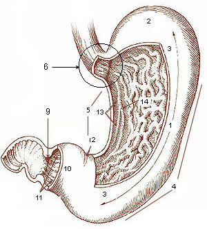
The stomach is a muscular, hollow organ in the gastrointestinal tract of humans and many other animals, including several invertebrates. The stomach has a dilated structure and functions as a vital organ in the digestive system. The stomach is involved in the gastric phase of digestion, following chewing. It performs a chemical breakdown by means of enzymes and hydrochloric acid. In humans and many other animals, the stomach is located between the oesophagus and the small intestine. The stomach secretes digestive enzymes and gastric acid to aid in food digestion. The pyloric sphincter controls the passage of partially digested food ( chyme) from the stomach into the duodenum, where peristalsis takes over to move this through the rest of intestines. Structure In the human digestive system, the stomach lies between the oesophagus and the duodenum (the first part of the small intestine). It is in the left upper quadrant of the abdominal cavity. The top of the stomach lies ag ... [...More Info...] [...Related Items...] OR: [Wikipedia] [Google] [Baidu] |
Liver
The liver is a major Organ (anatomy), organ only found in vertebrates which performs many essential biological functions such as detoxification of the organism, and the Protein biosynthesis, synthesis of proteins and biochemicals necessary for digestion and growth. In humans, it is located in the quadrant (anatomy), right upper quadrant of the abdomen, below the thoracic diaphragm, diaphragm. Its other roles in metabolism include the regulation of Glycogen, glycogen storage, decomposition of red blood cells, and the production of hormones. The liver is an accessory digestive organ that produces bile, an alkaline fluid containing cholesterol and bile acids, which helps the fatty acid degradation, breakdown of fat. The gallbladder, a small pouch that sits just under the liver, stores bile produced by the liver which is later moved to the small intestine to complete digestion. The liver's highly specialized biological tissue, tissue, consisting mostly of hepatocytes, regulates a w ... [...More Info...] [...Related Items...] OR: [Wikipedia] [Google] [Baidu] |


