|
Supramarginal Gyrus
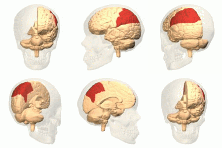
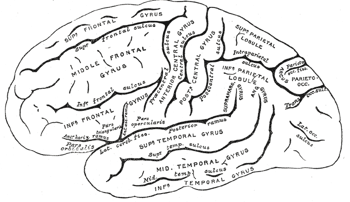
The supramarginal gyrus is a portion of the parietal lobe. This area of the brain is also known as Brodmann area 40 based on the brain map created by Korbinian Brodmann to define the structures in the cerebral cortex. It is probably involved with language perception and processing, and lesions in it may cause receptive aphasia. Important functions The supramarginal gyrus is part of the somatosensory association cortex, which interprets tactile sensory data and is involved in perception of space and limbs location. It is also involved in identifying postures and gestures of other people and is thus a part of the mirror neuron system. The right-hemisphere supramarginal gyrus appears to play a central role in controlling empathy towards other people. When this structure is not working properly or when individuals have to make very quick judgements, empathy becomes severely limited. Research has shown that disrupting the neurons in the right supramarginal gyrus causes humans to pr ... [...More Info...] [...Related Items...] OR: [Wikipedia] [Google] [Baidu] |
Parietal Lobe
The parietal lobe is one of the four major lobes of the cerebral cortex in the brain of mammals. The parietal lobe is positioned above the temporal lobe and behind the frontal lobe and central sulcus. The parietal lobe integrates sensory information among various modalities, including spatial sense and navigation (proprioception), the main sensory receptive area for the sense of touch in the somatosensory cortex which is just posterior to the central sulcus in the postcentral gyrus, and the dorsal stream of the visual system. The major sensory inputs from the skin (touch, temperature, and pain receptors), relay through the thalamus to the parietal lobe. Several areas of the parietal lobe are important in language processing. The somatosensory cortex can be illustrated as a distorted figure – the cortical homunculus (Latin: "little man") in which the body parts are rendered according to how much of the somatosensory cortex is devoted to them. The superior parietal lobule and in ... [...More Info...] [...Related Items...] OR: [Wikipedia] [Google] [Baidu] |
Brodmann Area 40
Brodmann area 40 (BA40) is part of the parietal cortex in the human brain. The inferior part of BA40 is in the area of the supramarginal gyrus, which lies at the posterior end of the lateral fissure, in the inferior lateral part of the parietal lobe. It is bounded approximately by the intraparietal sulcus, the inferior postcentral sulcus, the posterior subcentral sulcus and the lateral sulcus. It is bounded caudally by the angular area 39 (H), rostrally and dorsally by the caudal postcentral area 2, and ventrally by the subcentral area 43 and the superior temporal area 22 (Brodmann-1909). Cytoarchitectonically defined subregions of rostral BA40/the supramarginal gyrus are PF, PFcm, PFm, PFop, and PFt. Area PF is the homologue to macaque area PF, part of the mirror neuron system, and active in humans during imitation. The supramarginal gyrus part of Brodmann area 40 is the region in the inferior parietal lobe that is involved in reading both as regards meaning and phonolog ... [...More Info...] [...Related Items...] OR: [Wikipedia] [Google] [Baidu] |
Korbinian Brodmann
Korbinian Brodmann (17 November 1868 – 22 August 1918) was a German neurologist who became famous for mapping the cerebral cortex and defining 52 distinct regions, known as Brodmann areas, based on their cytoarchitectonic (histological) characteristics. Life and career Brodmann was born in Liggersdorf, Province of Hohenzollern. He studied medicine in Munich, Würzburg, Berlin, and Freiburg, where he received his medical diploma in 1895. Subsequently he studied at the Medical School in the University of Lausanne in Switzerland, and then worked in the University Clinic in Munich. He received a doctor of medicine degree from the University of Leipzig in 1898, with a thesis on chronic ependymal sclerosis. From 1900 to 1901, Brodmann also worked in the Psychiatric Clinic at the University of Jena, with Ludwig Binswanger, and in the Municipal Mental Asylum in Frankfurt. There, he met Alois Alzheimer, who was influential in his decision to pursue basic neuroscience research. Fol ... [...More Info...] [...Related Items...] OR: [Wikipedia] [Google] [Baidu] |
Receptive Aphasia
Wernicke's aphasia, also known as receptive aphasia, sensory aphasia or posterior aphasia, is a type of aphasia in which individuals have difficulty understanding written and spoken language. Patients with Wernicke's aphasia demonstrate fluent speech, which is characterized by typical speech rate, intact syntactic abilities and effortless speech output. Writing often reflects speech in that it tends to lack content or meaning. In most cases, motor deficits (i.e. hemiparesis) do not occur in individuals with Wernicke's aphasia. Therefore, they may produce a large amount of speech without much meaning. Individuals with Wernicke's aphasia are typically unaware of their errors in speech and do not realize their speech may lack meaning. They typically remain unaware of even their most profound language deficits. Like many acquired language disorders, Wernicke's aphasia can be experienced in many different ways and to many different degrees. Patients diagnosed with Wernicke's aphasia c ... [...More Info...] [...Related Items...] OR: [Wikipedia] [Google] [Baidu] |
Somatosensory System
In physiology, the somatosensory system is the network of neural structures in the brain and body that produce the perception of touch (haptic perception), as well as temperature (thermoception), body position (proprioception), and pain. It is a subset of the sensory nervous system, which also represents visual, auditory, olfactory, and gustatory stimuli. Somatosensation begins when mechano- and thermosensitive structures in the skin or internal organs sense physical stimuli such as pressure on the skin (see mechanotransduction, nociception). Activation of these structures, or receptors, leads to activation of peripheral sensory neurons that convey signals to the spinal cord as patterns of action potentials. Sensory information is then processed locally in the spinal cord to drive reflexes, and is also conveyed to the brain for conscious perception of touch and proprioception. Note, somatosensory information from the face and head enters the brain through periphera ... [...More Info...] [...Related Items...] OR: [Wikipedia] [Google] [Baidu] |
Mirror Neuron
A mirror neuron is a neuron that fires both when an animal acts and when the animal observes the same action performed by another. Thus, the neuron "mirrors" the behavior of the other, as though the observer were itself acting. Such neurons have been directly observed in human and primate species, and in birds. In humans, brain activity consistent with that of mirror neurons has been found in the premotor cortex, the supplementary motor area, the primary somatosensory cortex, and the inferior parietal cortex. The function of the mirror system in humans is a subject of much speculation. Birds have been shown to have imitative resonance behaviors and neurological evidence suggests the presence of some form of mirroring system. To date, no widely accepted neural or computational models have been put forward to describe how mirror neuron activity supports cognitive functions. The subject of mirror neurons continues to generate intense debate. In 2014, Philosophical Transactions o ... [...More Info...] [...Related Items...] OR: [Wikipedia] [Google] [Baidu] |
Anterior
Standard anatomical terms of location are used to unambiguously describe the anatomy of animals, including humans. The terms, typically derived from Latin or Greek roots, describe something in its standard anatomical position. This position provides a definition of what is at the front ("anterior"), behind ("posterior") and so on. As part of defining and describing terms, the body is described through the use of anatomical planes and anatomical axes. The meaning of terms that are used can change depending on whether an organism is bipedal or quadrupedal. Additionally, for some animals such as invertebrates, some terms may not have any meaning at all; for example, an animal that is radially symmetrical will have no anterior surface, but can still have a description that a part is close to the middle ("proximal") or further from the middle ("distal"). International organisations have determined vocabularies that are often used as standard vocabularies for subdisciplines of anatomy ... [...More Info...] [...Related Items...] OR: [Wikipedia] [Google] [Baidu] |
Angular Gyrus
The angular gyrus is a region of the brain lying mainly in the posteroinferior region of the parietal lobe, occupying the posterior part of the inferior parietal lobule. It represents the Brodmann area 39. Its significance is in transferring visual information to Wernicke's area, in order to make meaning out of visually perceived words. It is also involved in a number of processes related to language, number processing and spatial cognition, memory retrieval, attention, and theory of mind. Anatomy Connections Left and right angular gyri are connected by the dorsal splenium and isthmus of the corpus callosum. Boundaries * Anteriorly by the Supramarginal gyrus. * Superiorly by the Intraparietal sulcus. * Posteriorly by the Parieto-occipital sulcus. * Inferiorly the angular gyrus of the parietal lobe is continuous as the superior and middle temporal gyri. Also, the angular sulcus, which is capped by the angular gyrus, is continuous as the superior temporal sulcus inferio ... [...More Info...] [...Related Items...] OR: [Wikipedia] [Google] [Baidu] |
Inferior Parietal Lobule
The inferior parietal lobule (subparietal district) lies below the horizontal portion of the intraparietal sulcus, and behind the lower part of the postcentral sulcus. Also known as Geschwind's territory after Norman Geschwind, an American neurologist, who in the early 1960s recognised its importance. It is a part of the parietal lobe. Structure It is divided from rostral to caudal into two gyri: * One, the supramarginal gyrus, arches over the upturned end of the lateral fissure; it is continuous in front with the postcentral gyrus, and behind with the superior temporal gyrus. * The second, the angular gyrus, arches over the posterior end of the superior temporal sulcus, behind which it is continuous with the middle temporal gyrus. In macaque neuroanatomy, this region is often divided into caudal and rostral portions, cIPL and rIPL, respectively. The cIPL is further divided into areas Opt and PG whereas rIPL is divided into PFG and PF areas. Function Inferior parietal lobule has ... [...More Info...] [...Related Items...] OR: [Wikipedia] [Google] [Baidu] |
Lateral Sulcus
In neuroanatomy, the lateral sulcus (also called Sylvian fissure, after Franciscus Sylvius, or lateral fissure) is one of the most prominent features of the human brain. The lateral sulcus is a deep fissure in each hemisphere that separates the frontal and parietal lobes from the temporal lobe. The insular cortex lies deep within the lateral sulcus. Anatomy The lateral sulcus divides both the frontal lobe and parietal lobe above from the temporal lobe below. It is in both hemispheres of the brain. The lateral sulcus is one of the earliest-developing sulci of the human brain. It first appears around the fourteenth gestational week. The insular cortex lies deep within the lateral sulcus. The lateral sulcus has a number of side branches. Two of the most prominent and most regularly found are the ascending (also called vertical) ramus and the horizontal ramus of the lateral fissure, which subdivide the inferior frontal gyrus. The lateral sulcus also contains the transverse tempor ... [...More Info...] [...Related Items...] OR: [Wikipedia] [Google] [Baidu] |
Gyri
In neuroanatomy, a gyrus (pl. gyri) is a ridge on the cerebral cortex. It is generally surrounded by one or more sulci (depressions or furrows; sg. ''sulcus''). Gyri and sulci create the folded appearance of the brain in humans and other mammals. Structure The gyri are part of a system of folds and ridges that create a larger surface area for the human brain and other mammalian brains. Because the brain is confined to the skull, brain size is limited. Ridges and depressions create folds allowing a larger cortical surface area, and greater cognitive function, to exist in the confines of a smaller cranium. Development The human brain undergoes gyrification during fetal and neonatal development. In embryonic development, all mammalian brains begin as smooth structures derived from the neural tube. A cerebral cortex without surface convolutions is lissencephalic, meaning 'smooth-brained'. As development continues, gyri and sulci begin to take shape on the fetal brain, with ... [...More Info...] [...Related Items...] OR: [Wikipedia] [Google] [Baidu] |


