|
Brodmann Area 40
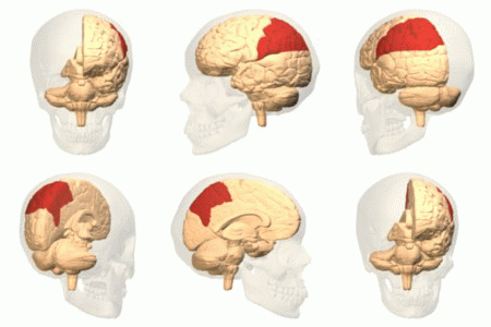
Brodmann area 40 (BA40) is part of the parietal cortex in the human brain. The inferior part of BA40 is in the area of the supramarginal gyrus, which lies at the posterior end of the lateral fissure, in the inferior lateral part of the parietal lobe. It is bounded approximately by the intraparietal sulcus, the inferior postcentral sulcus, the posterior subcentral sulcus and the lateral sulcus. It is bounded caudally by the angular area 39 (H), rostrally and dorsally by the caudal postcentral area 2, and ventrally by the subcentral area 43 and the superior temporal area 22 (Brodmann-1909). Cytoarchitectonically defined subregions of rostral BA40/the supramarginal gyrus are PF, PFcm, PFm, PFop, and PFt. Area PF is the homologue to macaque area PF, part of the mirror neuron system, and active in humans during imitation. The supramarginal gyrus part of Brodmann area 40 is the region in the inferior parietal lobe that is involved in reading both as regards meaning and p ... [...More Info...] [...Related Items...] OR: [Wikipedia] [Google] [Baidu] |
Parietal Lobe
The parietal lobe is one of the four major lobes of the cerebral cortex in the brain of mammals. The parietal lobe is positioned above the temporal lobe and behind the frontal lobe and central sulcus. The parietal lobe integrates sensory information among various modalities, including spatial sense and navigation (proprioception), the main sensory receptive area for the sense of touch in the somatosensory cortex which is just posterior to the central sulcus in the postcentral gyrus, and the dorsal stream of the visual system. The major sensory inputs from the skin ( touch, temperature, and pain receptors), relay through the thalamus to the parietal lobe. Several areas of the parietal lobe are important in language processing. The somatosensory cortex can be illustrated as a distorted figure – the cortical homunculus (Latin: "little man") in which the body parts are rendered according to how much of the somatosensory cortex is devoted to them. The superior parietal lobule a ... [...More Info...] [...Related Items...] OR: [Wikipedia] [Google] [Baidu] |
Caudal Postcentral Area 2
In neuroanatomy, the postcentral gyrus is a prominent gyrus in the lateral parietal lobe of the human brain. It is the location of the primary somatosensory cortex, the main sensory receptive area for the sense of touch. Like other sensory areas, there is a map of sensory space in this location, called the ''sensory homunculus''. The primary somatosensory cortex was initially defined from surface stimulation studies of Wilder Penfield, and parallel surface potential studies of Bard, Woolsey, and Marshall. Although initially defined to be roughly the same as Brodmann areas 3, 1 and 2, more recent work by Kaas has suggested that for homogeny with other sensory fields only area 3 should be referred to as "primary somatosensory cortex", as it receives the bulk of the thalamocortical projections from the sensory input fields. Structure The lateral postcentral gyrus is bounded by: *medial longitudinal fissure medially (to the middle) * central sulcus rostrally (in front) *postce ... [...More Info...] [...Related Items...] OR: [Wikipedia] [Google] [Baidu] |
List Of Regions In The Human Brain
The human brain anatomical regions are ordered following standard neuroanatomy hierarchies. Functional, connective, and developmental regions are listed in parentheses where appropriate. Hindbrain (rhombencephalon) Myelencephalon *Medulla oblongata **Medullary pyramids **Arcuate nucleus ** Olivary body *** Inferior olivary nucleus **Rostral ventrolateral medulla **Caudal ventrolateral medulla **Solitary nucleus (Nucleus of the solitary tract) **Respiratory center- Respiratory groups *** Dorsal respiratory group *** Ventral respiratory group or Apneustic centre ****Pre-Bötzinger complex **** Botzinger complex **** Retrotrapezoid nucleus **** Nucleus retrofacialis **** Nucleus retroambiguus **** Nucleus para-ambiguus ** Paramedian reticular nucleus ** Gigantocellular reticular nucleus **Parafacial zone ** Cuneate nucleus **Gracile nucleus ** Perihypoglossal nuclei *** Intercalated nucleus *** Prepositus nucleus *** Sublingual nucleus ** Area postrema **Medullary cr ... [...More Info...] [...Related Items...] OR: [Wikipedia] [Google] [Baidu] |
Brodmann Area
A Brodmann area is a region of the cerebral cortex, in the human or other primate brain, defined by its cytoarchitecture, or histological structure and organization of cells. History Brodmann areas were originally defined and numbered by the German anatomist Korbinian Brodmann based on the cytoarchitectural organization of neurons he observed in the cerebral cortex using the Nissl method of cell staining. Brodmann published his maps of cortical areas in humans, monkeys, and other species in 1909, along with many other findings and observations regarding the general cell types and laminar organization of the mammalian cortex. The same Brodmann area number in different species does not necessarily indicate homologous areas. A similar, but more detailed cortical map was published by Constantin von Economo and Georg N. Koskinas in 1925. Present importance Brodmann areas have been discussed, debated, refined, and renamed exhaustively for nearly a century and remain the most w ... [...More Info...] [...Related Items...] OR: [Wikipedia] [Google] [Baidu] |
Parietal Operculum
Parietal (literally: "pertaining or relating to walls") is an adjective used predominantly for the parietal lobe and other relevant anatomy Parietal may also refer to: Human anatomy Brain *The parietal lobe is found in all mammals. The human brain has a number of connected, related, and proximal suborgans and bones which contain the "parietal" in their names. **Inferior parietal lobule, below the horizontal portion of the intraparietal sulcus and behind the lower part of the postcentral sulcus ** Parietal operculum, portion of the parietal lobe on the outside surface of the brain ** Parietal pericardium, double-walled sac that contains the heart and the roots of the great vessel **Posterior parietal cortex, portion of parietal neocortex posterior to the primary somatosensory cortex **Superior parietal lobule, bounded in front by the upper part of the postcentral sulcus ** Parietal branch of superficial temporal artery, curves upward and backward on the side of the head * Parie ... [...More Info...] [...Related Items...] OR: [Wikipedia] [Google] [Baidu] |
Phonology
Phonology is the branch of linguistics that studies how languages or dialects systematically organize their sounds or, for sign languages, their constituent parts of signs. The term can also refer specifically to the sound or sign system of a particular language variety. At one time, the study of phonology related only to the study of the systems of phonemes in spoken languages, but may now relate to any linguistic analysis either: Sign languages have a phonological system equivalent to the system of sounds in spoken languages. The building blocks of signs are specifications for movement, location, and handshape. At first, a separate terminology was used for the study of sign phonology ('chereme' instead of 'phoneme', etc.), but the concepts are now considered to apply universally to all human languages. Terminology The word 'phonology' (as in ' phonology of English') can refer either to the field of study or to the phonological system of a given language. This is one ... [...More Info...] [...Related Items...] OR: [Wikipedia] [Google] [Baidu] |
Cytoarchitecture
Cytoarchitecture ( Greek '' κύτος''= "cell" + '' ἀρχιτεκτονική''= "architecture"), also known as cytoarchitectonics, is the study of the cellular composition of the central nervous system's tissues under the microscope. Cytoarchitectonics is one of the ways to parse the brain, by obtaining sections of the brain using a microtome and staining them with chemical agents which reveal where different neurons are located. The study of the parcellation of ''nerve fibers'' (primarily axons) into layers forms the subject of myeloarchitectonics ( History of the cerebral cytoarchitecture Defining cerebral cytoarchitecture began with the advent of —the science of slicin ...[...More Info...] [...Related Items...] OR: [Wikipedia] [Google] [Baidu] |
Superior Temporal Area 22
Brodmann area 22 is a Brodmann's area that is cytoarchitecturally located in the posterior superior temporal gyrus of the brain. In the left cerebral hemisphere, it is one portion of Wernicke's area. The left hemisphere BA22 helps with generation and understanding of individual words. On the right side of the brain, BA22 helps to discriminate pitch and sound intensity, both of which are necessary to perceive melody and prosody. Wernicke's area is active in processing language and consists of the left Brodmann area 22 and Brodmann area 40, the supramarginal gyrus. It is bounded rostrally by Brodmann area 38, medially by Brodmann area 42, ventrocaudally by Brodmann area 21, and dorsocaudally by Brodmann area 40, and Brodmann area 39. These cortical regions surround the lower left posterior Sylvian fissure. Language The Brodmann areas that are relevant to language include Broca's area (BA 44/45) and Wernicke's area (BA 42/22), where Broca's area is responsible for langu ... [...More Info...] [...Related Items...] OR: [Wikipedia] [Google] [Baidu] |
Subcentral Area 43
Brodmann area 43, the subcentral area, is a structurally distinct area of the cerebral cortex defined on the basis of cytoarchitecture. Along with Brodmann Area 1, 2, and 3, Brodmann area 43 is a subdivision of the postcentral region of the brain,Garey LJ. Brodmann's Localisation in the Cerebral Cortex. New York : Springer, 2006 () () suggesting a somatosensory ('feeling of the body') function. The histological structure of Area 43 was initially described by Korbinian Brodmann, but it was not labeled on his map of cortical areas.Brodmann, Korobian. Vergleichende Lokalisationslehre der Grosshirnrinde: in ihren Prinzipien dargestellt auf Grund des Zellenbaues. Ja Barth, 1909. Location In the human ''subcentral area 43'', a sub area of the cytoarchitecture is defined in the postcentral region of the cerebral cortex. It occupies the postcentral gyrus, which is between the ventrolateral extreme of the central sulcus and the depth of the lateral sulcus, at the insula. Its rostral an ... [...More Info...] [...Related Items...] OR: [Wikipedia] [Google] [Baidu] |
Angular Area 39
Brodmann area 39, or BA39, is part of the parietal cortex in the human brain. BA39 encompasses the angular gyrus, lying near to the junction of temporal, occipital and parietal lobes. This area is also known as angular area 39 (H). It corresponds to the angular gyrus surrounding the caudal tip of the superior temporal sulcus. It is bounded dorsally approximately by the intraparietal sulcus. In terms of its cytoarchitecture, it is bounded rostrally by the supramarginal area 40 (H), dorsally and caudally by the peristriate area 19, and ventrally by the occipitotemporal area 37 (H) (Brodmann-1909). Function Area 39 was regarded by Alexander Luria as a part of the parietal-temporal-occipital area, which includes Brodmann area 40, Brodmann area 19, and Brodmann area 37. Damage to the left Brodmann area 39 may result in dyslexia or in semantic aphasia. Albert Einstein had less neurones (relative to glial cells) in this (left sided) area than normal. The right Brodmann area 39 a ... [...More Info...] [...Related Items...] OR: [Wikipedia] [Google] [Baidu] |
Cerebral Cortex
The cerebral cortex, also known as the cerebral mantle, is the outer layer of neural tissue of the cerebrum of the brain in humans and other mammals. The cerebral cortex mostly consists of the six-layered neocortex, with just 10% consisting of allocortex. It is separated into two cortices, by the longitudinal fissure that divides the cerebrum into the left and right cerebral hemispheres. The two hemispheres are joined beneath the cortex by the corpus callosum. The cerebral cortex is the largest site of neural integration in the central nervous system. It plays a key role in attention, perception, awareness, thought, memory, language, and consciousness. The cerebral cortex is part of the brain responsible for cognition. In most mammals, apart from small mammals that have small brains, the cerebral cortex is folded, providing a greater surface area in the confined volume of the cranium. Apart from minimising brain and cranial volume, cortical folding is crucial for the br ... [...More Info...] [...Related Items...] OR: [Wikipedia] [Google] [Baidu] |
Lateral Sulcus
In neuroanatomy, the lateral sulcus (also called Sylvian fissure, after Franciscus Sylvius, or lateral fissure) is one of the most prominent features of the human brain. The lateral sulcus is a deep fissure in each hemisphere that separates the frontal and parietal lobes from the temporal lobe. The insular cortex lies deep within the lateral sulcus. Anatomy The lateral sulcus divides both the frontal lobe and parietal lobe above from the temporal lobe below. It is in both hemispheres of the brain. The lateral sulcus is one of the earliest-developing sulci of the human brain. It first appears around the fourteenth gestational week. The insular cortex lies deep within the lateral sulcus. The lateral sulcus has a number of side branches. Two of the most prominent and most regularly found are the ascending (also called vertical) ramus and the horizontal ramus of the lateral fissure, which subdivide the inferior frontal gyrus. The lateral sulcus also contains the transverse tempor ... [...More Info...] [...Related Items...] OR: [Wikipedia] [Google] [Baidu] |


