|
Striae Of Retzius
The striae of Retzius are incremental growth lines or bands seen in tooth enamel. They represent the incremental pattern of enamel, the successive apposition of different layers of enamel during crown formation. There are 3 types of incremental lines: Daily incremental lines (cross striation), striae of Retzius and neonatal lines. Appearance When viewed microscopically in cross-section, they appear as concentric rings. In a longitudinal section, they appear as a series of dark bands. The presence of the dark lines is similar to the annual rings on a tree. They are named after Swedish anatomist Anders Retzius. In the longitudinal section of a tooth. these lines appear near the dentin. They bend obliquely near the cervical region. They curve occlusally near the cuspal regions or the incisal regions. Produced during the second stage of enamel calcification, also known as the maturation stage, ameloblasts produce matrix and enamel at the rate of 4 micrometers per day; however e ... [...More Info...] [...Related Items...] OR: [Wikipedia] [Google] [Baidu] |
SKX 21841 Recto
SKX is a three-letter acronym that may stand for: * Saransk Airport (IATA: SKX), an airport near Saransk, Russia * Skyways (airline) (ICAO: SKX), a regional and domestic airline based in Linköping, Sweden ** Avia Express (ICAO: SKX), a charter airline based in Stockholm, Sweden formerly associated with Skyways * Taos Regional Airport (FAA LID: SKX), a public use airport near Taos, New Mexico, United States * The NYSE stock symbol for American shoe company Skechers * SKX, a three-letter acronym for designating a variant of Intel Skylake (microarchitecture), Skylake CPU microarchitecture that uses a different cache architecture (increased L2 cache size and reduced L3 cache size per core), adds support for some AVX-512 instructions, and uses a 2D-mesh network topology for inter-core communication {{disambig ... [...More Info...] [...Related Items...] OR: [Wikipedia] [Google] [Baidu] |
Amelogenesis
Amelogenesis is the formation of enamel on teeth and begins when the crown is forming during the advanced bell stage of tooth development after dentinogenesis forms a first layer of dentin. Dentin must be present for enamel to be formed. Ameloblasts must also be present for dentinogenesis to continue. A message is sent from the newly differentiated odontoblasts to the inner enamel epithelium (IEE) that causes epithelial cells to further differentiate into active secretory ameloblasts. Dentinogenesis is in turn dependent on signals from the differentiating IEE in order for the process to continue. This prerequisite is an example of the biological concept known as ''reciprocal induction'', in this instance between mesenchymal and epithelial cells. Stages Amelogenesis is considered to have three stages. The first stage is known as the inductive stage, the second is the secretory stage, and the third stage is known as the maturation stage. During the inductive stage, ameloblast di ... [...More Info...] [...Related Items...] OR: [Wikipedia] [Google] [Baidu] |
Perikymata
Perikymata (Greek language, Greek plural of περικύμα, perikyma) are incremental growth lines that appear on the surface of tooth enamel as a series of linear grooves. In anatomically modern humans, each perikyma takes approximately 6–12 days to form. Thus, the count of perikymata may be used to assess how long a tooth crown took to form. They may disappear as the enamel wears over time after the tooth erupts. Perikymata are the expression of striae of Retzius at the surface of enamel. They can be found on all teeth, but are usually the easiest to notice on anterior teeth (incisors and canines). References {{reflist Parts of tooth ... [...More Info...] [...Related Items...] OR: [Wikipedia] [Google] [Baidu] |
Dental-enamel Junction
The dentinoenamel junction or dentin-enamel junction (DEJ) is the boundary between the enamel and the underlying dentin that form the solid architecture of a tooth. It is also known as the amelo- dentinal junction, or ADJ. The dentinoenamel junction is thought to be of a scalloped structure which has occurred as an exaptation of the epithelial folding that is undergone during ontogeny. This scalloped exaptation has then provided stress relief during mastication and a reduction in dentin-enamel sliding and has thus, not been selected against, making it an accidental adaptation.T. Pievani & E. Serelli (2011) 'Exaptation in human evolution: how to test adaptive vs exaptive evolutionary hypotheses'. Journal of Anthropological Sciences The ''Journal of Anthropological Sciences'' is an annual peer-reviewed open-access scientific journal covering anthropology. It was established in 1893 as the ''Atti della Società Romana di Antropologia'', and was renamed the ''Rivista di Antropol ... ... [...More Info...] [...Related Items...] OR: [Wikipedia] [Google] [Baidu] |
Interrod Enamel
Interrod enamel is histologically identified on microscopic views of tooth enamel. Because interrod enamel is located around enamel rods, the areas of interrod enamel enhances the "keyhole" appearance of enamel rods by acting as its border. The location where the two areas of enamel meet is known as the rod sheath. All tooth enamel, including interrod enamel and enamel rods, is made by ameloblast Ameloblasts are cells present only during tooth development that deposit tooth enamel, which is the hard outermost layer of the tooth forming the surface of the crown. Structure Each ameloblast is a columnar cell approximately 4 micrometers in ...s. However, interrod enamel is formed slightly sooner than enamel rods. Interrod enamel has the same composition as enamel rods. A distinction is made between the two because they differ in the direction of their crystalline patterns. References *Cate, A.R. Ten. Oral Histology: development, structure, and function. 5th ed. 1998. . Pa ... [...More Info...] [...Related Items...] OR: [Wikipedia] [Google] [Baidu] |
Tomes' Process
Tomes's processes (also called Tomes processes) are a histologic landmark identified on an ameloblast, cells involved in the production of tooth enamel. During the synthesis of enamel, the ameloblast moves away from the enamel, forming a projection surrounded by the developing enamel. Tomes's processes are those projections and give the ameloblast a "picket-fence" appearance under a microscope. They are located on the secretory, basal, end of the ameloblast. Terminal bar apparatuses connect the Tomes's processes. Tonofilaments separate the developing enamel from the enamel organ. Gap junctions synchronize cell activation. The body of the cell between the processes first deposits enamel, which will become the periphery of the enamel prisms, then the Tomes's process will infill the main body of the enamel prism. More than one ameloblast contributes to a single prism. Tomes's processes are distinctly different from Tomes's fibers, which are odontoblastic processes that occupy den ... [...More Info...] [...Related Items...] OR: [Wikipedia] [Google] [Baidu] |
Enamel Rods
An enamel prism, or enamel rod, is the basic unit of tooth enamel. Measuring 3-6 μm in diameter, enamel prism are tightly packed hydroxyapatite crystals structures. The hydroxyapatite crystals are hexagonal in shape, providing rigidity to the prism and strengthening the enamel. In cross-section, it is best compared to a complex “keyhole” or a “fish-like” shape. The head, which is called the prism core, is oriented toward the tooth’s crown; The tail, which is called the prism sheath, is oriented toward the tooth cervical margin /sup>. The prism core has tightly packed hydroxyapatite crystals. On the other hand, the prism sheath has its crystals less tightly packed and has more space for organic components. These prism structures can usually be visualised within ground sections and/or with the use of a scanning electron microscope on enamel that has been acid etched .html" ;"title="/sup>">/sup>. The number of enamel prisms range approximately from 5 million to 12 mi ... [...More Info...] [...Related Items...] OR: [Wikipedia] [Google] [Baidu] |
Neonatal Line
The neonatal line is a particular band of incremental growth lines seen in histologic sections of both enamel and dentin of primary teeth. It belongs to a series of a growth lines in tooth enamel known as the Striae of Retzius denoting the prolonged rest period of enamel formation that occurs at the time of birth. The neonatal line is darker and larger than the rest of the striae of retzius. The neonatal line is the demarcation between the enamel formation before birth and after birth i.e., prenatal and postnatal enamel respectively. It is caused by the different physiologic changes at birth and is used to identify enamel formation before and after birth. The position of the neonatal line differs from tooth to tooth Formation The formation of the neonatal line is caused by changes in the direction and degree of tooth mineralization caused by the biological stress from passing into extra uterine life. Specific factors underlying its formation and width still remain unclear. Foren ... [...More Info...] [...Related Items...] OR: [Wikipedia] [Google] [Baidu] |
Fever
Fever, also referred to as pyrexia, is defined as having a body temperature, temperature above the human body temperature, normal range due to an increase in the body's temperature Human body temperature#Fever, set point. There is not a single agreed-upon upper limit for normal temperature with sources using values between in humans. The increase in set point triggers increased muscle tone, muscle contractions and causes a feeling of cold or chills. This results in greater heat production and efforts to conserve heat. When the set point temperature returns to normal, a person feels hot, becomes Flushing (physiology), flushed, and may begin to Perspiration, sweat. Rarely a fever may trigger a febrile seizure, with this being more common in young children. Fevers do not typically go higher than . A fever can be caused by many medical conditions ranging from non-serious to life-threatening. This includes viral infection, viral, bacterial infection, bacterial, and parasitic infect ... [...More Info...] [...Related Items...] OR: [Wikipedia] [Google] [Baidu] |
Tooth Enamel
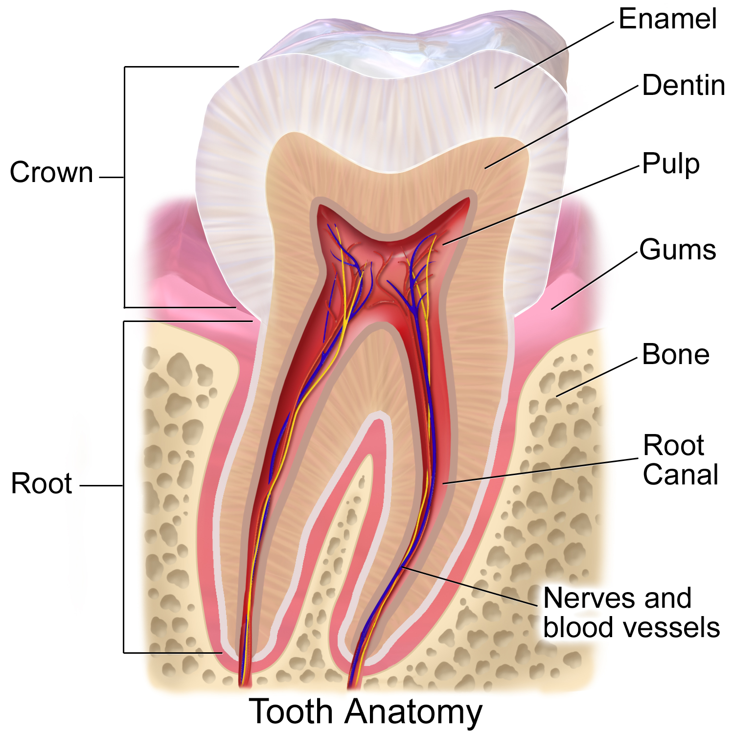
Tooth enamel is one of the four major Tissue (biology), tissues that make up the tooth in humans and many other animals, including some species of fish. It makes up the normally visible part of the tooth, covering the Crown (tooth), crown. The other major tissues are dentin, cementum, and Pulp (tooth), dental pulp. It is a very hard, white to off-white, highly mineralised substance that acts as a barrier to protect the tooth but can become susceptible to degradation, especially by acids from food and drink. Calcium hardens the tooth enamel. In rare circumstances enamel fails to form, leaving the underlying dentin exposed on the surface. Features Enamel is the hardest substance in the human body and contains the highest percentage of minerals (at 96%),Ross ''et al.'', p. 485 with water and organic material composing the rest.Ten Cate's Oral Histology, Nancy, Elsevier, pp. 70–94 The primary mineral is hydroxyapatite, which is a crystalline calcium phosphate. Enamel is formed o ... [...More Info...] [...Related Items...] OR: [Wikipedia] [Google] [Baidu] |
Perikyma
Perikymata (Greek plural of περικύμα, perikyma) are incremental growth lines that appear on the surface of tooth enamel as a series of linear grooves. In anatomically modern humans, each perikyma takes approximately 6–12 days to form. Thus, the count of perikymata may be used to assess how long a tooth crown took to form. They may disappear as the enamel wears over time after the tooth erupts. Perikymata are the expression of striae of Retzius The striae of Retzius are incremental growth lines or bands seen in tooth enamel. They represent the incremental pattern of enamel, the successive apposition of different layers of enamel during crown formation. There are 3 types of incremental ... at the surface of enamel. They can be found on all teeth, but are usually the easiest to notice on anterior teeth (incisors and canines). References {{reflist Parts of tooth ... [...More Info...] [...Related Items...] OR: [Wikipedia] [Google] [Baidu] |
Crown (tooth)
In dentistry, crown refers to the anatomical area of teeth, usually covered by enamel. The crown is usually visible in the mouth after developing below the gingiva The gums or gingiva (plural: ''gingivae'') consist of the mucosal tissue that lies over the mandible and maxilla inside the mouth. Gum health and disease can have an effect on general health. Structure The gums are part of the soft tissue lin ... and then erupting into place. If part of the tooth gets chipped or broken, a dentist can apply an artificial crown. Crowns are used most commonly to entirely cover a damaged tooth or cover an implant. Bridges are also used to cover a space if one or more teeth is missing. They are cemented to natural teeth or implants surrounding the space where the tooth once stood. There are various materials that can be used including a type of cement or stainless steel. The cement crowns look like regular teeth while the stainless steel crowns are silver or gold. References ... [...More Info...] [...Related Items...] OR: [Wikipedia] [Google] [Baidu] |

