|
Rhomboideus Major
The rhomboid major is a skeletal muscle on the back that connects the scapula with the vertebrae of the spinal column. In human anatomy, it acts together with the rhomboid minor to keep the scapula pressed against thoracic wall and to retract the scapula toward the vertebral column. Structure The rhomboid major arises from the spinous processes of the thoracic vertebrae T2 to T5 as well as the supraspinous ligament. It inserts on the medial border of the scapula, from about the level of the scapular spine to the scapula's inferior angle. The rhomboid major is considered a superficial back muscle. It is deep to the trapezius, and is located directly inferior to the rhomboid minor. As the word ''rhomboid'' suggests, the rhomboid major is diamond-shaped. The ''major'' in its name indicates that it is the larger of the two rhomboids. Variation The two rhomboids are sometimes fused into a single muscle. Nerve supply The rhomboid major, like the rhomboid minor, is innervated b ... [...More Info...] [...Related Items...] OR: [Wikipedia] [Google] [Baidu] |
Spinous Processes
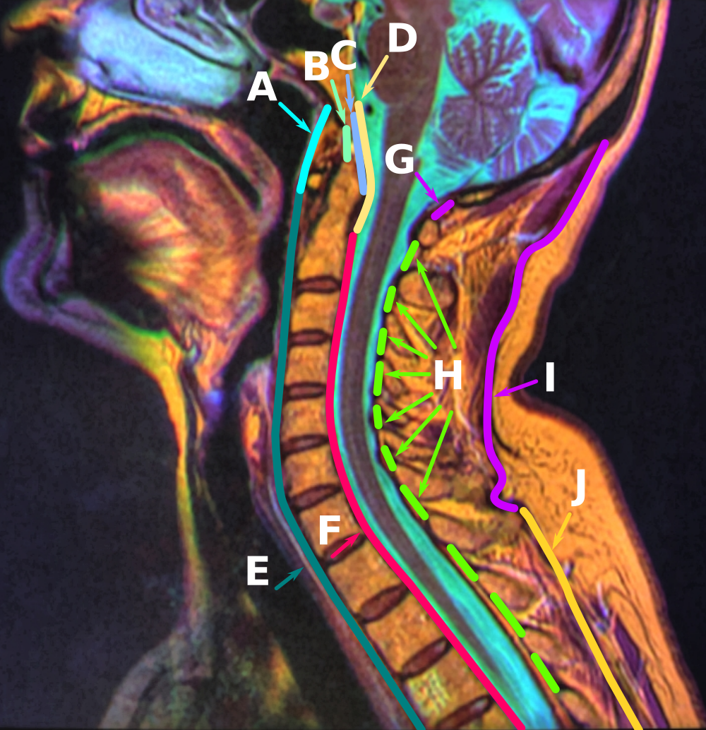
The spinal column, a defining synapomorphy shared by nearly all vertebrates,Hagfish are believed to have secondarily lost their spinal column is a moderately flexible series of vertebrae (singular vertebra), each constituting a characteristic irregular bone whose complex structure is composed primarily of bone, and secondarily of hyaline cartilage. They show variation in the proportion contributed by these two tissue types; such variations correlate on one hand with the cerebral/caudal rank (i.e., location within the backbone), and on the other with phylogenetic differences among the vertebrate taxa. The basic configuration of a vertebra varies, but the bone is its ''body'', with the central part of the body constituting the ''centrum''. The upper (closer to) and lower (further from), respectively, the cranium and its central nervous system surfaces of the vertebra body support attachment to the intervertebral discs. The posterior part of a vertebra forms a vertebral arch ... [...More Info...] [...Related Items...] OR: [Wikipedia] [Google] [Baidu] |
Supraspinous Ligament
The supraspinous ligament, also known as the supraspinal ligament, is a ligament found along the vertebral column. Structure The supraspinous ligament connects the tips of the spinous processes from the seventh cervical vertebra to the sacrum. Above the seventh cervical vertebra, the supraspinous ligament is continuous with the nuchal ligament. Between the spinous processes it is continuous with the interspinous ligaments. It is thicker and broader in the lumbar than in the thoracic region, and intimately blended, in both situations, with the neighboring fascia. The most superficial fibers of this ligament extend over three or four vertebrae; those more deeply seated pass between two or three vertebrae while the deepest connect the spinous processes of neighboring vertebrae. Development Function The supraspinous ligament, along with the posterior longitudinal ligament, interspinous ligaments and ligamentum flavum, help to limit hyperflexion of the vertebral column. Clinical ... [...More Info...] [...Related Items...] OR: [Wikipedia] [Google] [Baidu] |
Yoga
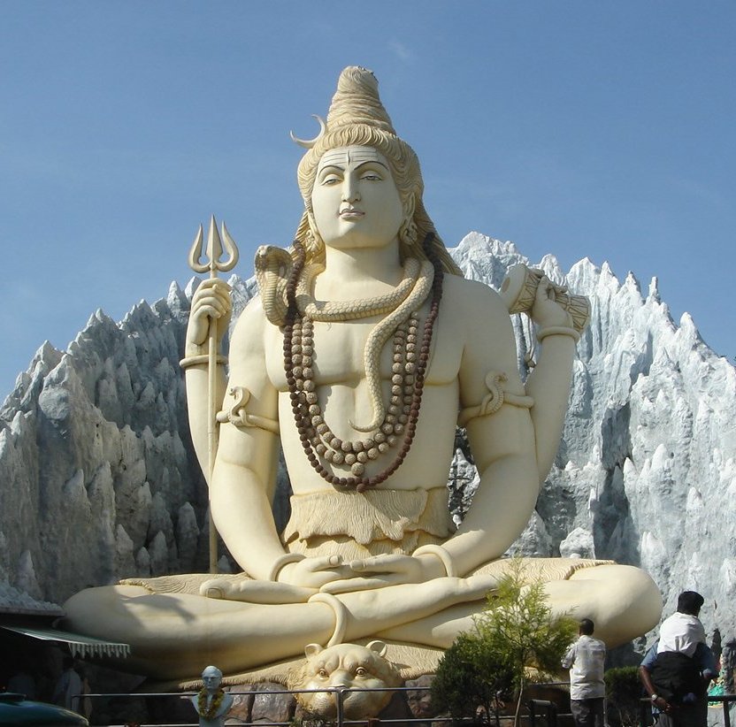
Yoga (; sa, योग, lit=yoke' or 'union ) is a group of physical, mental, and spiritual practices or disciplines which originated in ancient India and aim to control (yoke) and still the mind, recognizing a detached witness-consciousness untouched by the mind ('' Chitta'') and mundane suffering (''Duḥkha''). There is a wide variety of schools of yoga, practices, and goals in Hinduism, Buddhism, and Jainism,Stuart Ray Sarbacker, ''Samādhi: The Numinous and Cessative in Indo-Tibetan Yoga''. SUNY Press, 2005, pp. 1–2.Tattvarthasutra .1 see Manu Doshi (2007) Translation of Tattvarthasutra, Ahmedabad: Shrut Ratnakar p. 102. and traditional and modern yoga is practiced worldwide. Two general theories exist on the origins of yoga. The linear model holds that yoga originated in the Vedic period, as reflected in the Vedic textual corpus, and influenced Buddhism; according to author Edward Fitzpatrick Crangle, this model is mainly supported by Hindu scholars. Accordi ... [...More Info...] [...Related Items...] OR: [Wikipedia] [Google] [Baidu] |
Pilates
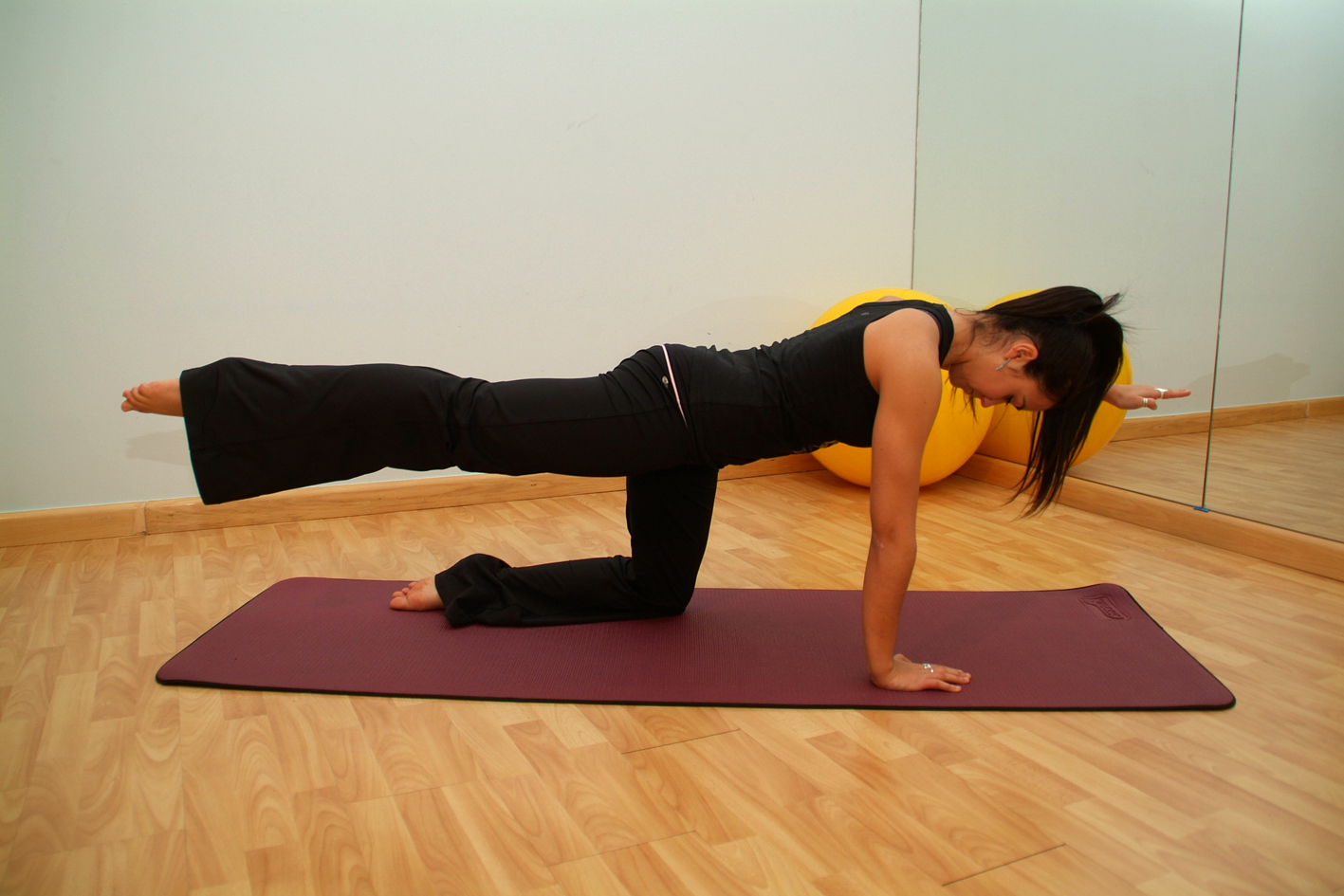
Pilates (; ) is a type of mind-body exercise developed in the early 20th century by German physical trainer Joseph Pilates, after whom it was named. Pilates called his method "Contrology". It is practiced worldwide, especially in countries such as Australia, Canada, South Korea, the United States and the United Kingdom. As of 2005, there were 11 million people practicing the discipline regularly and 14,000 instructors in the United States. Pilates developed in the aftermath of the late 19th century physical culture of exercising in order to alleviate ill health. There is however only limited evidence to support the use of Pilates to alleviate problems such as lower back pain. Evidence from studies show that while Pilates improves balance, it has not been shown to be an effective treatment for any medical condition other than evidence that regular Pilates sessions can help muscle conditioning in healthy adults, when compared to doing no exercise. History Pilates was develop ... [...More Info...] [...Related Items...] OR: [Wikipedia] [Google] [Baidu] |
Physical Therapy
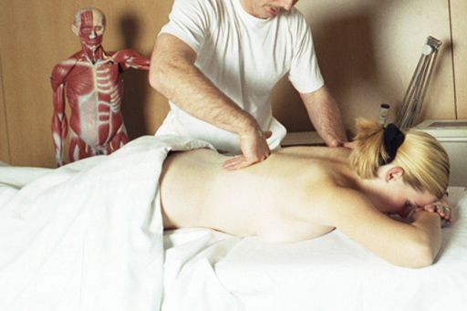
Physical therapy (PT), also known as physiotherapy, is one of the allied health professions. It is provided by physical therapists who promote, maintain, or restore health through physical examination, diagnosis, management, prognosis, patient education, physical intervention, rehabilitation, disease prevention, and health promotion. Physical therapists are known as physiotherapists in many countries. In addition to clinical practice, other aspects of physical therapist practice include research, education, consultation, and health administration. Physical therapy is provided as a primary care treatment or alongside, or in conjunction with, other medical services. In some jurisdictions, such as the United Kingdom, physical therapists have the authority to prescribe medication. Overview Physical therapy addresses the illnesses or injuries that limit a person's abilities to move and perform functional activities in their daily lives. PTs use an individual's history a ... [...More Info...] [...Related Items...] OR: [Wikipedia] [Google] [Baidu] |
Winged Scapula
A winged scapula (scapula alata) is a skeletal medical condition in which the shoulder blade protrudes from a person's back in an abnormal position. In rare conditions it has the potential to lead to limited functional activity in the upper extremity to which it is adjacent. It can affect a person's ability to lift, pull, and push weighty objects. In some serious cases, the ability to perform activities of daily living such as changing one's clothes and washing one's hair may be hindered. The name of this condition comes from its appearance, a wing-like resemblance, due to the medial border of the scapula sticking straight out from the back. Scapular winging has been observed to disrupt scapulohumeral rhythm, contributing to decreased flexion and abduction of the upper extremity, as well as a loss in power and the source of considerable pain. A winged scapula is considered normal posture in young children, but not older children and adults. Signs and symptoms The severity ... [...More Info...] [...Related Items...] OR: [Wikipedia] [Google] [Baidu] |
Levator Scapulae Muscle
The levator scapulae is a slender skeletal muscle situated at the back and side of the neck. As the Latin name suggests, its main function is to lift the scapula. Anatomy Attachments The muscle descends diagonally from its origin to its insertion. Origin The levator scapulae originates from the posterior tubercles of the transverse processes of cervical vertebrae C1-4. At its origin, it attaches via tendinous slips. Insertion It inserts onto the medial border of the scapula (with its site of insertion extending between the superior angle of the scapula superiorly, and the junction of spine of scapula and medial border of scapula inferiorly). Relations One of the muscles within the floor of the posterior triangle of the neck, the superior part of levator scapulae is covered by sternocleidomastoid and its inferior part by the trapezius. It is bounded in front by the scalenus medius and behind by splenius cervicis. The spinal accessory nerve crosses laterally in the midd ... [...More Info...] [...Related Items...] OR: [Wikipedia] [Google] [Baidu] |
Vertebral Column
The vertebral column, also known as the backbone or spine, is part of the axial skeleton. The vertebral column is the defining characteristic of a vertebrate in which the notochord (a flexible rod of uniform composition) found in all chordates has been replaced by a segmented series of bone: vertebrae separated by intervertebral discs. Individual vertebrae are named according to their region and position, and can be used as anatomical landmarks in order to guide procedures such as lumbar punctures. The vertebral column houses the spinal canal, a cavity that encloses and protects the spinal cord. There are about 50,000 species of animals that have a vertebral column. The human vertebral column is one of the most-studied examples. Many different diseases in humans can affect the spine, with spina bifida and scoliosis being recognisable examples. The general structure of human vertebrae is fairly typical of that found in mammals, reptiles, and birds. The shape of the verteb ... [...More Info...] [...Related Items...] OR: [Wikipedia] [Google] [Baidu] |
Pectoralis Minor
Pectoralis minor muscle () is a thin, triangular muscle, situated at the upper part of the chest, beneath the pectoralis major in the human body. Structure Attachments Pectoralis minor muscle arises from the upper margins and outer surfaces of the third, fourth, and fifth ribs, near their costal cartilages and from the aponeuroses covering the intercostalis. The fibers pass superior and lateral and converge to form a flat tendon. This tendon inserts onto the medial border and upper surface of the coracoid process of the scapula. Relations Pectoralis minor muscle forms part of the anterior wall of the axilla. It is covered anteriorly (superficially) by the clavipectoral fascia. The medial pectoral nerve pierces the pectoralis minor and the clavipectoral fascia. In attaching to the coracoid process, the pectoralis minor forms a 'bridge' - structures passing into the upper limb from the thorax will pass directly underneath.http://www.teachmeanatomy.com/muscles-of-the-pecto ... [...More Info...] [...Related Items...] OR: [Wikipedia] [Google] [Baidu] |
Serratus Anterior
The serratus anterior is a muscle that originates on the surface of the 1st to 8th ribs at the side of the chest and inserts along the entire anterior length of the medial border of the scapula. The serratus anterior acts to pull the scapula forward around the thorax. The muscle is named from Latin: ''serrare'' = to saw, referring to the shape, ''anterior'' = on the front side of the body. Structure Serratus anterior normally originates by nine or ten muscle slips – branches from either the first to ninth ribs or the first to eighth ribs. Because two slips usually arise from the second rib, the number of slips is greater than the number of ribs from which they originate. The muscle is inserted along the medial border of the scapula between the superior and inferior angles along with being inserted along the thoracic vertebrae. The muscle is divided into three named parts depending on their points of insertions: #the serratus anterior superior is inserted near the superior ... [...More Info...] [...Related Items...] OR: [Wikipedia] [Google] [Baidu] |
Rhomboid Minor
In human anatomy, the rhomboid minor is a small skeletal muscle on the back that connects the scapula with the vertebrae of the spinal column. Located inferior to levator scapulae and superior to rhomboid major, it acts together with the latter to keep the scapula pressed against the thoracic wall. It lies deep to trapezius but superficial to the long spinal muscles. Origin and insertion The rhomboid minor arises from the inferior border of the nuchal ligament, from the spinous processes of the seventh cervical and first thoracic vertebrae, and from the intervening supraspinous ligaments. It is inserted into a small area of the medial border of the scapula at the level of the scapular spine. Action Together with the rhomboid major, the rhomboid minor retracts the scapula when trapezius is contracted. Acting as a synergist to the trapezius, the rhomboid major and minor elevate the medial border of the scapula medially and upward, working in tandem with the levator sca ... [...More Info...] [...Related Items...] OR: [Wikipedia] [Google] [Baidu] |


