|
Pioneer Neuron
A pioneer neuron is a cell that is a derivative of the preplate in the early stages of corticogenesis of the brain. Pioneer neurons settle in the marginal zone of the cortex and project to sub-cortical levels. In the rat, pioneer neurons are only present in prenatal brains. Unlike Cajal-Retzius cells, these neurons are reelin-negative. Pioneer neurons are born in the ventricular neuroepithelium all over the cortical primordium. In the rat cortex, they appear at embryonic day (E) 11.5 in the lateral aspect of the telencephalic vesicle and cover its whole surface on E12. These cells, which show intense immunoreactivity for calbindin and calretinin, are characterized by their large size and axonal projection. They remain in the marginal zone after the formation of the cortical plate; they project first into the ventricular zone, and then into the subplate and the internal capsule. Therefore, these cells are the origin of the earliest efferent pathway of the developing cortex. Funct ... [...More Info...] [...Related Items...] OR: [Wikipedia] [Google] [Baidu] |
Corticogenesis
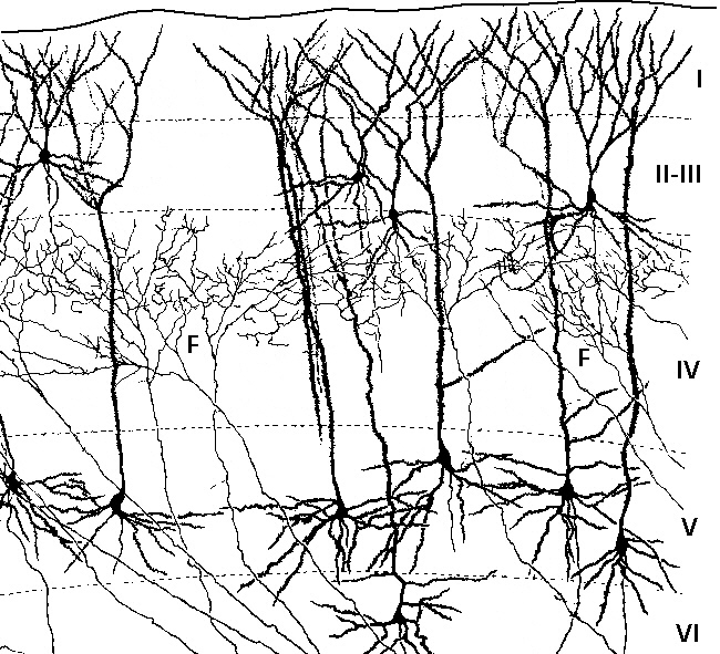
Corticogenesis is the process during which the cerebral cortex of the brain is formed as part of the development of the nervous system of mammals including development of the nervous system in humans, its development in humans. The cortex is the outer layer of the brain and is composed of up to Cerebral cortex#Layers of neocortex, six layers. Neurons formed in the ventricular zone migrate to their final locations in one of the six layers of the cortex. The process occurs from embryonic day 10 to 17 in mice and between gestational weeks seven to 18 in humans. The cortex is the outermost layer of the brain and consists primarily of Grey matter, gray matter, or neuronal cell bodies. Interior areas of the brain consist of Myelin, myelinated Axon, axons and appear as white matter. Cortical plates Preplate The preplate is the first stage in corticogenesis prior to the development of the cortical plate. The preplate is located between the pia mater and the ventricular zone. According ... [...More Info...] [...Related Items...] OR: [Wikipedia] [Google] [Baidu] |
Reelin
Reelin, encoded by the ''RELN'' gene, is a large secreted extracellular matrix glycoprotein that helps regulate processes of neuronal migration and positioning in the developing brain by controlling cell–cell interactions. Besides this important role in early development, reelin continues to work in the adult brain. It modulates synaptic plasticity by enhancing the induction and maintenance of long-term potentiation. It also stimulates dendrite and dendritic spine development and regulates the continuing migration of neuroblasts generated in adult neurogenesis sites like the subventricular and subgranular zones. It is found not only in the brain but also in the liver, thyroid gland, adrenal gland, Fallopian tube, breast and in comparatively lower levels across a range of anatomical regions. Reelin has been suggested to be implicated in pathogenesis of several brain diseases. The expression of the protein has been found to be significantly lower in schizophrenia and psycho ... [...More Info...] [...Related Items...] OR: [Wikipedia] [Google] [Baidu] |
Neuroepithelium
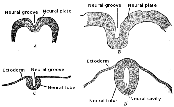
Neuroepithelial cells, or neuroectodermal cells, form the wall of the closed neural tube in early embryonic development. The neuroepithelial cells span the thickness of the tube's wall, connecting with the pial surface and with the ventricular or lumenal surface. They are joined at the lumen of the tube by junctional complexes, where they form a pseudostratified layer of epithelium called neuroepithelium. Neuroepithelial cells are the stem cells of the central nervous system, known as neural stem cells, and generate the intermediate progenitor cells known as radial glial cells, that differentiate into neurons and glia in the process of neurogenesis. Embryonic neural development Brain development During the third week of embryonic growth the brain begins to develop in the early fetus in a process called morphogenesis. Neuroepithelial cells of the ectoderm begin multiplying rapidly and fold in forming the neural plate, which invaginates during the fourth week of embryonic growth ... [...More Info...] [...Related Items...] OR: [Wikipedia] [Google] [Baidu] |
Primordium
A primordium (; plural: primordia; synonym: anlage) in embryology, is an organ or tissue in its earliest recognizable stage of development. Cells of the primordium are called primordial cells. A primordium is the simplest set of cells capable of triggering growth of the would-be organ and the initial foundation from which an organ is able to grow. In flowering plants, a floral primordium gives rise to a flower. Although it is a frequently used term in plant biology, the word is used in describing the biology of all multicellular organisms (for example: a tooth primordium in animals, a leaf primordium in plants or a sporophore primordium in fungi.) Primordium development in plants Plants produce both leaf and flower primordia cells at the shoot apical meristem (SAM). Primordium development in plants is critical to the proper positioning and development of plant organs and cells. The process of primordium development is intricately regulated by a set of genes that affect the pos ... [...More Info...] [...Related Items...] OR: [Wikipedia] [Google] [Baidu] |
Telencephalic
The cerebrum, telencephalon or endbrain is the largest part of the brain containing the cerebral cortex (of the two cerebral hemispheres), as well as several subcortical structures, including the hippocampus, basal ganglia, and olfactory bulb. In the human brain, the cerebrum is the uppermost region of the central nervous system. The cerebrum prenatal development, develops prenatally from the forebrain (prosencephalon). In mammals, the Dorsum (biology), dorsal telencephalon, or Pallium (neuroanatomy), pallium, develops into the cerebral cortex, and the ventral telencephalon, or Pallium (neuroanatomy), subpallium, becomes the basal ganglia. The cerebrum is also divided into approximately symmetric Lateralization of brain function, left and right cerebral hemispheres. With the assistance of the cerebellum, the cerebrum controls all voluntary actions in the human body. Structure The cerebrum is the largest part of the brain. Depending upon the position of the animal it lies eithe ... [...More Info...] [...Related Items...] OR: [Wikipedia] [Google] [Baidu] |
Calbindin
Calbindins are three different calcium-binding proteins: calbindin, calretinin and S100G. They were originally described as vitamin D-dependent calcium-binding proteins in the intestine and kidney in the chick and mammals. They are now classified in different subfamilies as they differ in the number of Ca2+ binding EF hands. Calbindin 1 Calbindin 1 or simply calbindin was first shown to be present in the intestine in birds and then found in the mammalian kidney. It is also expressed in a number of neuronal and endocrine cells, particularly in the cerebellum. It is a 28 kDa protein encoded in humans by the ''CALB1'' gene. Calbindin contains 4 active calcium-binding domains, and 2 modified domains that have lost their calcium-binding capacity. Calbindin acts as a calcium buffer and calcium sensor and can hold four Ca2+ in the EF-hands of loops EF1, EF3, EF4 and EF5. The structure of rat calbindin was originally solved by nuclear magnetic resonance and was one of the largest prote ... [...More Info...] [...Related Items...] OR: [Wikipedia] [Google] [Baidu] |
Calretinin
Calretinin, also known as calbindin 2 (formerly 29 kDa calbindin), is a calcium-binding protein involved in calcium signaling. In humans, the calretinin protein is encoded by the ''CALB2'' gene. Function This gene encodes an intracellular calcium-binding protein belonging to the troponin C superfamily. Members of this protein family have six EF-hand domains which bind calcium. This protein plays a role in diverse cellular functions, including message targeting and intracellular calcium buffering. Calretinin is abundantly expressed in neurons including retina (which gave it the name) and cortical interneurons. Expression was found in different neurons than that of the similar vitamin D-dependent calcium-binding protein, calbindin-28kDa. Calretinin has an important role as a modulator of neuronal excitability including the induction of long-term potentiation. Loss of expression of calretinin in hippocampal interneurons has been suggested to be relevant in temporal lobe epilep ... [...More Info...] [...Related Items...] OR: [Wikipedia] [Google] [Baidu] |
Cortical Plate
The cerebral cortex, also known as the cerebral mantle, is the outer layer of neural tissue of the cerebrum of the brain in humans and other mammals. The cerebral cortex mostly consists of the six-layered neocortex, with just 10% consisting of allocortex. It is separated into two cortices, by the longitudinal fissure that divides the cerebrum into the left and right cerebral hemispheres. The two hemispheres are joined beneath the cortex by the corpus callosum. The cerebral cortex is the largest site of neural integration in the central nervous system. It plays a key role in attention, perception, awareness, thought, memory, language, and consciousness. The cerebral cortex is part of the brain responsible for cognition. In most mammals, apart from small mammals that have small brains, the cerebral cortex is folded, providing a greater surface area in the confined volume of the cranium. Apart from minimising brain and cranial volume, cortical folding is crucial for the br ... [...More Info...] [...Related Items...] OR: [Wikipedia] [Google] [Baidu] |
Subplate
The subplate, also called the subplate zone, together with the marginal zone and the cortical plate, in the fetus represents the developmental anlage of the mammalian cerebral cortex. It was first described, as a separate transient fetal zone by Ivica Kostović and Mark E. Molliver in 1974. During the midfetal period of fetal development the subplate zone is the largest zone in the developing telencephalon. It serves as a waiting compartment for growing cortical afferents; its cells are involved in the establishment of pioneering cortical efferent projections and transient fetal circuitry, and apparently have a number of other developmental roles. The subplate zone is a phylogenetically recent structure and it is most developed in the human brain. Subplate neurons (SPNs) are among the first generated neurons in the mammalian cerebral cortex . These neurons disappear during postnatal development and are important in establishing the correct wiring and functional maturati ... [...More Info...] [...Related Items...] OR: [Wikipedia] [Google] [Baidu] |
Internal Capsule
The internal capsule is a white matter structure situated in the inferomedial part of each cerebral hemisphere of the brain. It carries information past the basal ganglia, separating the caudate nucleus and the thalamus from the putamen and the globus pallidus. The internal capsule contains both ascending and descending axons, going to and coming from the cerebral cortex. It also separates the caudate nucleus and the putamen in the dorsal striatum, a brain region involved in motor and reward pathways. The corticospinal tract constitutes a large part of the internal capsule, carrying motor information from the primary motor cortex to the lower motor neurons in the spinal cord. Above the basal ganglia the corticospinal tract is a part of the corona radiata. Below the basal ganglia the tract is called cerebral crus (a part of the cerebral peduncle) and below the pons it is referred to as the corticospinal tract. Structure The internal capsule consists of three parts and is V-shap ... [...More Info...] [...Related Items...] OR: [Wikipedia] [Google] [Baidu] |


