|
Orbital Cellulitis
Orbital cellulitis is inflammation of eye tissues behind the orbital septum. It is most commonly caused by an acute spread of infection into the eye socket from either the adjacent sinuses or through the blood. It may also occur after trauma. When it affects the rear of the eye, it is known as retro-orbital cellulitis. It should not be confused with periorbital cellulitis, which refers to cellulitis anterior to the septum. Without proper treatment, orbital cellulitis may lead to serious consequences, including permanent loss of vision or even death. Signs and symptoms Orbital cellulitis commonly presents with painful eye movement, sudden vision loss, chemosis, bulging of the infected eye, and limited eye movement. Along with these symptoms, patients typically have redness and swelling of the eyelid, pain, discharge, inability to open the eye, occasional fever and lethargy. Complications Complications include hearing loss, blood infection, meningitis, cavernous sinus thromb ... [...More Info...] [...Related Items...] OR: [Wikipedia] [Google] [Baidu] |
Periorbital Cellulitis
Periorbital cellulitis, or preseptal cellulitis (not to be confused with orbital cellulitis, which is posterior to the orbital septum), is an inflammation and infection of the eyelid and portions of skin around the eye anterior to the orbital septum. It may be caused by breaks in the skin around the eye, and subsequent spread to the eyelid; infection of the sinuses around the nose (sinusitis); or from spread of an infection elsewhere through the blood. Signs and symptoms Periorbital cellulitis must be differentiated from orbital cellulitis, which is an emergency and requires intravenous (IV) antibiotics. In contrast to orbital cellulitis, patients with periorbital cellulitis do not have bulging of the eye (proptosis), limited eye movement (ophthalmoplegia), pain on eye movement, or loss of vision. If any of these features is present, one must assume that the patient has orbital cellulitis and begin treatment with IV antibiotics. CT scan may be done to delineate the extension of th ... [...More Info...] [...Related Items...] OR: [Wikipedia] [Google] [Baidu] |
Inflammation
Inflammation (from la, wikt:en:inflammatio#Latin, inflammatio) is part of the complex biological response of body tissues to harmful stimuli, such as pathogens, damaged cells, or Irritation, irritants, and is a protective response involving immune cells, blood vessels, and molecular mediators. The function of inflammation is to eliminate the initial cause of cell injury, clear out necrotic cells and tissues damaged from the original insult and the inflammatory process, and initiate tissue repair. The five cardinal signs are heat, pain, redness, swelling, and Functio laesa, loss of function (Latin ''calor'', ''dolor'', ''rubor'', ''tumor'', and ''functio laesa''). Inflammation is a generic response, and therefore it is considered as a mechanism of innate immune system, innate immunity, as compared to adaptive immune system, adaptive immunity, which is specific for each pathogen. Too little inflammation could lead to progressive tissue destruction by the harmful stimulus (e.g. b ... [...More Info...] [...Related Items...] OR: [Wikipedia] [Google] [Baidu] |
Cavernous Sinus Thrombosis
The cavernous sinus within the human head is one of the dural venous sinuses creating a cavity called the lateral sellar compartment bordered by the temporal bone of the skull and the sphenoid bone, lateral to the sella turcica. Structure The cavernous sinus is one of the dural venous sinuses of the head. It is a network of veins that sit in a cavity. It sits on both sides of the sphenoidal bone and pituitary gland, approximately 1 × 2 cm in size in an adult. The carotid siphon of the internal carotid artery, and cranial nerves III, IV, V (branches V1 and V2) and VI all pass through this blood filled space. Both sides of cavernous sinus is connected to each other via intercavernous sinuses. The cavernous sinus lies in between the inner and outer layers of dura mater. Nearby structures * Above: optic tract, optic chiasma, internal carotid artery. * Inferiorly: foramen lacerum, and the junction of the body and greater wing of sphenoid bone. * Medially: pituitary gla ... [...More Info...] [...Related Items...] OR: [Wikipedia] [Google] [Baidu] |
Staphylococcus Epidermidis
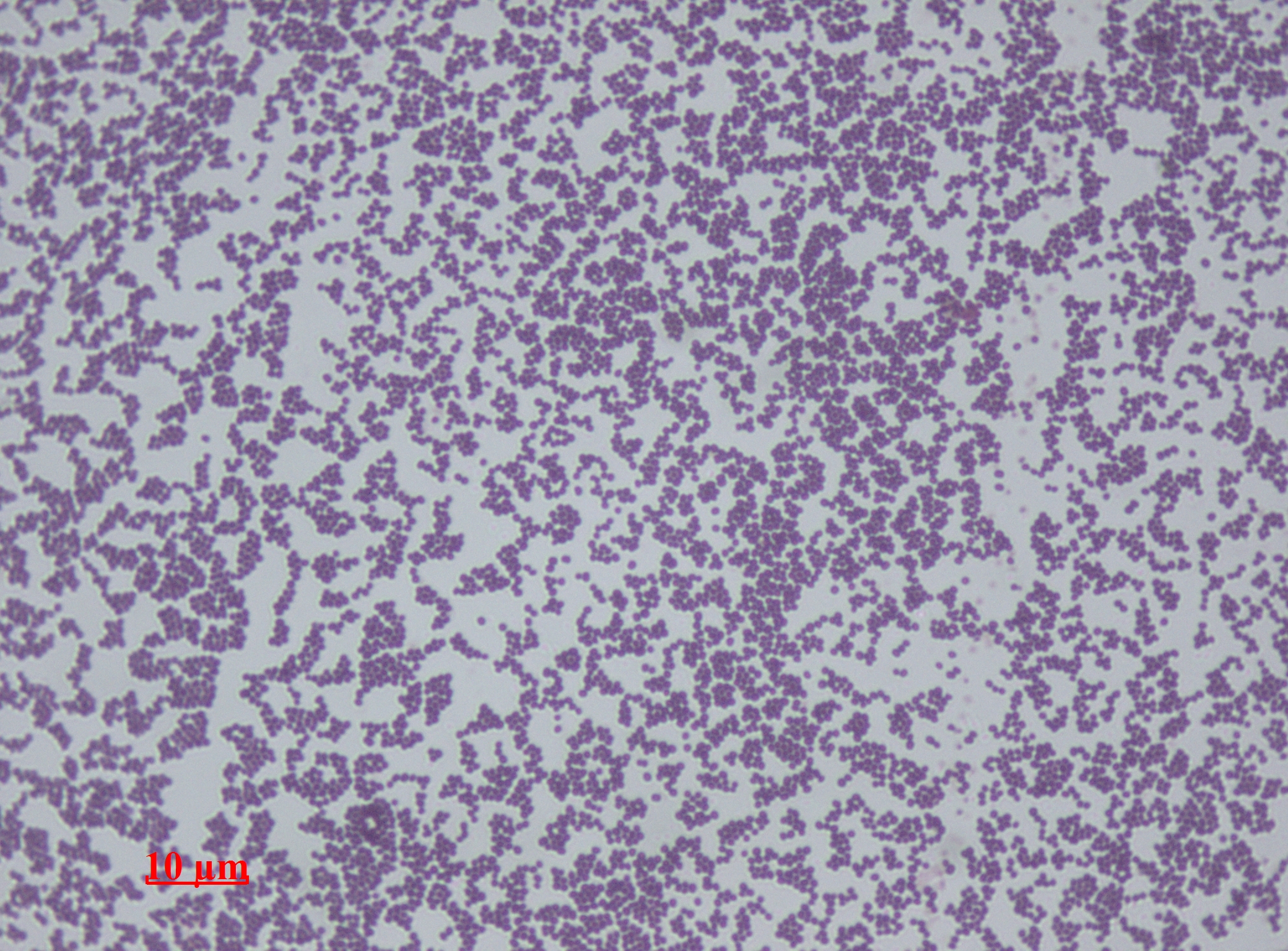
''Staphylococcus epidermidis'' is a Gram-positive bacterium, and one of over 40 species belonging to the genus '' Staphylococcus''. It is part of the normal human microbiota, typically the skin microbiota, and less commonly the mucosal microbiota and also found in marine sponges. It is a facultative anaerobic bacteria. Although ''S. epidermidis'' is not usually pathogenic, patients with compromised immune systems are at risk of developing infection. These infections are generally hospital-acquired. ''S. epidermidis'' is a particular concern for people with catheters or other surgical implants because it is known to form biofilms that grow on these devices. Being part of the normal skin microbiota, ''S. epidermidis'' is a frequent contaminant of specimens sent to the diagnostic laboratory. Some strains of ''S. epidermidis'' are highly salt tolerant and commonly found in marine environment. S.I. Paul et al. (2021) isolated and identified salt tolerant strains of ''S. epiderm ... [...More Info...] [...Related Items...] OR: [Wikipedia] [Google] [Baidu] |
Gram Stain
In microbiology and bacteriology, Gram stain (Gram staining or Gram's method), is a method of staining used to classify bacterial species into two large groups: gram-positive bacteria and gram-negative bacteria. The name comes from the Danish bacteriologist Hans Christian Gram, who developed the technique in 1884. Gram staining differentiates bacteria by the chemical and physical properties of their cell walls. Gram-positive cells have a thick layer of peptidoglycan in the cell wall that retains the primary stain, crystal violet. Gram-negative cells have a thinner peptidoglycan layer that allows the crystal violet to wash out on addition of ethanol. They are stained pink or red by the counterstain, commonly safranin or fuchsine. Lugol's iodine solution is always added after addition of crystal violet to strengthen the bonds of the stain with the cell membrane. Gram staining is almost always the first step in the preliminary identification of a bacterial organism. While Gram s ... [...More Info...] [...Related Items...] OR: [Wikipedia] [Google] [Baidu] |
Virulence
Virulence is a pathogen's or microorganism's ability to cause damage to a host. In most, especially in animal systems, virulence refers to the degree of damage caused by a microbe to its host. The pathogenicity of an organism—its ability to cause disease—is determined by its virulence factors. In the specific context of gene for gene systems, often in plants, virulence refers to a pathogen's ability to infect a resistant host. The noun ''virulence'' derives from the adjective ''virulent'', meaning disease severity. The word ''virulent'' derives from the Latin word ''virulentus'', meaning "a poisoned wound" or "full of poison." From an ecological standpoint, virulence is the loss of fitness induced by a parasite upon its host. Virulence can be understood in terms of proximate causes—those specific traits of the pathogen that help make the host ill—and ultimate causes—the evolutionary pressures that lead to virulent traits occurring in a pathogen strain. Virulent ba ... [...More Info...] [...Related Items...] OR: [Wikipedia] [Google] [Baidu] |
Gram Positive Bacteria
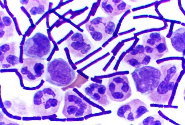
In bacteriology, gram-positive bacteria are bacteria that give a positive result in the Gram stain test, which is traditionally used to quickly classify bacteria into two broad categories according to their type of cell wall. Gram-positive bacteria take up the crystal violet stain used in the test, and then appear to be purple-coloured when seen through an optical microscope. This is because the thick peptidoglycan layer in the bacterial cell wall retains the stain after it is washed away from the rest of the sample, in the decolorization stage of the test. Conversely, gram-negative bacteria cannot retain the violet stain after the decolorization step; alcohol used in this stage degrades the outer membrane of gram-negative cells, making the cell wall more porous and incapable of retaining the crystal violet stain. Their peptidoglycan layer is much thinner and sandwiched between an inner cell membrane and a bacterial outer membrane, causing them to take up the counterstain (safr ... [...More Info...] [...Related Items...] OR: [Wikipedia] [Google] [Baidu] |
Group A Beta Hemolytic Streptococcus
''Streptococcus pyogenes'' is a species of Gram-positive, aerotolerant bacteria in the genus ''Streptococcus''. These bacteria are extracellular, and made up of non-motile and non-sporing cocci (round cells) that tend to link in chains. They are clinically important for humans, as they are an infrequent, but usually pathogenic, part of the skin microbiota that can cause Group A streptococcal infection. ''S. pyogenes'' is the predominant species harboring the Lancefield group A antigen, and is often called group A ''Streptococcus'' (GAS). However, both ''Streptococcus dysgalactiae'' and the '' Streptococcus anginosus'' group can possess group A antigen as well. Group A streptococci, when grown on blood agar, typically produce small (2–3 mm) zones of beta-hemolysis, a complete destruction of red blood cells. The name group A (beta-hemolytic) ''Streptococcus'' (GABHS) is thus also used. The species name is derived from Greek words meaning 'a chain' () of berries ( at ... [...More Info...] [...Related Items...] OR: [Wikipedia] [Google] [Baidu] |
Streptococcus Pneumoniae
''Streptococcus pneumoniae'', or pneumococcus, is a Gram-positive, spherical bacteria, alpha-hemolytic (under aerobic conditions) or beta-hemolytic (under anaerobic conditions), aerotolerant anaerobic member of the genus Streptococcus. They are usually found in pairs (diplococci) and do not form spores and are non motile. As a significant human pathogenic bacterium ''S. pneumoniae'' was recognized as a major cause of pneumonia in the late 19th century, and is the subject of many humoral immunity studies. ''Streptococcus pneumoniae'' resides asymptomatically in healthy carriers typically colonizing the respiratory tract, sinuses, and nasal cavity. However, in susceptible individuals with weaker immune systems, such as the elderly and young children, the bacterium may become pathogenic and spread to other locations to cause disease. It spreads by direct person-to-person contact via respiratory droplets and by auto inoculation in persons carrying the bacteria in their upper res ... [...More Info...] [...Related Items...] OR: [Wikipedia] [Google] [Baidu] |
Staphylococcus Aureus
''Staphylococcus aureus'' is a Gram-positive spherically shaped bacterium, a member of the Bacillota, and is a usual member of the microbiota of the body, frequently found in the upper respiratory tract and on the skin. It is often positive for catalase and nitrate reduction and is a facultative anaerobe that can grow without the need for oxygen. Although ''S. aureus'' usually acts as a commensal of the human microbiota, it can also become an opportunistic pathogen, being a common cause of skin infections including abscesses, respiratory infections such as sinusitis, and food poisoning. Pathogenic strains often promote infections by producing virulence factors such as potent protein toxins, and the expression of a cell-surface protein that binds and inactivates antibodies. ''S. aureus'' is one of the leading pathogens for deaths associated with antimicrobial resistance and the emergence of antibiotic-resistant strains, such as methicillin-resistant ''S. aureus'' (MRSA ... [...More Info...] [...Related Items...] OR: [Wikipedia] [Google] [Baidu] |
Sinusitis
Sinusitis, also known as rhinosinusitis, is inflammation of the nasal mucosa, mucous membranes that line the paranasal sinuses, sinuses resulting in symptoms that may include thick Mucus#Respiratory system, nasal mucus, a nasal congestion, plugged nose, and Orofacial pain, facial pain. Other signs and symptoms may include fever, headaches, a hyposmia, poor sense of smell, sore throat, a feeling that phlegm is oozing out from the back of the nose to the throat along with a necessity to clear the throat frequently and frequent attacks of cough. Generally sinusitis starts off as a common viral infection like common cold. This infection generally subsides within 5 to 7 days. During this time the nasal structures can swell and facilitate the stagnation of fluids in sinuses that leads to acute (medicine), acute sinusitis which lasts from 6th day of the infection to 15th day. From the 15th day to 45th day of the infection comes the subacute stage followed by chronic (medicine), chronic ... [...More Info...] [...Related Items...] OR: [Wikipedia] [Google] [Baidu] |
Paranasal Sinuses
Paranasal sinuses are a group of four paired air-filled spaces that surround the nasal cavity. The maxillary sinuses are located under the eyes; the frontal sinuses are above the eyes; the ethmoidal sinuses are between the eyes and the sphenoidal sinuses are behind the eyes. The sinuses are named for the facial bones in which they are located. Structure Humans possess four pairs of paranasal sinuses, divided into subgroups that are named according to the bones within which the sinuses lie. They are all innervated by branches of the trigeminal nerve (CN V). * The maxillary sinuses, the largest of the paranasal sinuses, are under the eyes, in the maxillary bones (open in the back of the semilunar hiatus of the nose). They are innervated by the maxillary nerve (CN V2). * The frontal sinuses, superior to the eyes, in the frontal bone, which forms the hard part of the forehead. They are innervated by the ophthalmic nerve (CN V1). * The ethmoidal sinuses, which are formed from sever ... [...More Info...] [...Related Items...] OR: [Wikipedia] [Google] [Baidu] |


