|
Obturator
Obturator may refer to: Medicine Anatomical structures * Obturator foramen * Obturator fascia * Obturator canal * Obturator vessels (other) ** Obturator artery ** Obturator veins * Obturator nerve ** Anterior branch of obturator nerve ** Posterior branch of obturator nerve ** Cutaneous branch of the obturator nerve * Obturator internus nerve * Accessory obturator nerve * Obturator membrane * Obturator crest * Obturator muscles (other) ** Obturator internus muscle ** Obturator externus muscle * Obturator externus groove * Obturator process (in archosaurs) Diseases and disorders * Obturator hernia Medical procedures * Obturator sign Medical devices * Part of a trocar device * A device used as a guide during tracheostomy tube insertion * Palatal obturator, a dental prosthesis used to seal an opening in the palate, i.e. cleft palate Botany * Part of the ovary The ovary is an organ in the female reproductive system that produces an ovum. ... [...More Info...] [...Related Items...] OR: [Wikipedia] [Google] [Baidu] |
Obturator Externus Muscle
The external obturator muscle, obturator externus muscle (; OE) is a flat, triangular muscle, which covers the outer surface of the anterior wall of the pelvis. It is sometimes considered part of the medial compartment of thigh, and sometimes considered part of the gluteal region. Structure It arises from the margin of bone immediately around the medial side of the obturator membrane and surrounding bone, viz., from the inferior pubic ramus, and the ramus of the ischium; it also arises from the medial two-thirds of the outer surface of the obturator membrane, and from the tendinous arch which completes the canal for the passage of the obturator vessels and nerves. The fibers springing from the pubic arch extend on to the inner surface of the bone, where they obtain a narrow origin between the margin of the foramen and the attachment of the obturator membrane. The fibers converge and pass posterolateral and upward, and end in a tendon which runs across the back of the neck of t ... [...More Info...] [...Related Items...] OR: [Wikipedia] [Google] [Baidu] |
Obturator Artery
The obturator artery is a branch of the internal iliac artery that passes antero-inferiorly (forwards and downwards) on the lateral wall of the pelvis, to the upper part of the obturator foramen, and, escaping from the pelvic cavity through the obturator canal, it divides into both an anterior and a posterior branch. Structure In the pelvic cavity this vessel is in relation, laterally, with the obturator fascia; medially, with the ureter, ductus deferens, and peritoneum; while a little below it is the obturator nerve. The obturator artery usually arises from the internal iliac artery. Inside the pelvis the obturator artery gives off iliac branches to the iliac fossa, which supply the bone and the Iliacus, and anastomose with the ilio-lumbar artery; a vesical branch, which runs backward to supply the bladder; and a pubic branch, which is given off from the vessel just before it leaves the pelvic cavity. The pubic branch ascends upon the back of the pubis, communicating with the cor ... [...More Info...] [...Related Items...] OR: [Wikipedia] [Google] [Baidu] |
Obturator Nerve
The obturator nerve in human anatomy arises from the ventral divisions of the second, third, and fourth lumbar nerves in the lumbar plexus; the branch from the third is the largest, while that from the second is often very small. Structure The obturator nerve originates from the anterior divisions of the L2, L3, and L4 spinal nerve roots. It descends through the fibers of the psoas major, and emerges from its medial border near the brim of the pelvis. It then passes behind the common iliac arteries, and on the lateral side of the internal iliac artery and vein, and runs along the lateral wall of the lesser pelvis, above and in front of the obturator vessels, to the upper part of the obturator foramen. Here it enters the thigh, through the obturator canal, and divides into an anterior and a posterior branch, which are separated at first by some of the fibers of the obturator externus, and lower down by the adductor brevis. An accessory obturator nerve may be present in approx ... [...More Info...] [...Related Items...] OR: [Wikipedia] [Google] [Baidu] |
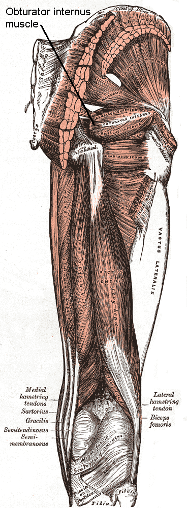
Obturator Internus Muscle
The internal obturator muscle or obturator internus muscle originates on the medial surface of the obturator membrane, the ischium near the membrane, and the rim of the pubis. It exits the pelvic cavity through the lesser sciatic foramen. The internal obturator is situated partly within the lesser pelvis, and partly at the back of the hip-joint. It functions to help laterally rotate femur with hip extension and abduct femur with hip flexion, as well as to steady the femoral head in the acetabulum. Structure Origin The internal obturator muscle arises from the inner surface of the antero-lateral wall of the pelvis. It surrounds the obturator foramen. It is attached to the inferior pubic ramus and ischium, and at the side to the inner surface of the hip bone below and behind the pelvic brim. It reaches from the upper part of the greater sciatic foramen above and behind to the obturator foramen below and in front. It also arises from the pelvic surface of the obturator membran ... [...More Info...] [...Related Items...] OR: [Wikipedia] [Google] [Baidu] |
Palatal Obturator
{{no footnotes, date=February 2017 The Latham Device Post Latham Nasal Alveolar Molding Device Post Insertion A palatal obturator is a prosthesis that totally occludes an opening such as an oronasal fistula (in the roof of the mouth). They are similar to dental retainers, but without the front wire. Palatal obturators are typically short-term prosthetics used to close defects of the hard/soft palate that may affect speech production or cause nasal regurgitation during feeding. Following surgery, there may remain a residual orinasal opening on the palate, alveolar ridge, or vestibule of the larynx. A palatal obturator may be used to compensate for hypernasality and to aid in speech therapy targeting correction of compensatory articulation caused by the cleft palate. In simpler terms, a palatal obturator covers any fistulas (or "holes") in the roof of the mouth that lead to the nasal cavity, providing the wearer with a plastic/acrylic, removable roof of the mouth, which aids in ... [...More Info...] [...Related Items...] OR: [Wikipedia] [Google] [Baidu] |
Obturator Foramen
The obturator foramen (Latin foramen obturatum) is the large opening created by the ischium and pubis (bone), pubis bones of the pelvis through which nerves and blood vessels pass. Structure It is bounded by a thin, uneven margin, to which a strong membrane is attached, and presents, superiorly, a deep groove, the obturator groove, which runs from the pelvis obliquely medialward and downward. This groove is converted into the obturator canal by a ligamentous band, a specialized part of the obturator membrane, attached to two tubercles: * one, the posterior obturator tubercle, on the medial border of the ischium, just in front of the acetabular notch * the other, the anterior obturator tubercle, on the obturator crest of the superior pubic ramus, superior ramus of the pubis (bone), pubis Variation Reflecting the overall sex differences in human physiology, sex differences between male and female pelvises, the obturator foramina are oval in the male and wider and more triangular ... [...More Info...] [...Related Items...] OR: [Wikipedia] [Google] [Baidu] |
Obturator Canal
The obturator canal is a passageway formed in the obturator foramen by part of the obturator membrane and the pelvis. It connects the pelvis to the thigh. Structure The obturator canal is formed between the obturator membrane and the pelvis. The obturator artery, obturator vein, and obturator nerve all travel through the canal. Clinical significance An obturator hernia is a type of hernia involving an intrusion into the obturator canal. The obturator nerve can be compressed in the obturator canal. The obturator canal may be compressed during pregnancy and major traumatic injuries, causing obturator syndrome. See also * Obturator fascia The obturator fascia, or fascia of the internal obturator muscle, covers the pelvic surface of that muscle and is attached around the margin of its origin. Above, it is loosely connected to the back part of the arcuate line, and here it is conti ... References External links Pelvis {{Anatomy-stub ... [...More Info...] [...Related Items...] OR: [Wikipedia] [Google] [Baidu] |
Obturator Sign
The obturator sign, also called Cope's obturator test, is an indicator of irritation to the obturator internus muscle. The technique for detecting the obturator sign, called the ''obturator test'', is carried out on each leg in succession. The patient lies on her/his back with the hip and knee both flexed at ninety degrees. The examiner holds the patient's ankle with one hand and knee with the other hand. The examiner internally rotates the hip by moving the patient's ankle away from the patient's body while allowing the knee to move only inward. This is flexion and internal rotation of the hip. In the clinical context, it is performed when acute appendicitis is suspected. In this condition, the appendix becomes inflamed and enlarged. The appendix may come into physical contact with the obturator internus muscle, which will be stretched when this maneuver is performed on the right leg. This causes pain and is evidence in support of an inflamed appendix. The principles of the obtu ... [...More Info...] [...Related Items...] OR: [Wikipedia] [Google] [Baidu] |
Obturator Fascia
The obturator fascia, or fascia of the internal obturator muscle, covers the pelvic surface of that muscle and is attached around the margin of its origin. Above, it is loosely connected to the back part of the arcuate line, and here it is continuous with the iliac fascia. In front of this, as it follows the line of origin of the internal obturator, it gradually separates from the iliac fascia and the continuity between the two is retained only through the periosteum. It arches beneath the obturator vessels and nerve, completing the obturator canal, and at the front of the pelvis is attached to the back of the superior ramus of the pubis. Below, the obturator fascia is attached to the falciform process of the sacrotuberous ligament and to the pubic arch, where it becomes continuous with the superior fascia of the urogenital diaphragm. Behind, it is prolonged into the gluteal region. The internal pudendal vessels and pudendal nerve cross the pelvic surface of the internal o ... [...More Info...] [...Related Items...] OR: [Wikipedia] [Google] [Baidu] |
Obturator Membrane , i. e., within the margin.
Both obturator muscles are connected with this membrane ...
The obturator membrane is a thin fibrous sheet, which almost completely closes the obturator foramen. Its fibers are arranged in interlacing bundles mainly transverse in direction; the uppermost bundle is attached to the obturator tubercles and completes the obturator canal for the passage of the obturator vessels and nerve. The membrane is attached to the sharp margin of the obturator foramen except at its lower lateral angle, where it is fixed to the pelvic surface of the inferior ramus of the ischium The ischium () form ... [...More Info...] [...Related Items...] OR: [Wikipedia] [Google] [Baidu] |
Obturator Internus Nerve
The nerve to obturator internus, also known as the obturator internus nerve, is a nerve that innervates the obturator internus and gemellus superior muscles. Structure The nerve to obturator internus originates in the lumbosacral plexus. It arises from the ventral divisions of the fifth lumbar and first and second sacral nerves. It leaves the pelvis through the greater sciatic foramen below the piriformis muscle, and gives off the branch to the gemellus superior, which enters the upper part of the posterior surface of the muscle. It then crosses the ischial spine, re-enters the pelvis through the lesser sciatic foramen, and pierces the pelvic surface of the obturator internus The internal obturator muscle or obturator internus muscle originates on the medial surface of the obturator membrane, the ischium near the membrane, and the rim of the pubis. It exits the pelvic cavity through the lesser sciatic foramen. The i .... See also * Obturator nerve * Nerve to quadra ... [...More Info...] [...Related Items...] OR: [Wikipedia] [Google] [Baidu] |
Accessory Obturator Nerve
In human anatomy, the accessory obturator nerve is an accessory nerve in the lumbar region present in about 29% of cases. It is of small size, and arises from the ventral divisions of the third and fourth lumbar nerves. Recent evidence support that this nerve arises from Dorsal divisions. It descends along the medial border of the psoas major, crosses the superior ramus of the pubis, and passes under the pectineus, where it divides into numerous branches. One of these supplies the pectineus, penetrating its deep surface, another is distributed to the hip-joint; while a third communicates with the anterior branch of the obturator nerve The obturator nerve in human anatomy arises from the ventral divisions of the second, third, and fourth lumbar nerves in the lumbar plexus; the branch from the third is the largest, while that from the second is often very small. Structure The ob .... Occasionally the accessory obturator nerve is very small and is lost in the capsule of the h ... [...More Info...] [...Related Items...] OR: [Wikipedia] [Google] [Baidu] |

