|
Obturator Sign
The obturator sign, also called Cope's obturator test, is an indicator of irritation to the obturator internus muscle. The technique for detecting the obturator sign, called the ''obturator test'', is carried out on each leg in succession. The patient lies on her/his back with the hip and knee both flexed at ninety degrees. The examiner holds the patient's ankle with one hand and knee with the other hand. The examiner internally rotates the hip by moving the patient's ankle away from the patient's body while allowing the knee to move only inward. This is flexion and internal rotation of the hip. In the clinical context, it is performed when acute appendicitis is suspected. In this condition, the appendix becomes inflamed and enlarged. The appendix may come into physical contact with the obturator internus muscle, which will be stretched when this maneuver is performed on the right leg. This causes pain and is evidence in support of an inflamed appendix. The principles of the obtu ... [...More Info...] [...Related Items...] OR: [Wikipedia] [Google] [Baidu] |
Irritation
Irritation, in biology and physiology, is a state of inflammation or painful reaction to allergy or cell-lining damage. A stimulus or agent which induces the state of irritation is an irritant. Irritants are typically thought of as chemical agents (for example phenol and capsaicin) but mechanical, thermal (heat), and radiative stimuli (for example ultraviolet light or ionising radiations) can also be irritants. Irritation also has non-clinical usages referring to bothersome physical or psychological pain or discomfort. Irritation can also be induced by some allergic response due to exposure of some allergens for example contact dermatitis, irritation of mucosal membranes and pruritus. Mucosal membrane is the most common site of irritation because it contains secretory glands that release mucous which attracts the allergens due to its sticky nature. Chronic irritation is a medical term signifying that afflictive health conditions have been present for a while. There are many dis ... [...More Info...] [...Related Items...] OR: [Wikipedia] [Google] [Baidu] |
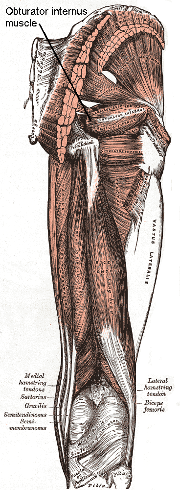
Obturator Internus Muscle
The internal obturator muscle or obturator internus muscle originates on the medial surface of the obturator membrane, the ischium near the membrane, and the rim of the pubis. It exits the pelvic cavity through the lesser sciatic foramen. The internal obturator is situated partly within the lesser pelvis, and partly at the back of the hip-joint. It functions to help laterally rotate femur with hip extension and abduct femur with hip flexion, as well as to steady the femoral head in the acetabulum. Structure Origin The internal obturator muscle arises from the inner surface of the antero-lateral wall of the pelvis. It surrounds the obturator foramen. It is attached to the inferior pubic ramus and ischium, and at the side to the inner surface of the hip bone below and behind the pelvic brim. It reaches from the upper part of the greater sciatic foramen above and behind to the obturator foramen below and in front. It also arises from the pelvic surface of the obturator membran ... [...More Info...] [...Related Items...] OR: [Wikipedia] [Google] [Baidu] |
Abdominal Examination
An abdominal examination is a portion of the physical examination which a physician or nurse uses to clinically observe the abdomen of a patient for signs of disease. The physical examination typically occurs after a thorough medical history is taken, that is, after the physician asks the patient the course of their symptoms. The abdominal examination is conventionally split into four different stages: first, inspection of the patient and the visible characteristics of their abdomen. Auscultation (listening) of the abdomen with a stethoscope. Palpation of the patient's abdomen. Finally, percussion (tapping) of the patient's abdomen and abdominal organs. Depending on the need to test for specific diseases such as ascites, special tests may be performed as a part of the physical examination. An abdominal examination may be performed because the physician suspects a disease of the organs inside the abdominal cavity (including the liver, spleen, large or small intestines), or simpl ... [...More Info...] [...Related Items...] OR: [Wikipedia] [Google] [Baidu] |
Acute Appendicitis
Appendicitis is inflammation of the appendix. Symptoms commonly include right lower abdominal pain, nausea, vomiting, and decreased appetite. However, approximately 40% of people do not have these typical symptoms. Severe complications of a ruptured appendix include widespread, painful inflammation of the inner lining of the abdominal wall and sepsis. Appendicitis is caused by a blockage of the hollow portion of the appendix. This is most commonly due to a calcified "stone" made of feces. Inflamed lymphoid tissue from a viral infection, parasites, gallstone, or tumors may also cause the blockage. This blockage leads to increased pressures in the appendix, decreased blood flow to the tissues of the appendix, and bacterial growth inside the appendix causing inflammation. The combination of inflammation, reduced blood flow to the appendix and distention of the appendix causes tissue injury and tissue death. If this process is left untreated, the appendix may burst, releasing ba ... [...More Info...] [...Related Items...] OR: [Wikipedia] [Google] [Baidu] |
Psoas Sign
The psoas sign, also known as Cope's sign (or Cope's psoas test) or Obraztsova's sign, is a medical sign that indicates irritation to the iliopsoas group of hip flexors in the abdomen, and consequently indicates that the inflamed appendix is retrocaecal in orientation (as the iliopsoas muscle is retroperitoneal). The technique for detecting the psoas sign is carried out on the patient's right leg. The patient lies on his/her left side with the knees extended. The examiner holds the patient's right thigh and passively extends the hip. Alternatively, the patient lies on their back, and the examiner asks the patient to actively flex the right hip against the examiner's hand. If abdominal pain results, it is a "positive psoas sign". The pain results because the psoas borders the peritoneal cavity, so stretching (by hyperextension at the hip) or contraction (by flexion of the hip) of the muscles causes friction against nearby inflamed tissues. In particular, the right iliopsoas muscl ... [...More Info...] [...Related Items...] OR: [Wikipedia] [Google] [Baidu] |
Zachary Cope
Sir Vincent Zachary Cope MD MS FRCS (14 February 1881 – 28 December 1974) was an English physician, surgeon, author, historian and poet perhaps best known for authoring the book ''Cope's Early Diagnosis of the Acute Abdomen'' from 1921 until 1971. The work remains a respected and standard text of general surgery, and new editions continue being published by editors long after his death, the most recent one being the 22nd edition, published in 2010. Cope also wrote widely on the history of medicine and of public dispensaries. Early life Cope was the youngest of ten children of a minister, Thomas John Cope and his wife Celia Anne Crowle. He was head boy at Westminster City School where he was awarded a gold medal in 1899 and then a scholarship to go to St Mary's Hospital Medical School. He passed surgery and forensic medicine with distinction in 1905 and became house physician to David Lees, author of ''The Abdominal Inflammations.'' Lees influenced Cope in his lifelong in ... [...More Info...] [...Related Items...] OR: [Wikipedia] [Google] [Baidu] |
Markle's Sign
Markle's sign, or jar tenderness, is a clinical sign in which pain in the right lower quadrant of the abdomen is elicited by the heel-drop test (dropping to the heels, from standing on the toes, with a jarring landing). It is found in patients with localised peritonitis due to acute appendicitis. It is similar to rebound tenderness, but may be easier to elicit when the patient has firm abdominal wall In anatomy, the abdominal wall represents the boundaries of the abdominal cavity. The abdominal wall is split into the anterolateral and posterior walls. There is a common set of layers covering and forming all the walls: the deepest being the v ... muscles. Abdominal pain on walking or running is an equivalent sign. It was first described by the George Bushar Markle IV (1921–1999), an American surgeon, in 1985. References Medical signs {{med-sign-stub ... [...More Info...] [...Related Items...] OR: [Wikipedia] [Google] [Baidu] |
Psoas Sign
The psoas sign, also known as Cope's sign (or Cope's psoas test) or Obraztsova's sign, is a medical sign that indicates irritation to the iliopsoas group of hip flexors in the abdomen, and consequently indicates that the inflamed appendix is retrocaecal in orientation (as the iliopsoas muscle is retroperitoneal). The technique for detecting the psoas sign is carried out on the patient's right leg. The patient lies on his/her left side with the knees extended. The examiner holds the patient's right thigh and passively extends the hip. Alternatively, the patient lies on their back, and the examiner asks the patient to actively flex the right hip against the examiner's hand. If abdominal pain results, it is a "positive psoas sign". The pain results because the psoas borders the peritoneal cavity, so stretching (by hyperextension at the hip) or contraction (by flexion of the hip) of the muscles causes friction against nearby inflamed tissues. In particular, the right iliopsoas muscl ... [...More Info...] [...Related Items...] OR: [Wikipedia] [Google] [Baidu] |
Rovsing's Sign
Rovsing's sign, named after the Danish surgeon Niels Thorkild Rovsing (1862–1927), is a sign of appendicitis. If palpation of the left lower quadrant of a person's abdomen increases the pain felt in the right lower quadrant, the patient is said to have a positive Rovsing's sign and may have appendicitis. The phenomenon was first described by Swedish surgeon Emil Samuel Perman (1856-1945) writing in the journal ''Hygiea'' in 1904. In acute appendicitis, palpation in the left iliac fossa may produce pain in the right iliac fossa. Referral of pain This anomaly occurs because the pain nerves deep in the intestines do not localize well to an exact spot on the abdominal wall, unlike pain nerves in muscles. Pain from a stomach ulcer or gallstone can be interpreted by the brain as pain from the stomach, liver, gall bladder, duodenum, or first part of the small intestine. It will "refer" pain often to the mid upper abdomen, the epigastrum. Because the appendix is a piece of inte ... [...More Info...] [...Related Items...] OR: [Wikipedia] [Google] [Baidu] |
McBurney's Point
McBurney's point is the name given to the point over the right side of the abdomen that is one-third of the distance from the anterior superior iliac spine to the umbilicus (navel). This is near the most common location of the appendix. Location McBurney's point is located one third of the distance from the right anterior superior iliac spine to the umbilicus (navel). This point roughly corresponds to the most common location of the base of the appendix, where it is attached to the cecum. Clinical significance Appendicitis Deep tenderness at McBurney's point, known as McBurney's sign, is a sign of acute appendicitis. The clinical sign of referred pain in the epigastrium when pressure is applied is also known as Aaron's sign. Specific localization of tenderness to McBurney's point indicates that inflammation is no longer limited to the lumen of the bowel (which localizes pain poorly), and is irritating the lining of the peritoneum at the place where the peritoneum comes ... [...More Info...] [...Related Items...] OR: [Wikipedia] [Google] [Baidu] |


