|
Obturator Internus Nerve
The nerve to obturator internus, also known as the obturator internus nerve, is a nerve that innervates the obturator internus and gemellus superior muscles. Structure The nerve to obturator internus originates in the lumbosacral plexus. It arises from the ventral divisions of the fifth lumbar and first and second sacral nerves. It leaves the pelvis through the greater sciatic foramen below the piriformis muscle, and gives off the branch to the gemellus superior, which enters the upper part of the posterior surface of the muscle. It then crosses the ischial spine, re-enters the pelvis through the lesser sciatic foramen, and pierces the pelvic surface of the obturator internus. See also * Obturator nerve * Nerve to quadratus femoris The nerve to quadratus femoris is a nerve that provides innervation to the quadratus femoris muscle and gemellus inferior muscle. Structure The nerve to quadratus femoris is a sacral plexus nerve. It arises from the ventral divisions of th ... [...More Info...] [...Related Items...] OR: [Wikipedia] [Google] [Baidu] |
Sacral Plexus
In human anatomy, the sacral plexus is a nerve plexus which provides motor and sensory nerves for the posterior thigh, most of the lower leg and foot, and part of the pelvis. It is part of the lumbosacral plexus and emerges from the lumbar vertebrae and sacral vertebrae (L4-S4).''Thieme Atlas of Anatomy'' (2006), pp 470-471 A sacral plexopathy is a disorder affecting the nerves of the sacral plexus, usually caused by trauma, nerve compression, vascular disease, or infection. Symptoms may include pain, loss of motor control, and sensory deficits. Structure The sacral plexus is formed by: * the lumbosacral trunk * the anterior division of the first sacral nerve * portions of the anterior divisions of the second and third sacral nerves The nerves forming the sacral plexus converge toward the lower part of the greater sciatic foramen, and unite to form a flattened band, from the anterior and posterior surfaces of which several branches arise. The band itself is continued as the sc ... [...More Info...] [...Related Items...] OR: [Wikipedia] [Google] [Baidu] |
Pudendal Plexus (nerves)
The pudendal plexus is a term used for a compound structure consisting of sacral spinal nerves. Some sources describe it as S2-S4 of the lumbosacral plexus. In some older texts, it is a rough approximation of the coccygeal plexus The coccygeal plexus is a nerve plexus near the coccyx bone. Structure This plexus is formed by the ventral rami The ventral ramus (pl. ''rami'') (Latin for ''branch'') is the anterior division of a spinal nerve. The ventral rami supply the ant .... References Nerve plexus {{neuroanatomy-stub ... [...More Info...] [...Related Items...] OR: [Wikipedia] [Google] [Baidu] |
Obturator Internus
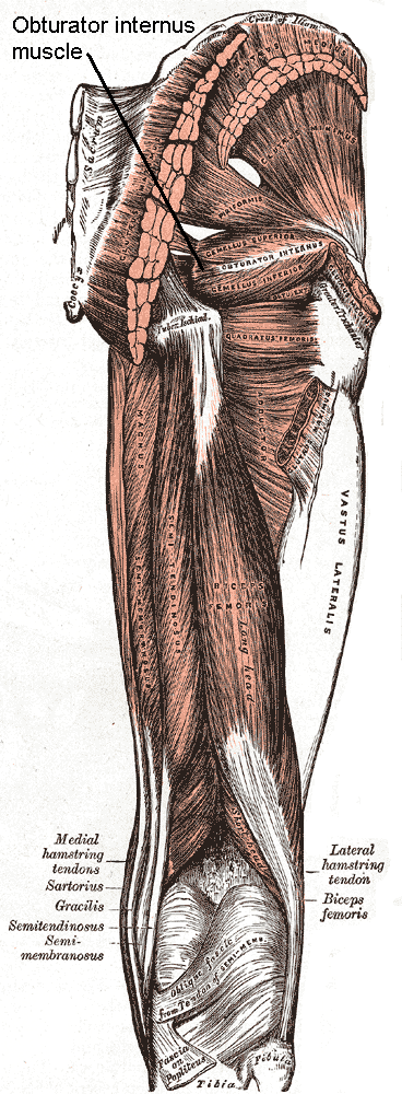
The internal obturator muscle or obturator internus muscle originates on the medial surface of the obturator membrane, the ischium near the membrane, and the rim of the pubis. It exits the pelvic cavity through the lesser sciatic foramen. The internal obturator is situated partly within the lesser pelvis, and partly at the back of the hip-joint. It functions to help laterally rotate femur with hip extension and abduct femur with hip flexion, as well as to steady the femoral head in the acetabulum. Structure Origin The internal obturator muscle arises from the inner surface of the antero-lateral wall of the pelvis. It surrounds the obturator foramen. It is attached to the inferior pubic ramus and ischium, and at the side to the inner surface of the hip bone below and behind the pelvic brim. It reaches from the upper part of the greater sciatic foramen above and behind to the obturator foramen below and in front. It also arises from the pelvic surface of the obturator membra ... [...More Info...] [...Related Items...] OR: [Wikipedia] [Google] [Baidu] |
Gemellus Superior
The gemelli muscles are the inferior gemellus muscle and the superior gemellus muscle, two small accessory fasciculi to the tendon of the internal obturator muscle. The gemelli muscles belong to the lateral rotator group of six muscles of the hip that rotate the femur in the hip joint. Superior gemellus muscle The gemelli muscles are two small muscular fasciculi, accessories to the tendon of the internal obturator muscle which is received into a groove between them. The superior gemellus muscle is the higher placed gemellus muscle that arises from the outer (gluteal) surface of the ischial spine, and blends with the upper part of the tendon of the internal obturator. It is smaller than the inferior gemellus. In some people, the fibres of the gemellus superior extend further than average, and are prolonged onto the medial surface of the greater trochanter of the femur. The superior and inferior gemelli are supplied by the inferior gluteal artery. Nerve supply to the superior geme ... [...More Info...] [...Related Items...] OR: [Wikipedia] [Google] [Baidu] |
Obturator Internus
The internal obturator muscle or obturator internus muscle originates on the medial surface of the obturator membrane, the ischium near the membrane, and the rim of the pubis. It exits the pelvic cavity through the lesser sciatic foramen. The internal obturator is situated partly within the lesser pelvis, and partly at the back of the hip-joint. It functions to help laterally rotate femur with hip extension and abduct femur with hip flexion, as well as to steady the femoral head in the acetabulum. Structure Origin The internal obturator muscle arises from the inner surface of the antero-lateral wall of the pelvis. It surrounds the obturator foramen. It is attached to the inferior pubic ramus and ischium, and at the side to the inner surface of the hip bone below and behind the pelvic brim. It reaches from the upper part of the greater sciatic foramen above and behind to the obturator foramen below and in front. It also arises from the pelvic surface of the obturator membra ... [...More Info...] [...Related Items...] OR: [Wikipedia] [Google] [Baidu] |
Sacral Plexus
In human anatomy, the sacral plexus is a nerve plexus which provides motor and sensory nerves for the posterior thigh, most of the lower leg and foot, and part of the pelvis. It is part of the lumbosacral plexus and emerges from the lumbar vertebrae and sacral vertebrae (L4-S4).''Thieme Atlas of Anatomy'' (2006), pp 470-471 A sacral plexopathy is a disorder affecting the nerves of the sacral plexus, usually caused by trauma, nerve compression, vascular disease, or infection. Symptoms may include pain, loss of motor control, and sensory deficits. Structure The sacral plexus is formed by: * the lumbosacral trunk * the anterior division of the first sacral nerve * portions of the anterior divisions of the second and third sacral nerves The nerves forming the sacral plexus converge toward the lower part of the greater sciatic foramen, and unite to form a flattened band, from the anterior and posterior surfaces of which several branches arise. The band itself is continued as the sc ... [...More Info...] [...Related Items...] OR: [Wikipedia] [Google] [Baidu] |
Nerve
A nerve is an enclosed, cable-like bundle of nerve fibers (called axons) in the peripheral nervous system. A nerve transmits electrical impulses. It is the basic unit of the peripheral nervous system. A nerve provides a common pathway for the electrochemical nerve impulses called action potentials that are transmitted along each of the axons to peripheral organs or, in the case of sensory nerves, from the periphery back to the central nervous system. Each axon, within the nerve, is an extension of an individual neuron, along with other supportive cells such as some Schwann cells that coat the axons in myelin. Within a nerve, each axon is surrounded by a layer of connective tissue called the endoneurium. The axons are bundled together into groups called fascicles, and each fascicle is wrapped in a layer of connective tissue called the perineurium. Finally, the entire nerve is wrapped in a layer of connective tissue called the epineurium. Nerve cells (often called neurons) are f ... [...More Info...] [...Related Items...] OR: [Wikipedia] [Google] [Baidu] |
Lumbosacral Plexus
The anterior divisions of the lumbar nerves, sacral nerves, and coccygeal nerve form the lumbosacral plexus, the first lumbar nerve being frequently joined by a branch from the twelfth thoracic. For descriptive purposes this plexus is usually divided into three parts: * lumbar plexus * sacral plexus * pudendal plexus Injuries to the lumbosacral plexus are predominantly witnessed as bone injuries. Lumbosacral trunk and sacral plexus In human anatomy, the sacral plexus is a nerve plexus which provides motor and sensory nerves for the posterior thigh, most of the lower leg and foot, and part of the pelvis. It is part of the lumbosacral plexus and emerges from the lumbar verte ... palsies are common injury patterns. References External links * - "Lumbosacral Plexus" Additional Images File:Slide2Anat.JPG, Lumbosacral plexus Deep dissection. File:Slide4Anat.JPG, Lumbosacral plexus Deep dissection. Nerve plexus Nerves of the lower limb and lower torso {{Neuroana ... [...More Info...] [...Related Items...] OR: [Wikipedia] [Google] [Baidu] |
Lumbar
In tetrapod anatomy, lumbar is an adjective that means ''of or pertaining to the abdominal segment of the torso, between the diaphragm and the sacrum.'' The lumbar region is sometimes referred to as the lower spine, or as an area of the back in its proximity. In human anatomy the five lumbar vertebrae (vertebrae in the lumbar region of the back) are the largest and strongest in the movable part of the spinal column, and can be distinguished by the absence of a foramen in the transverse process, and by the absence of facets on the sides of the body. In most mammals, the lumbar region of the spine curves outward. The actual spinal cord terminates between vertebrae one and two of this series, called L1 and L2. The nervous tissue that extends below this point are individual strands that collectively form the cauda equina. In between each lumbar vertebra a nerve root exits, and these nerve roots come together again to form the largest single nerve in the human body, the sciatic n ... [...More Info...] [...Related Items...] OR: [Wikipedia] [Google] [Baidu] |
Sacral Nerves
A spinal nerve is a mixed nerve, which carries motor, sensory, and autonomic signals between the spinal cord and the body. In the human body there are 31 pairs of spinal nerves, one on each side of the vertebral column. These are grouped into the corresponding cervical, thoracic, lumbar, sacral and coccygeal regions of the spine. There are eight pairs of cervical nerves, twelve pairs of thoracic nerves, five pairs of lumbar nerves, five pairs of sacral nerves, and one pair of coccygeal nerves. The spinal nerves are part of the peripheral nervous system. Structure Each spinal nerve is a mixed nerve, formed from the combination of nerve fibers from its dorsal and ventral roots. The dorsal root is the afferent sensory root and carries sensory information to the brain. The ventral root is the efferent motor root and carries motor information from the brain. The spinal nerve emerges from the spinal column through an opening (intervertebral foramen) between adjacent vertebrae. Th ... [...More Info...] [...Related Items...] OR: [Wikipedia] [Google] [Baidu] |
Piriformis
The piriformis muscle () is a flat, pyramidally-shaped muscle in the buttock, gluteal region of the lower limbs. It is one of the six muscles in the lateral rotator group. The piriformis muscle has its origin upon the front surface of the sacrum, and inserts onto the greater trochanter of the femur. Depending upon the given position of the leg, it acts either as external (lateral) rotator of the thigh or as abductor of the thigh. It is innervated by the piriformis nerve. Structure The piriformis is a flat muscle, and is pyramidal in shape. Origin The piriformis muscle originates from the Pelvic surface of sacrum, anterior (front) surface of the sacrum by three fleshy digitations attached to the Sacrum, second, third, and fourth sacral vertebra. It also arises from the superior margin of the greater sciatic notch, the gluteal surface of the Ilium (bone), ilium (near the posterior inferior iliac spine), the sacroiliac joint capsule, and (sometimes) the sacrotuberous ligament (m ... [...More Info...] [...Related Items...] OR: [Wikipedia] [Google] [Baidu] |
Ischial Spine
The ischial spine is part of the posterior border of the body of the ischium bone of the pelvis. It is a thin and pointed triangular eminence, more or less elongated in different subjects. Structure The pudendal nerve travels close to the ischial spine. Clinical significance The ischial spine can serve as a landmark in pudendal anesthesia, as the pudendal nerve The pudendal nerve is the main nerve of the perineum. It carries sensation from the external genitalia of both sexes and the skin around the anus and perineum, as well as the motor supply to various pelvic muscles, including the male or fem ... lies close to the ischial spine. Additional images File:Sciatic notches.png, Right hip bone, external surface, showing the greater and lesser sciatic notches, separated by the ischial spine. File:Gray319.png, Articulations of pelvis. Anterior view. File:Slide3ADA.JPG, PELVIS. ANTERIOR VIEW. References External links * - "The Female Perineum: Osteology" * - "Th ... [...More Info...] [...Related Items...] OR: [Wikipedia] [Google] [Baidu] |


