|
Musculocutaneous Nerve
The musculocutaneous nerve arises from the lateral cord of the brachial plexus, opposite the lower border of the pectoralis major, its fibers being derived from C5, C6 and C7. Structure The musculocutaneous nerve arises from the lateral cord of the brachial plexus, courses through the anterior part of the arm, and terminates at 2 cm above elbow as the lateral cutaneous nerve of the forearm. Musculocutaneous nerve arises from the lateral cord of the brachial plexus with root value of C5 to C7 of the spinal cord. It follows the course of the third part of the axillary artery (part of the axillary artery distal to the pectoralis minor) laterally and enters the frontal aspect of the arm where it penetrates the coracobrachialis muscle. It then passes downwards and laterally between the biceps brachii (above) and the brachialis muscles (below), to the lateral side of the arm; at 2 cm above the elbow it pierces the deep fascia lateral to the tendon of the biceps brachii ... [...More Info...] [...Related Items...] OR: [Wikipedia] [Google] [Baidu] |
Anterior Compartment Of The Arm
The fascial compartments of arm refers to the specific anatomical term of the compartments within the upper segment of the upper limb (the arm) of the body. The upper limb is divided into two segments, the arm and the forearm. Each of these segments is further divided into two compartments which are formed by deep fascia – tough connective tissue septa (walls). Each compartment encloses specific muscles and nerves. The compartments of the arm are the anterior compartment of the arm and the posterior compartment of the arm, divided by the lateral and the medial intermuscular septa. The compartments of the forearm are the anterior compartment of the forearm and posterior compartment of the forearm. Intermuscular septa The lateral intermuscular septum extends from the lower part of the crest of the greater tubercle of the humerus, along the lateral supracondylar ridge, to the lateral epicondyle; it is blended with the tendon of the deltoid muscle, gives attachment to the tricep ... [...More Info...] [...Related Items...] OR: [Wikipedia] [Google] [Baidu] |
Brachialis
The brachialis (brachialis anticus), also known as the Teichmann muscle, is a muscle in the upper arm that flexes the elbow. It lies deeper than the biceps brachii, and makes up part of the floor of the region known as the cubital fossa (elbow pit). The brachialis is the prime mover of elbow flexion generating about 50% more power than the biceps.Saladin, Kenneth S, Stephen J. Sullivan, and Christina A. Gan. Anatomy & Physiology: The Unity of Form and Function. 2015. Print. Structure The brachialis originates from the anterior surface of the distal half of the humerus, near the insertion of the deltoid muscle, which it embraces by two angular processes. Its origin extends below to within 2.5 cm of the margin of the articular surface of the humerus at the elbow joint. Its fibers converge to a thick tendon, which is inserted into the tuberosity of the ulna and the rough depression on the anterior surface of the coronoid process of the ulna. Blood supply The brachialis is supp ... [...More Info...] [...Related Items...] OR: [Wikipedia] [Google] [Baidu] |
Neurolysis
Neurolysis is the application of physical or chemical agents to a nerve in order to cause a temporary degeneration of targeted nerve fibers. When the nerve fibers degenerate, it causes an interruption in the transmission of nerve signals. In the medical field, this is most commonly and advantageously used to alleviate pain in cancer patients. The different types of neurolysis include celiac plexus neurolysis, endoscopic ultrasound guided neurolysis, and lumbar sympathetic neurolysis. Chemodenervation and nerve blocks are also associated with neurolysis. Additionally, there is external neurolysis. Peripheral nerves move (glide) across bones and muscles. A peripheral nerve can be trapped by scarring of surrounding tissue which may lead to potential nerve damage or pain. An external neurolysis is when scar tissue is removed from around the nerve without entering the nerve itself. Background Neurolysis is a chemical ablation technique that is used to alleviate pain. Neurolysis is only ... [...More Info...] [...Related Items...] OR: [Wikipedia] [Google] [Baidu] |
Electromyography
Electromyography (EMG) is a technique for evaluating and recording the electrical activity produced by skeletal muscles. EMG is performed using an instrument called an electromyograph to produce a record called an electromyogram. An electromyograph detects the electric potential generated by muscle cells when these cells are electrically or neurologically activated. The signals can be analyzed to detect abnormalities, activation level, or recruitment order, or to analyze the biomechanics of human or animal movement. Needle EMG is an electrodiagnostic medicine technique commonly used by neurologists. Surface EMG is a non-medical procedure used to assess muscle activation by several professionals, including physiotherapists, kinesiologists and biomedical engineers. In Computer Science, EMG is also used as middleware in gesture recognition towards allowing the input of physical action to a computer as a form of human-computer interaction. Clinical uses EMG testing has a variety of ... [...More Info...] [...Related Items...] OR: [Wikipedia] [Google] [Baidu] |
Abduction (anatomy)
Motion, the process of movement, is described using specific anatomical terms. Motion includes movement of organs, joints, limbs, and specific sections of the body. The terminology used describes this motion according to its direction relative to the anatomical position of the body parts involved. Anatomists and others use a unified set of terms to describe most of the movements, although other, more specialized terms are necessary for describing unique movements such as those of the hands, feet, and eyes. In general, motion is classified according to the anatomical plane it occurs in. ''Flexion'' and ''extension'' are examples of ''angular'' motions, in which two axes of a joint are brought closer together or moved further apart. ''Rotational'' motion may occur at other joints, for example the shoulder, and are described as ''internal'' or ''external''. Other terms, such as ''elevation'' and ''depression'', describe movement above or below the horizontal plane. Many anatomic ... [...More Info...] [...Related Items...] OR: [Wikipedia] [Google] [Baidu] |
Nerve Point Of Neck
The nerve point of the neck, also known as Erb's point is a site at the upper trunk of the brachial plexus located 2–3 cm above the clavicle. It is named for Wilhelm Heinrich Erb. Taken together, there are six types of nerves that meet at this point. "Erb's point" is also a term used in head and neck surgery to describe the point on the posterior border of the sternocleidomastoid muscle where the four superficial branches of the cervical plexus—the greater auricular, lesser occipital, transverse cervical, and supraclavicular nerves—emerge from behind the muscle. This point is located approximately at the junction of the upper and middle thirds of this muscle. From here, the accessory nerve courses through the posterior triangle of the neck to enter the anterior border of the trapezius muscle at a point located approximately at the junction of the middle and lower thirds of the anterior border of this muscle. The spinal accessory nerve can often be found 1 cm abov ... [...More Info...] [...Related Items...] OR: [Wikipedia] [Google] [Baidu] |
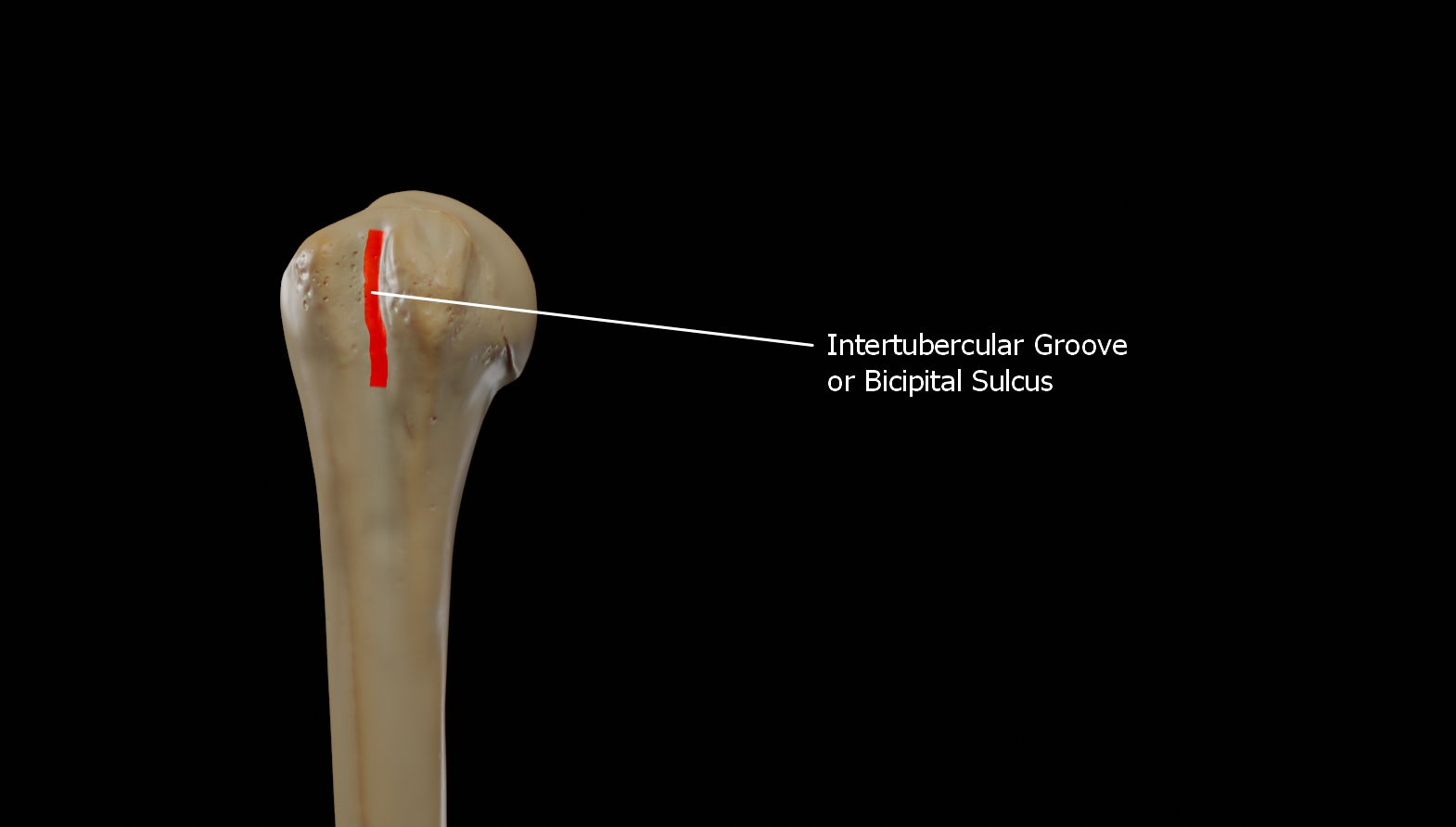
Bicipital Groove
The bicipital groove (intertubercular groove, sulcus intertubercularis) is a deep groove on the humerus that separates the greater tubercle from the lesser tubercle. It allows for the long tendon of the biceps brachii muscle to pass. Structure The bicipital groove separates the greater tubercle from the lesser tubercle. It is usually around 8 cm long and 1 cm wide in adults. It lodges the long tendon of the biceps brachii muscle between the tendon of the pectoralis major muscle on the lateral lip and the tendon of the teres major muscle on the medial lip. It also transmits a branch of the anterior humeral circumflex artery to the shoulder joint. The insertion of the latissimus dorsi muscle is found along the floor of the bicipital groove. The teres major muscle inserts on the medial lip of the groove. It runs obliquely downward, and ends near the junction of the upper with the middle third of the bone. It is the lateral wall of the axilla. Function The bicipital groove all ... [...More Info...] [...Related Items...] OR: [Wikipedia] [Google] [Baidu] |
Tinel's Sign
Tinel's sign (also Hoffmann-Tinel sign) is a way to detect irritated nerves. It is performed by lightly tapping ( percussing) over the nerve to elicit a sensation of tingling or " pins and needles" in the distribution of the nerve. Percussion is usually performed moving distal to proximal. It is named after Jules Tinel.Tinel, J. (1978) The "tingling sign" in peripheral nerve lesions (Translated by EB Kaplan). In: M. Spinner M (Ed.), Injuries to the Ma jor Branches of Peripheral Nerves of the Forearm. (2nd ed.) (pp 8–13). Philadelphia: WD Saunders CoTinel, J. (1915) Le signe du fourmillement dans les lésions des nerfs périphériques. Presse médicale, 47, 388–389Tinel, J., Nerve wounds. London: Baillère, Tindall and Cox, 1917 It is a potential sign of carpal tunnel syndrome, cubital tunnel syndrome Ulnar nerve entrapment is a condition where the ulnar nerve becomes physically trapped or pinched, resulting in pain, numbness, or weakness, primarily affecting the little fing ... [...More Info...] [...Related Items...] OR: [Wikipedia] [Google] [Baidu] |
Coracoid Process
The coracoid process (from Greek κόραξ, raven) is a small hook-like structure on the lateral edge of the superior anterior portion of the scapula (hence: coracoid, or "like a raven's beak"). Pointing laterally forward, it, together with the acromion, serves to stabilize the shoulder joint. It is palpable in the deltopectoral groove between the deltoid and pectoralis major muscles. Structure The coracoid process is a thick curved process attached by a broad base to the upper part of the neck of the scapula; it runs at first upward and medialward; then, becoming smaller, it changes its direction, and projects forward and lateralward. Anatomically it is divided into intervals of: base of coracoid process, angle of coracoid process, shaft and the apex of the coracoid process. The coracoglenoid notch is an indentation localized between the coracoid process and the glenoid. As the coracoid process projects laterally, it house underneath it the subcoracoid space. The ''ascend ... [...More Info...] [...Related Items...] OR: [Wikipedia] [Google] [Baidu] |
Pronator Teres
The pronator teres is a muscle (located mainly in the forearm) that, along with the pronator quadratus, serves to pronate the forearm (turning it so that the palm faces posteriorly when from the anatomical position). It has two attachments, to the medial humeral supracondylar ridge and the ulnar tuberosity, and inserts near the middle of the radius. Structure The pronator teres has two heads—humeral and ulnar. * The humeral head, the larger and more superficial, arises from the medial supracondylar ridge immediately superior to the medial epicondyle of the humerus, and from the common flexor tendon (which arises from the medial epicondyle). * The ulnar head (or ulnar tuberosity) is a thin fasciculus, which arises from the medial side of the coronoid process of the ulna, and joins the preceding at an acute angle. The median nerve enters the forearm between the two heads of the muscle, and is separated from the ulnar artery by the ulnar head. The muscle passes obliquely across ... [...More Info...] [...Related Items...] OR: [Wikipedia] [Google] [Baidu] |
Lateral Cutaneous Nerve Of The Forearm
The lateral antebrachial cutaneous nerve (or lateral cutaneous nerve of forearm) (branch of musculocutaneous nerve, also sometimes spelled "antebrachial") passes behind the cephalic vein, and divides, opposite the elbow-joint, into a volar and a dorsal branch. Volar branch The volar branch (ramus volaris; anterior branch) descends along the radial border of the forearm to the wrist, and supplies the skin over the lateral half of its volar surface. At the wrist-joint it is placed in front of the radial artery, and some filaments, piercing the deep fascia, accompany that vessel to the dorsal surface of the carpus. The nerve then passes downward to the ball of the thumb, where it ends in cutaneous filaments. It communicates with the superficial branch of the radial nerve, and with the palmar cutaneous branch of the median nerve. Dorsal branch The dorsal branch (ramus dorsalis; posterior branch) descends, along the dorsal surface of the radial side of the forearm to the wrist. ... [...More Info...] [...Related Items...] OR: [Wikipedia] [Google] [Baidu] |
Antebrachial Fascia
The antebrachial fascia (antibrachial fascia or deep fascia of forearm) continuous above with the brachial fascia, is a dense, membranous investment, which forms a general sheath for the muscles in this region; it is attached, behind, to the olecranon and dorsal border of the ulna, and gives off from its deep surface numerous intermuscular septa, which enclose each muscle separately. Over the flexor muscles tendons as they approach the wrist it is especially thickened, and forms the volar carpal ligament. This is continuous with the transverse carpal ligament, and forms a sheath for the tendon of the palmaris longus which passes over the transverse carpal ligament to be inserted into the palmar aponeurosis. Behind, near the wrist-joint, it is thickened by the addition of many transverse fibers, and forms the dorsal carpal ligament. It is much thicker on the dorsal than on the volar surface, and at the lower than at the upper part of the forearm, and is strengthened above by ... [...More Info...] [...Related Items...] OR: [Wikipedia] [Google] [Baidu] |


