|
Antebrachial Fascia
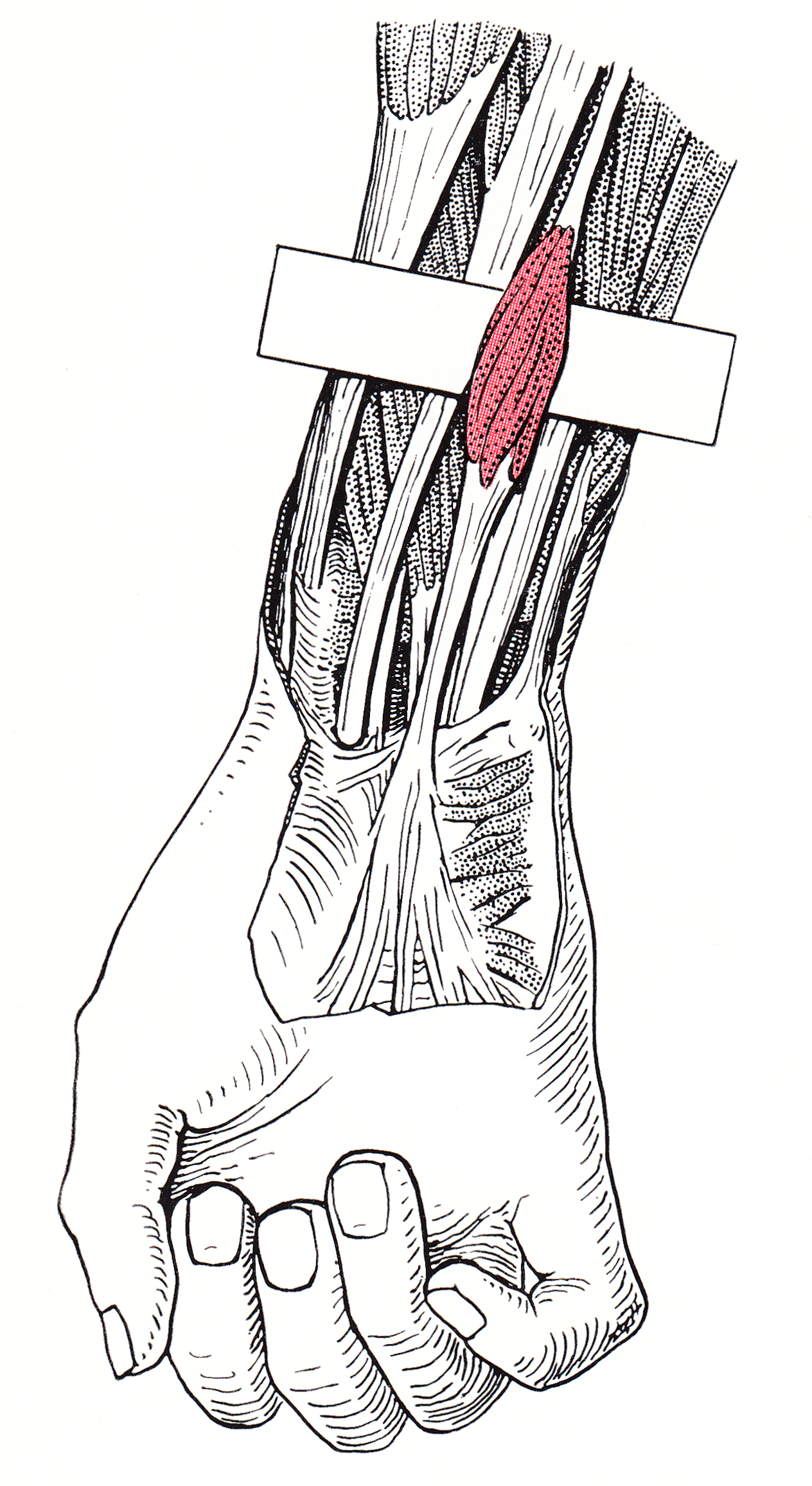
The antebrachial fascia (antibrachial fascia or deep fascia of forearm) continuous above with the brachial fascia, is a dense, membranous investment, which forms a general sheath for the muscles in this region; it is attached, behind, to the olecranon and dorsal border of the ulna, and gives off from its deep surface numerous intermuscular septa, which enclose each muscle separately. Over the flexor muscles tendons as they approach the wrist it is especially thickened, and forms the volar carpal ligament. This is continuous with the transverse carpal ligament, and forms a sheath for the tendon of the palmaris longus which passes over the transverse carpal ligament to be inserted into the palmar aponeurosis. Behind, near the wrist-joint, it is thickened by the addition of many transverse fibers, and forms the dorsal carpal ligament. It is much thicker on the dorsal than on the volar surface, and at the lower than at the upper part of the forearm, and is strengthened above by ... [...More Info...] [...Related Items...] OR: [Wikipedia] [Google] [Baidu] |
Brachial Fascia
The brachial fascia (deep fascia of the arm) is continuous with that covering the deltoideus and the pectoralis major muscle, by means of which it is attached, above, to the clavicle, acromion, and spine of the scapula; it forms a thin, loose, membranous sheath for the muscles of the arm, and sends septa between them; it is composed of fibers disposed in a circular or spiral direction, and connected together by vertical and oblique fibers. It differs in thickness at different parts, being thin over the biceps brachii, but thicker where it covers the triceps brachii, and over the epicondyles of the humerus: it is strengthened by fibrous aponeuroses, derived from the pectoralis major and latissimus dorsi medially, and from the deltoideus laterally. On either side it gives off a strong intermuscular septum, which is attached to the corresponding supracondylar ridge and epicondyle An epicondyle () is a rounded eminence on a bone that lies upon a condyle ('' epi-'', "upon" + ''con ... [...More Info...] [...Related Items...] OR: [Wikipedia] [Google] [Baidu] |
Olecranon
The olecranon (, ), is a large, thick, curved bony eminence of the ulna, a long bone in the forearm that projects behind the elbow. It forms the most pointed portion of the elbow and is opposite to the cubital fossa or elbow pit. The olecranon serves as a lever for the extensor muscles that straighten the elbow joint. Structure The olecranon is situated at the proximal end of the ulna, one of the two bones in the forearm. When the hand faces forward (supination) the olecranon faces towards the back (posteriorly). It is bent forward at the summit so as to present a prominent lip which is received into the olecranon fossa of the humerus during extension of the forearm. Its base is contracted where it joins the body and the narrowest part of the upper end of the ulna. Its posterior surface, directed backward, is triangular, smooth, subcutaneous, and covered by a bursa. Its superior surface is of quadrilateral form, marked behind by a rough impression for the insertion of the Tr ... [...More Info...] [...Related Items...] OR: [Wikipedia] [Google] [Baidu] |
Ulna
The ulna (''pl''. ulnae or ulnas) is a long bone found in the forearm that stretches from the elbow to the smallest finger, and when in anatomical position, is found on the medial side of the forearm. That is, the ulna is on the same side of the forearm as the little finger. It runs parallel to the radius, the other long bone in the forearm. The ulna is usually slightly longer than the radius, but the radius is thicker. Therefore, the radius is considered to be the larger of the two. Structure The ulna is a long bone found in the forearm that stretches from the elbow to the smallest finger, and when in anatomical position, is found on the medial side of the forearm. It is broader close to the elbow, and narrows as it approaches the wrist. Close to the elbow, the ulna has a bony process, the olecranon process, a hook-like structure that fits into the olecranon fossa of the humerus. This prevents hyperextension and forms a hinge joint with the trochlea of the humerus. There is ... [...More Info...] [...Related Items...] OR: [Wikipedia] [Google] [Baidu] |
Lateral Intermuscular Septum Of Arm
The fascial compartments of arm refers to the specific anatomical term of the compartments within the upper segment of the upper limb (the arm) of the body. The upper limb is divided into two segments, the arm and the forearm. Each of these segments is further divided into two compartments which are formed by deep fascia – tough connective tissue septa (walls). Each compartment encloses specific muscles and nerves. The compartments of the arm are the anterior compartment of the arm and the posterior compartment of the arm, divided by the lateral and the medial intermuscular septa. The compartments of the forearm are the anterior compartment of the forearm and posterior compartment of the forearm. Intermuscular septa The lateral intermuscular septum extends from the lower part of the crest of the greater tubercle of the humerus, along the lateral supracondylar ridge, to the lateral epicondyle; it is blended with the tendon of the deltoid muscle, gives attachment to the tricep ... [...More Info...] [...Related Items...] OR: [Wikipedia] [Google] [Baidu] |
Anterior Compartment Of The Forearm
The anterior compartment of the forearm (or flexor compartment) contains the following muscles: The muscles are largely involved with extension and supination. The superficial muscles have their origin on the common flexor tendon. The ulnar nerve and artery are also contained within this compartment. The flexor digitorum superficialis lies in between the other four muscles of the superficial group and the three muscles of the deep group. This is why it is also classified as the intermediate group. See also * Compartment syndrome * Posterior compartment of the forearm References External links * Topographical Anatomy of the Upper Limb - Listed Alphabetically University of Arkansas Additional images Image:Gray421.png, Transverse section across distal ends of radius and ulna. Image:Gray422.png, Transverse section across the wrist In human anatomy, the wrist is variously defined as (1) the carpus or carpal bones, the complex of eight bones forming the proximal skelet ... [...More Info...] [...Related Items...] OR: [Wikipedia] [Google] [Baidu] |
Tendon
A tendon or sinew is a tough, high-tensile-strength band of dense fibrous connective tissue that connects muscle to bone. It is able to transmit the mechanical forces of muscle contraction to the skeletal system without sacrificing its ability to withstand significant amounts of tension. Tendons are similar to ligaments; both are made of collagen. Ligaments connect one bone to another, while tendons connect muscle to bone. Structure Histologically, tendons consist of dense regular connective tissue. The main cellular component of tendons are specialized fibroblasts called tendon cells (tenocytes). Tenocytes synthesize the extracellular matrix of tendons, abundant in densely packed collagen fibers. The collagen fibers are parallel to each other and organized into tendon fascicles. Individual fascicles are bound by the endotendineum, which is a delicate loose connective tissue containing thin collagen fibrils and elastic fibres. Groups of fascicles are bounded by the epitenon, ... [...More Info...] [...Related Items...] OR: [Wikipedia] [Google] [Baidu] |
Volar Carpal Ligament
The palmar carpal ligament (also volar carpal ligament or ''Guyon's Tunnel'') is the thickened portion of antebrachial fascia on the anterior of the wrist. It is officially unnamed.Moore, Keith L., Arthur F. Dalley II: ''Clinically Oriented Anatomy'', 4th ed. Lippincott, Williams & Wilkins,1999. The palmar carpal ligament is a different structure than the flexor retinaculum of the hand, but the two are frequently confused. The palmar carpal ligament lies superficial and proximal to the flexor retinaculum. The ulnar nerve and the ulnar artery run through the ulnar canal, which is deep to the palmar carpal ligament and superficial to the flexor retinaculum. The palmar carpal ligament is continuous with the extensor retinaculum of the hand, which is located on the posterior side of the wrist. References See also * Flexor retinaculum of the hand * Extensor retinaculum of the hand * Antebrachial fascia The antebrachial fascia (antibrachial fascia or deep fascia of forearm) conti ... [...More Info...] [...Related Items...] OR: [Wikipedia] [Google] [Baidu] |
Transverse Carpal Ligament
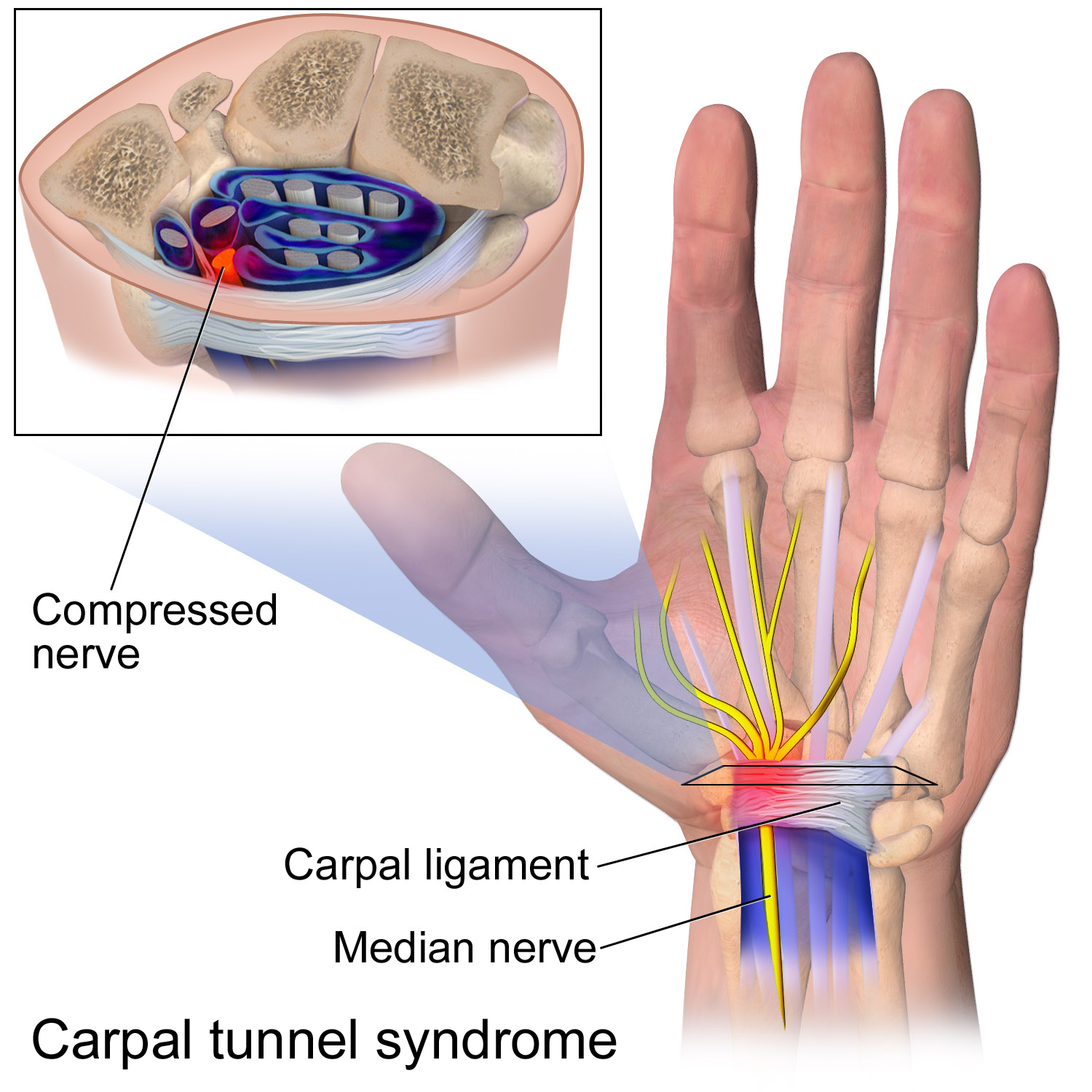
The flexor retinaculum (transverse carpal ligament, or anterior annular ligament) is a fibrous band on the palmar side of the hand near the wrist. It arches over the carpal bones of the hands, covering them and forming the carpal tunnel. Structure The flexor retinaculum is a strong, fibrous band that covers the carpal bones on the palmar side of the hand near the wrist. It attaches to the bones near the radius (bone), radius and ulna. On the ulnar side, the flexor retinaculum attaches to the pisiform bone and the hamate, hook of the hamate bone. On the radial side, it attaches to the tubercle of the scaphoid bone, and to the medial part of the palmar surface and the ridge of the trapezium (bone), trapezium bone. The flexor retinaculum is continuous with the palmar carpal ligament, and deeper with the palmar aponeurosis. The ulnar artery and ulnar nerve, and the cutaneous branches of the median and ulnar nerves, pass on top of the flexor retinaculum. On the radial side of the retin ... [...More Info...] [...Related Items...] OR: [Wikipedia] [Google] [Baidu] |
Palmaris Longus
The palmaris longus is a muscle visible as a small tendon located between the flexor carpi radialis and the flexor carpi ulnaris, although it is not always present. It is absent in about 14 percent of the population; this number can vary in African, Asian, and Native American populations, however. Absence of the palmaris longus does not have an effect on grip strength. The lack of palmaris longus muscle does result in decreased pinch strength in fourth and fifth fingers. The absence of palmaris longus muscle is more prevalent in females than males. The palmaris longus muscle can be seen by touching the pads of the fourth finger and thumb and flexing the wrist. The tendon, if present, will be visible in the midline of the anterior wrist. Structure Palmaris longus is a slender, elongated, spindle shaped muscle, lying on the medial side of the flexor carpi radialis. It is widest in the middle, and narrowest at the proximal and distal attachments.'' Gray's Anatomy'' (1918), see infob ... [...More Info...] [...Related Items...] OR: [Wikipedia] [Google] [Baidu] |
Palmar Aponeurosis
The palmar aponeurosis (palmar fascia) invests the muscles of the palm, and consists of central, lateral, and medial portions. Structure The central portion occupies the middle of the palm, is triangular in shape, and of great strength Its apex is continuous with the lower margin of the transverse carpal ligament, and receives the expanded tendon of the palmaris longus. Its base divides below into four slips, one for each finger. Each slip gives off superficial fibers to the skin of the palm and finger, those to the palm joining the skin at the furrow corresponding to the metacarpophalangeal articulations, and those to the fingers passing into the skin at the transverse fold at the bases of the fingers. The deeper part of each slip subdivides into two processes, which are inserted into the fibrous sheaths of the flexor tendons. From the sides of these processes offsets are attached to the transverse metacarpal ligament. By this arrangement short channels are formed on the front ... [...More Info...] [...Related Items...] OR: [Wikipedia] [Google] [Baidu] |
Dorsal Carpal Ligament
The extensor retinaculum (dorsal carpal ligament, or posterior annular ligament) is an anatomical term for the thickened part of the antebrachial fascia that holds the tendons of the extensor muscles in place. It is located on the back of the forearm, just proximal to the hand. It is continuous with the palmar carpal ligament, which is located on the anterior side of the forearm. Structure The extensor retinaculum is a strong, fibrous band, extending obliquely downward and medialward across the back of the wrist. It consists of part of the deep fascia of the back of the forearm, strengthened by the addition of some transverse fibers. The extensor retinaculum is attached laterally to the lateral margin of the radius. However, it is not attached to the ulna, as the distance between these two bones varies with supination and pronation of the forearm. Instead the medial attachment is to the pisiform bone and triquetral bone. Other authors may state the medial attachment of extensor ... [...More Info...] [...Related Items...] OR: [Wikipedia] [Google] [Baidu] |
Biceps Brachii
The biceps or biceps brachii ( la, musculus biceps brachii, "two-headed muscle of the arm") is a large muscle that lies on the front of the upper arm between the shoulder and the elbow. Both heads of the muscle arise on the scapula and join to form a single muscle belly which is attached to the upper forearm. While the biceps crosses both the shoulder and elbow joints, its main function is at the elbow where it flexes the forearm and supinates the forearm. Both these movements are used when opening a bottle with a corkscrew: first biceps screws in the cork (supination), then it pulls the cork out (flexion). Structure The biceps is one of three muscles in the anterior compartment of the upper arm, along with the brachialis muscle and the coracobrachialis muscle, with which the biceps shares a nerve supply. The biceps muscle has two heads, the short head and the long head, distinguished according to their origin at the coracoid process and supraglenoid tubercle of the sca ... [...More Info...] [...Related Items...] OR: [Wikipedia] [Google] [Baidu] |


