|
Middle Mediastinum
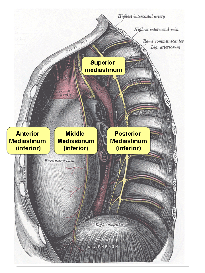
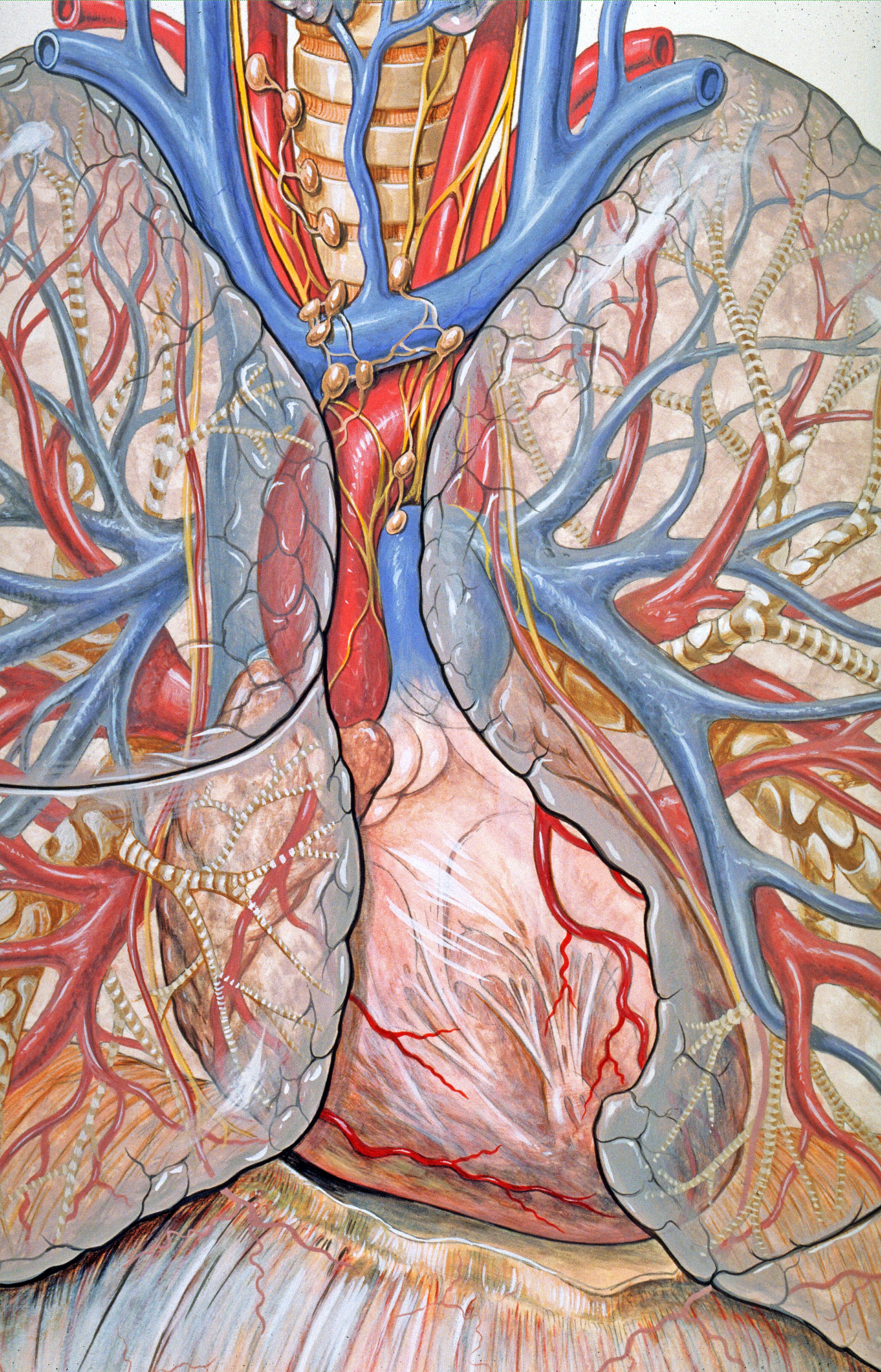
The mediastinum (from ) is the central compartment of the thoracic cavity. Surrounded by loose connective tissue, it is an undelineated region that contains a group of structures within the thorax, namely the heart and its vessels, the esophagus, the trachea, the phrenic and cardiac nerves, the thoracic duct, the thymus and the lymph nodes of the central chest. Anatomy The mediastinum lies within the thorax and is enclosed on the right and left by pleurae. It is surrounded by the chest wall in front, the lungs to the sides and the spine at the back. It extends from the sternum in front to the vertebral column behind. It contains all the organs of the thorax except the lungs. It is continuous with the loose connective tissue of the neck. The mediastinum can be divided into an upper (or superior) and lower (or inferior) part: * The superior mediastinum starts at the superior thoracic aperture and ends at the thoracic plane. * The inferior mediastinum from this level to the ... [...More Info...] [...Related Items...] OR: [Wikipedia] [Google] [Baidu] |
3D Rendering
3D rendering is the 3D computer graphics process of converting 3D modeling, 3D models into 2D computer graphics, 2D images on a computer. 3D renders may include photorealistic rendering, photorealistic effects or non-photorealistic rendering, non-photorealistic styles. Rendering methods Rendering (computer graphics), Rendering is the final process of creating the actual 2D image or animation from the prepared scene. This can be compared to taking a photo or filming the scene after the setup is finished in real life. Several different, and often specialized, rendering methods have been developed. These range from the distinctly non-realistic Wire frame model, wireframe rendering through polygon-based rendering, to more advanced techniques such as: scanline rendering, Ray tracing (graphics), ray tracing, or Radiosity (computer graphics), radiosity. Rendering may take from fractions of a second to days for a single image/frame. In general, different methods are better suited for e ... [...More Info...] [...Related Items...] OR: [Wikipedia] [Google] [Baidu] |
Lung
The lungs are the primary organs of the respiratory system in humans and most other animals, including some snails and a small number of fish. In mammals and most other vertebrates, two lungs are located near the backbone on either side of the heart. Their function in the respiratory system is to extract oxygen from the air and transfer it into the bloodstream, and to release carbon dioxide from the bloodstream into the atmosphere, in a process of gas exchange. Respiration is driven by different muscular systems in different species. Mammals, reptiles and birds use their different muscles to support and foster breathing. In earlier tetrapods, air was driven into the lungs by the pharyngeal muscles via buccal pumping, a mechanism still seen in amphibians. In humans, the main muscle of respiration that drives breathing is the diaphragm. The lungs also provide airflow that makes vocal sounds including human speech possible. Humans have two lungs, one on the left and on ... [...More Info...] [...Related Items...] OR: [Wikipedia] [Google] [Baidu] |
Anatomical Plane
An anatomical plane is a hypothetical plane used to transect the body, in order to describe the location of structures or the direction of movements. In human and animal anatomy, three principal planes are used: * The sagittal plane or lateral plane (''longitudinal, anteroposterior'') is a plane parallel to the sagittal suture. It divides the body into left and right. * The coronal plane or frontal plane (''vertical'') divides the body into dorsal and ventral (back and front, or posterior and anterior) portions. * The transverse plane or axial plane (''horizontal'') divides the body into cranial and caudal (head and tail) portions. Terminology There could be any number of sagittal planes; however, there is only one cardinal sagittal plane. The term ''cardinal'' refers to the one plane that divides the body into equal segments, with exactly one half of the body on either side of the cardinal plane. The term ''cardinal plane'' appears in some texts as the ''principal plane''. The ... [...More Info...] [...Related Items...] OR: [Wikipedia] [Google] [Baidu] |
Radiologists
Radiology ( ) is the medical discipline that uses medical imaging to diagnose diseases and guide their treatment, within the bodies of humans and other animals. It began with radiography (which is why its name has a root referring to radiation), but today it includes all imaging modalities, including those that use no electromagnetic radiation (such as ultrasonography and magnetic resonance imaging), as well as others that do, such as computed tomography (CT), fluoroscopy, and nuclear medicine including positron emission tomography (PET). Interventional radiology is the performance of usually minimally invasive medical procedures with the guidance of imaging technologies such as those mentioned above. The modern practice of radiology involves several different healthcare professions working as a team. The radiologist is a medical doctor who has completed the appropriate post-graduate training and interprets medical images, communicates these findings to other physicians by me ... [...More Info...] [...Related Items...] OR: [Wikipedia] [Google] [Baidu] |
Surgeon
In modern medicine, a surgeon is a medical professional who performs surgery. Although there are different traditions in different times and places, a modern surgeon usually is also a licensed physician or received the same medical training as physicians before specializing in surgery. There are also surgeons in podiatry, dentistry, and veterinary medicine. It is estimated that surgeons perform over 300 million surgical procedures globally each year. History The first person to document a surgery was the 6th century BC Indian physician-surgeon, Sushruta. He specialized in cosmetic plastic surgery and even documented an open rhinoplasty procedure.Ira D. Papel, John Frodel, ''Facial Plastic and Reconstructive Surgery'' His magnum opus ''Suśruta-saṃhitā'' is one of the most important surviving ancient treatises on medicine and is considered a foundational text of both Ayurveda and surgery. The treatise addresses all aspects of general medicine, but the translator G. D. Si ... [...More Info...] [...Related Items...] OR: [Wikipedia] [Google] [Baidu] |
Anatomist
Anatomy () is the branch of biology concerned with the study of the structure of organisms and their parts. Anatomy is a branch of natural science that deals with the structural organization of living things. It is an old science, having its beginnings in prehistoric times. Anatomy is inherently tied to developmental biology, embryology, comparative anatomy, evolutionary biology, and phylogeny, as these are the processes by which anatomy is generated, both over immediate and long-term timescales. Anatomy and physiology, which study the structure and function of organisms and their parts respectively, make a natural pair of related disciplines, and are often studied together. Human anatomy is one of the essential basic sciences that are applied in medicine. The discipline of anatomy is divided into macroscopic and microscopic. Macroscopic anatomy, or gross anatomy, is the examination of an animal's body parts using unaided eyesight. Gross anatomy also includes the branch of ... [...More Info...] [...Related Items...] OR: [Wikipedia] [Google] [Baidu] |
Pericardium
The pericardium, also called pericardial sac, is a double-walled sac containing the heart and the roots of the great vessels. It has two layers, an outer layer made of strong connective tissue (fibrous pericardium), and an inner layer made of serous membrane (serous pericardium). It encloses the pericardial cavity, which contains pericardial fluid, and defines the middle mediastinum. It separates the heart from interference of other structures, protects it against infection and blunt trauma, and lubricates the heart's movements. The English name originates from the Ancient Greek prefix "''peri-''" (περί; "around") and the suffix "''-cardion''" (κάρδιον; "heart"). Anatomy The pericardium is a tough fibroelastic sac which covers the heart from all sides except at the cardiac root (where the great vessels join the heart) and the bottom (where only the serous pericardium exists to cover the upper surface of the central tendon of diaphragm). The fibrous pericardiu ... [...More Info...] [...Related Items...] OR: [Wikipedia] [Google] [Baidu] |
Thoracic Diaphragm
The thoracic diaphragm, or simply the diaphragm ( grc, διάφραγμα, diáphragma, partition), is a sheet of internal Skeletal striated muscle, skeletal muscle in humans and other mammals that extends across the bottom of the thoracic cavity. The diaphragm is the most important Muscles of respiration, muscle of respiration, and separates the thoracic cavity, containing the heart and lungs, from the abdominal cavity: as the diaphragm contracts, the volume of the thoracic cavity increases, creating a negative pressure there, which draws air into the lungs. Its high oxygen consumption is noted by the many mitochondria and capillaries present; more than in any other skeletal muscle. The term ''diaphragm'' in anatomy, created by Gerard of Cremona, can refer to other flat structures such as the urogenital diaphragm or Pelvic floor, pelvic diaphragm, but "the diaphragm" generally refers to the thoracic diaphragm. In humans, the diaphragm is slightly asymmetric—its right half is h ... [...More Info...] [...Related Items...] OR: [Wikipedia] [Google] [Baidu] |
Thoracic Plane
The mediastinum (from ) is the central compartment of the thoracic cavity. Surrounded by loose connective tissue, it is an undelineated region that contains a group of structures within the thorax, namely the heart and its vessels, the esophagus, the trachea, the phrenic and cardiac nerves, the thoracic duct, the thymus and the lymph nodes of the central chest. Anatomy The mediastinum lies within the thorax and is enclosed on the right and left by pleurae. It is surrounded by the chest wall in front, the lungs to the sides and the spine at the back. It extends from the sternum in front to the vertebral column behind. It contains all the organs of the thorax except the lungs. It is continuous with the loose connective tissue of the neck. The mediastinum can be divided into an upper (or superior) and lower (or inferior) part: * The superior mediastinum starts at the superior thoracic aperture and ends at the thoracic plane. * The inferior mediastinum from this level to the ... [...More Info...] [...Related Items...] OR: [Wikipedia] [Google] [Baidu] |
Superior Thoracic Aperture
The superior thoracic aperture, also known as the thoracic outlet, or thoracic inlet refers to the opening at the top of the thoracic cavity. It is also clinically referred to as the thoracic outlet, in the case of thoracic outlet syndrome. A lower thoracic opening is the ''inferior thoracic aperture''. Structure The superior thoracic aperture is essentially a hole surrounded by a bony ring, through which several vital structures pass. It is bounded by: the first thoracic vertebra (T1) ''posteriorly''; the first pair of ribs ''laterally'', forming lateral C-shaped curves posterior to anterior; and the costal cartilage of the first rib and the superior border of the manubrium ''anteriorly''. Dimensions The adult thoracic outlet is around 6.5 cm antero-posteriorly and 11 cm transversely. Because of the obliquity of the first pair of ribs, the aperture slopes antero-inferiorly. Relations The clavicle articulates with the manubrium to form the anterior border of the thoracic out ... [...More Info...] [...Related Items...] OR: [Wikipedia] [Google] [Baidu] |
Mediastinum
The mediastinum (from ) is the central compartment of the thoracic cavity. Surrounded by loose connective tissue, it is an undelineated region that contains a group of structures within the thorax, namely the heart and its vessels, the esophagus, the trachea, the phrenic nerve, phrenic and cardiac nerves, the thoracic duct, the thymus and the lymph nodes of the central chest. Anatomy The mediastinum lies within the thorax and is enclosed on the right and left by pulmonary pleurae, pleurae. It is surrounded by the chest wall in front, the lungs to the sides and the Spine (anatomy), spine at the back. It extends from the sternum in front to the vertebral column behind. It contains all the organs of the thorax except the lungs. It is continuous with the loose connective tissue of the neck. The mediastinum can be divided into an upper (or superior) and lower (or inferior) part: * The superior mediastinum starts at the superior thoracic aperture and ends at the #Thoracic plane, t ... [...More Info...] [...Related Items...] OR: [Wikipedia] [Google] [Baidu] |
Neck
The neck is the part of the body on many vertebrates that connects the head with the torso. The neck supports the weight of the head and protects the nerves that carry sensory and motor information from the brain down to the rest of the body. In addition, the neck is highly flexible and allows the head to turn and flex in all directions. The structures of the human neck are anatomically grouped into four compartments; vertebral, visceral and two vascular compartments. Within these compartments, the neck houses the cervical vertebrae and cervical part of the spinal cord, upper parts of the respiratory and digestive tracts, endocrine glands, nerves, arteries and veins. Muscles of the neck are described separately from the compartments. They bound the neck triangles. In anatomy, the neck is also called by its Latin names, or , although when used alone, in context, the word ''cervix'' more often refers to the uterine cervix, the neck of the uterus. Thus the adjective ''cervical'' ma ... [...More Info...] [...Related Items...] OR: [Wikipedia] [Google] [Baidu] |


